Catalyst Questions What are the five layers of


 Lines • Cleavage (tension) lines: elastic and collagen fibers oriented in some")


- Slides: 21
Catalyst Questions • • What are the five layers of the epidermis? List the functions of the skin. How does the skin respond to aging? What is the rule of nines?
Lets Review… • Resistance to trauma and infection, water retention, vitamin D synthesis, sensation, thermoregulation, and nonverbal communication. • Stratum basale, stratum spinsum, stran granulosum, stranum lucidum, and stratum corneum.
Epidermal Layers and Keratinization 5 -3
Effects of Aging on the Integumentary System • Skin more easily damaged because epidermis thins and amount of collagen decreases • Skin infections more likely • Wrinkling occurs due to decrease in elastic fibers • Skin becomes drier • Decrease in blood supply causes poor ability to regulate body temperature • Functioning melanocytes decrease or increase; age spots • Sunlight ages skin more rapidly 5 -4
The Rule of Nines • Used to estimate amount of body that is burned. • Note differing proportions in adult and child. 5 -5
Class Updates… • Review day on Wednesday August 24 th, plus in class project! • Test corrections on due on Monday, September 22 nd. • Test will be on Friday, 26 th !
Review Sheets • A completed review sheet will be worth +10 on the exam.
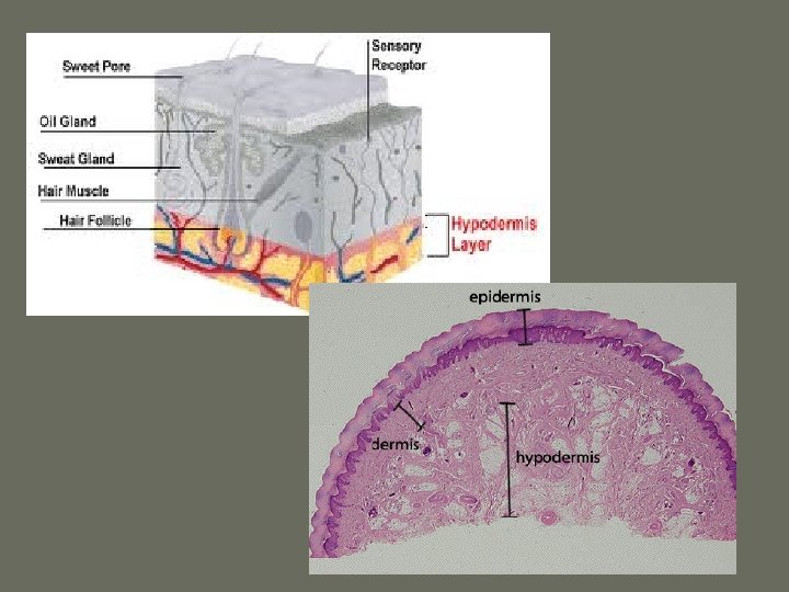
Dermis, Hypodermis, and Structures of the Integumentary System
Objectives • You will be able to describe the histological structure of the epidermis, and subcutaneous tissue • You will be able to describe the function of each of the individual parts in the epidermis, hypodermis
Dermis • Gives structural strength. C. T. with many fibers, fibroblasts, macrophages. Some adipocytes and blood vessels. • Contains nerves, blood vessels, hair follicles, smooth muscles, glands, and lymphatic vessels. • Sensory functions: pain, itch, tickle, temperature, touch, pressure, two-point discrimination. 5 -10
Two Layers of the Dermis • Two layers variable in thickness – Papillary. Superficial (outer) 1/5. Areolar with lots of elastic fibers. Dermal papillae, capillary beds. Fingerprints. Whorls of ridges. Touch receptors (Meissner’s), free nerve endings sensing pain – Reticular: Deep (inner) 4/5. Dense irregular C. T. Collagen and elastic fibers. In the figure see: some adipose, hair follicles, nerves, oil glands, ducts of sweat glands, heat sensors. 5 -11
Dermis • The dermis is directly below the epidermis and is a connective tissue layer. • It is composed mainly of collagen but also contains other tissues like elastic and reticular fibers, fibroblasts, and other fibrous connective tissue cells. • It is well supplied with blood vessels, sweat glands, sebaceous glands, and nerve endings. • The hair follicles and nail roots are embedded in the dermis.
Dermis, cont’d • Smooth muscles associated with hair follicles contract in response to such stimuli as cold, fear, and touch. • In the face, skeletal muscles attach to dermal collagen fibers and produce such expression as a smile, a wrinkle of the forehead, and the lifting of an eyebrow.
Dermis, cont’d • The boundary between the epidermis and dermis is very obvious and wavy. The upward waves are like finger-like extensions of the dermis called dermal papillae. – NOTE: DO NOT CONFUSE THIS WITH THE DERMAL PAPILLA OF THE HAIR!! • The downward waves are like extensions of the epidermis called epidermal ridges.
Dermis, cont’d • There are two zones of the dermis called the papillary and reticular layers. • The papillary layer is a thin zone of areolar tissue in and near the dermal papillae. – The loosely organized tissue of the papillary layer allows for mobility of leukocytes (defense cells) and other defenses against organisms introduced through breaks in the epidermis. • The reticular layer of the dermis is deeper and much thicker. – It consists of dense, irregular connective tissue.
Hypodermis • Beneath the skin is a layer called the hypodermis, aka the subcutaneous tissue or superficial fascia. • The boundary between the dermis and hypodermis is indistinct, but the hypodermis generally has more areolar and adipose tissue (fat). • The hypodermis binds the skin to the underlying tissues and pads the body. • Subcutaneous fat is hypodermis composed predominantly of adipose tissue. – This fat serves as an energy reservoir and thermal insulation.
Cleavage (Tension) Lines • Cleavage (tension) lines: elastic and collagen fibers oriented in some directions more than in others • Important in surgery – If incision parallel to lines, there is less gapping, faster healing, less scar tissue • If skin is overstretched, striae (stretch marks) occur 5 -18
Structures • • -dermal papillae -hair follicle -sebaceous gland -hair receptor -apocrine sweat gland -hair bulb -sensory nerve fibers • -piloerector muscle • -lamellated corpusclethese are receptors for deep pressure • -Tactile corpusclesreceptors of light touch and texture • -merocrine sweat gland • -blood capillaries • -hypodermis • -epidermis
Structures of Integumentary System
Effects of Aging on Tissues Continued. . Cells divide more slowly • • Collagen fibers become more irregular in structure, though they may increase in number – Tendons and ligaments become less flexible and more fragile • Elastic fibers fragment, bind to calcium ions, and become less elastic – Arterial walls and elastic ligaments become less elastic • Changes in collagen and elastin result in – Atherosclerosis and reduced blood supply to tissues – Wrinkling of the skin – Increased tendency for bones to break • Rate of blood cell synthesis declines in the elderly 4 -21 • Injuries don’t heal as readily