Integumentary System Hypodermis Integumentary System Skin Hair Nails

")
")

 · Main layers – superficial to deep ·")


 ·")


 · Exposure to allergen/irritant (ie. poison ivy) cause allergic reaction")
 Reddish discoloration, scaling, crusting Treat with")


- Slides: 70
Integumentary System
(Hypodermis)
Integumentary System · Skin · Hair · Nails · Associated Structures (vessels, nerves, glands)
Membranes n Epithelial membranes – Cutaneous – Mucous – Serous § Parietal vs visceral § Pleura, pericardium, peritoneum
Integumentary System · Skin (cutaneous membrane) · Main layers – superficial to deep · Epidermis · Dermis · Hypodermis – not always considered part of skin
Functions of Skin · Protection · Sensation · Movement without energy · Excretion · Vitamin D production – needed to absorb calcium Sun+Skin Vit D blood kidney/liver regulates calcium & phosphorous · Immunity · Healing Wounds · Body temperature homeostasis · vasoconstriction & vasodilation calcitriol blood
Skin Structure
Epidermis · Epidermis – outer layer · Keratinized stratified squamous epithelium · Avascular (hardened by keratin) · Renews itself ~ every 45 days
Epidermis – cell types · Keratinocytes · produce keratin – waterproofing protein · Originate in deeper layers & get pushed to surface – becomes keratin filled & dies · Connected to each other by desmosomes & tight junctions · Cell production & keratinization are accelerated in areas of friction ·Callus – thickened skin
Epidermis – cell types • Melanocytes • Produce melanin • Prevents DNA mutation from the UV radiation • UV increases melanin production • Same number in everyone, but different amount of pigment produced • Accumulation of melanin results in freckles and moles
Epidermis – Skin Color · Determined by three factors: · Types of pigments present · Melanin – brown, black, or yellow pigment · Carotene · Orange-yellow pigment from some vegetables · Vitamin A precurser – vitamin A forms retinal which is needed for sight · Accumulates in adipose and stratum corneum cells · Hemoglobin · Red, oxygen-carrying pigment in erythrocytes · More obviously detected in fair skin · Blood circulation · Stratum corneum thickness
Skin as a Diagnostic · Skin color is influenced by emotional & disease states: You should know the states that cause these. · Cyanosis – bluish color - lack of oxygen · Erythema – redness – heat, inflammation, fever · Albinism – genetically black, but white – no melanin produced from melanocytes · Pallor – paleness – lack of blood flow · Jaundice – yellowish color – liver damage; accumulation of bilirubin · Bronzing – bronze (tan) – Addison’s disease · Hematomas – black & blue – blood under skin
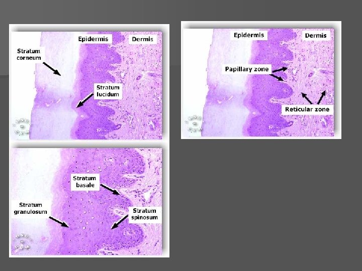
5 strata of the Epidermis – Deep to Superficial · Stratum basale · highly mitotic (produces new skin layer) · ~ 25% melanocytes · Stratum spinosum · Slightly mitotic · Contains Langerhan’s macrophages · Several layers of many sided cells (looks spiny) · Stratum granulosum · Also contains Langerhans cell · contains keratohyalin (helps form keratin) · Stratum lucidum · ONLY found in thicker epidermis – palms, soles, callus · Completely keratinized (and dead!) · contains closely packed, clear cells that contain gel-like substance eleiden
5 strata of the Epidermis · Stratum corneum · Outermost layer · Also completely keratinized · Dead cells · Tough, waterproofing protection
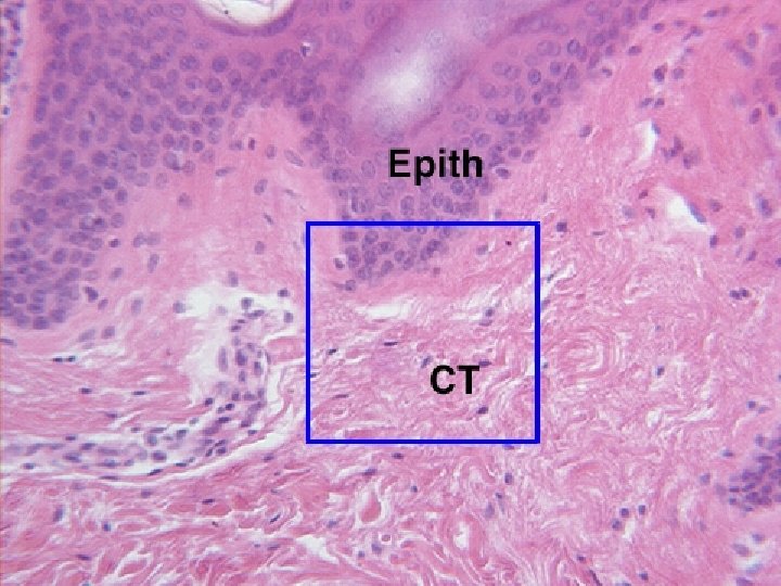
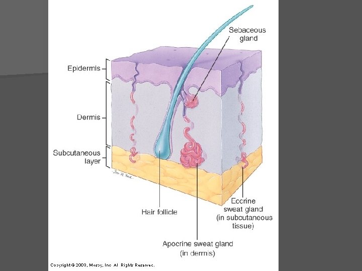
Dermis · Middle layer of skin · Contains hair folllicles, glands, nerves, vessels, muscle · All four tissue types present · Mainly strong, flexible CT - Two layers
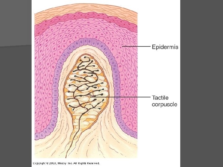
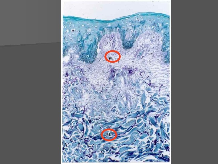
Dermis · Papillary layer · Contains Areolar CT · Dermal papillae ·Indent into epidermis ·forms fingerprints ·Important for grip ·Contains blood vessels ·Meissner’s Corpuscles – nerve (touch) receptors
Dermis · Reticular layer · Dense irregular CT · contains blood vessels, nerves, glands, adipose · Pacinian Corpuscles – nerve endings responsible for sensitivity to deep pressure touch and high frequency vibration · Collagen – prevents overstretching and tearing of skin · Elastin – allows skin to stretch ·stretch marks – dermal tears
Hypodermis · Not usually considered part of the skin · Also called subcutaneous layer · Site of subcutaneous injections – absorbed directly into blood stream · Anchors skin to underlying organs, shock absorption, insulation · Composed mostly of adipose tissue · Very vascular
Skin Appendages
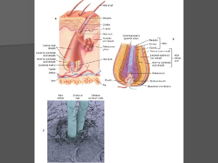
Appendages of the Skin Hair · Minor protective functions (retain heat, decrease sunburn, eyelashes protect eyes) · Structure · shaft – projects from skin · follicle – extends into dermis · root – lies within the follicle · bulb – contains CT, vessels and nerves · sebaceous gland – lubricates hair · arrector pili muscle – attached to follicle and contracts to move hair (hair growth, goosebumps)
Hair
Appendages of the Skin · Hair Growth · influenced by: (in this order) · nutrition - main influence · hormones · blood flow · baldness ( alopecia ) · male pattern baldness - sex linked recessive genetic trait · thinning – can be caused by medications, nutrition, stress, etc. · Hair Pigment · caused by proportions of 3 melanin types · dark hair – true melanin · blonde and red – melanin with iron and sulfur · gray/white hair - melanin replaced by air bubbles in shaft
Appendages of the Skin Nails · Scale-like modifications of the epidermis · Heavily keratinized · Stratum basale extends beneath the nail bed to form nail matrix · Responsible for growth ( matrix region) · Lack of pigment makes them colorless · Lunula “little moon” – area of cell growth (white semicircle at base of nail) · Cuticle – area of skin that covers base of nail
Nail Structures
Sweat Glands · Eccrine glands · Widely distributed in skin: abundant on palms, soles, forehead · Sweat composition: mostly water with a slightly acidic 4 -6 p. H · Function: thermoregulation • Apocrine glands · · Ducts empty into hair follicles Found mainly in anogenital & axillary region Begin to function at puberty due to hormones / pheromones Organic contents: Fatty acids and proteins – can have a yellowish color that stains clothes · Odor is from associated bacteria · Ceruminous glands · Modified apocrine gland · Found in outer 1/3 of ear canal · Produce ear wax to trap “invaders”
Appendages of the Skin · Sebaceous glands · all over except palms and soles of feet · Produce oil for waterproofing · Lubricant for skin & kills bacteria · Most with ducts that empty into hair follicles · Glands are activated at puberty: stimulated by hormones · Acne – active infection of sebaceous glands
Burns
Burns · Protein denaturation and cell death caused by heat, electricity, UV radiation, or chemicals · 2 main dangers · Dehydration–Loss of fluids & Electrolytes lead to: ·Renal Shutdown ·Circulatory shock · Infection ·Skin (mechanical) barrier lost ·Immune system depresses
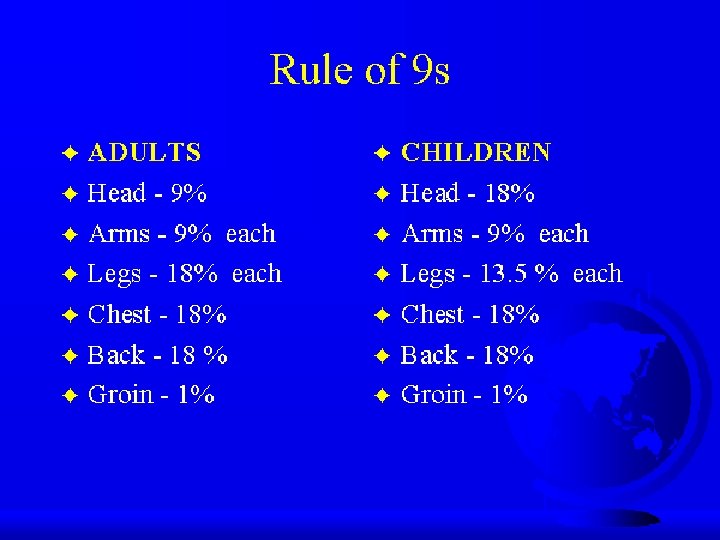
Rules of Nines · Way to determine the extent of burns · Primary importance is to estimate fluids needed for rehydration · Body is divided into 11 areas for quick estimation · Each area represents about 9%
Rule of nines diagram
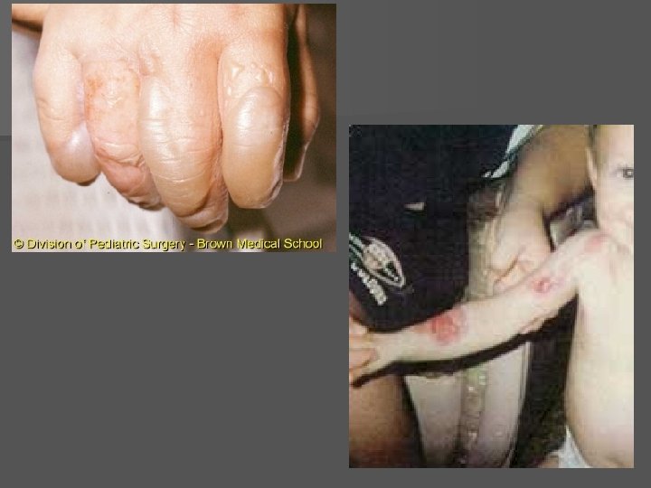
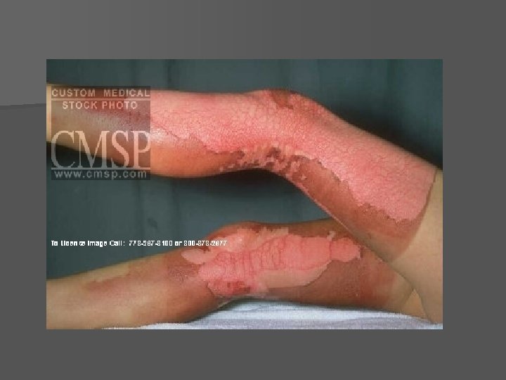
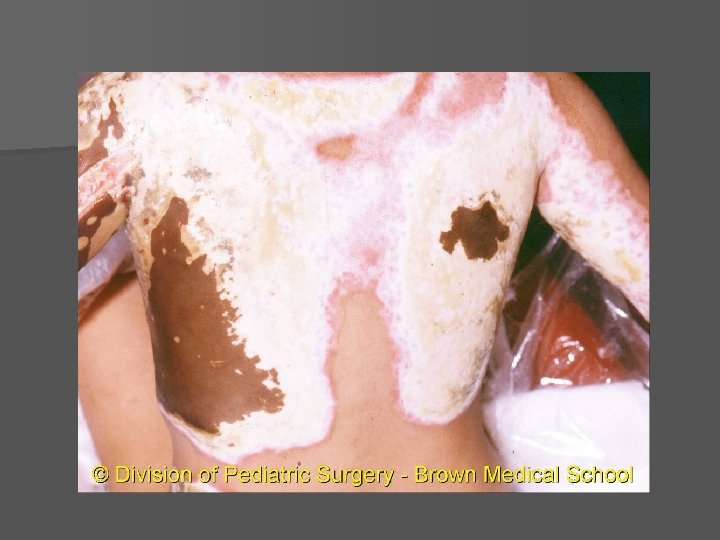
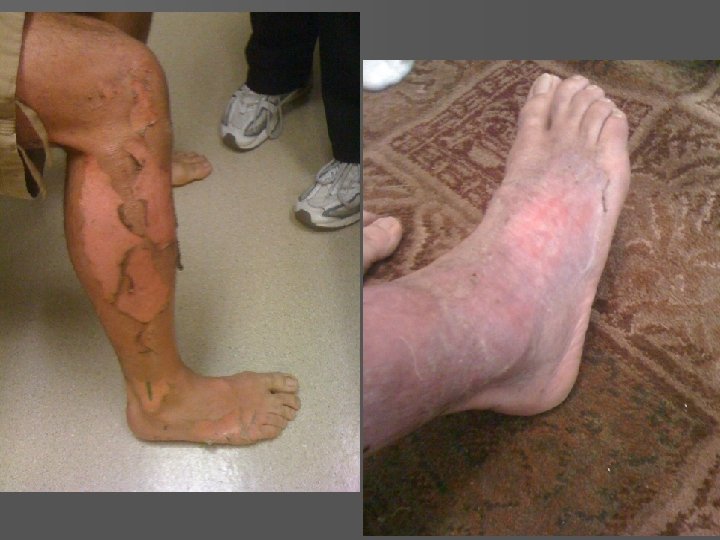
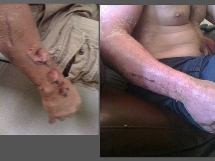
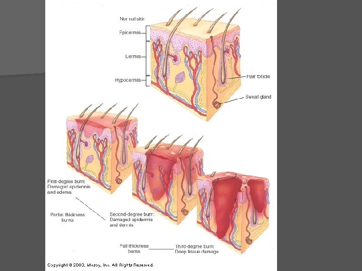
Partial Thickness Burns · First-degree burns · Only epidermis is damaged · Local redness, swelling, & pain · Usually heal in 2 -3 days (short time period) with NO scarring Slide 4. 27
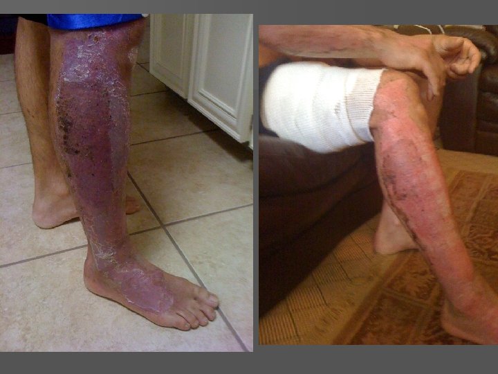
Partial Thickness Burns · Second degree burns · Epidermis and dermis & structures within dermis are damaged · Appearance of blisters of any size · Skin regeneration in 3 -4 weeks with some scarring · There is a danger of infection
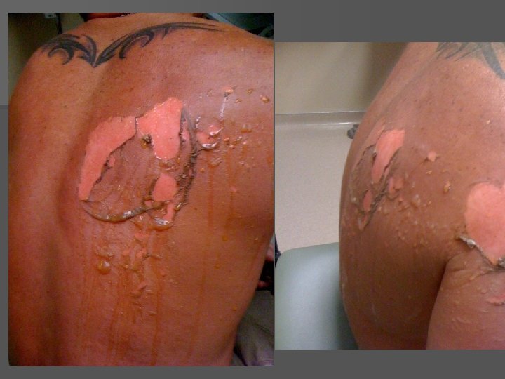
Full Thickness Burns · Third-degree burns · Epidermis, Dermis, Hypodermis and all structures within are completely destroyed · Usually painless at site of burn due to destruction of sense receptors · Burn is gray-white, tan, brown, black, or deep cherry red · Surrounded by areas of 1 st & 2 nd degree burns that will be painful · Treatments are numerous but will involve skin grafting of some sort, fluid replacement and debridement
All degrees of burns
Skin Cancer Skin cancer is the most common type of cancer 2 out of 5 cancers are skin cancers
Skin Cancer · Cancer – uncontrolled cell growth · Caused by damage to the DNA usually through chemicals or radiation · Two types · Benign · Does not spread (encapsulated) · Malignant · Metastasized (moves) to other parts of the body
Skin Cancer Types Basal cell carcinoma · Least malignant · Most common type (90% of skin cancers) · Arises from stratum basale · cannot produce keratin · Boundary lost between dermis and epidermis · Seldom metastasizes – treated surgically or by radiation – 99% cure rate if caught early · Signs · Pale marks · Reddish patches · Round, smooth growth with raised edge · Shiny bumps · Sores that don’t heal
Basal Cell Carcinoma
Basal Cell Carcinoma
Skin Cancer Types · Squamous cell carcinoma · 2 nd most common skin cancer · Highest risk – fair skin, light hair, blue/green eyes · Arises from stratum spinosum · Metastasizes to lymph nodes if left untreated · 1500 -2000 deaths in US per year · Early removal allows a good chance of cure · Signs are same as basal cell carcinoma
Squamous Cell Carcinoma
Cumulative Effects IMPORTANT TO KNOW n Basal cell & squamous cell carcinomas are due to cumulative effects of the sun’s radiation (or chemical exposures as well) n These tend to develop in ages 30 -40 s after years of daily sun exposure
Skin Cancer Types · Malignant melanoma · Least common · most deadly of skin cancers · Originates melanocytes · Metastasizes rapidly to lymph and blood vessels · Early detection is critical – see notes for survival rates
Intensive Effects n Malignant Melanoma tends to occur in younger ages (as well as older people) n It is due to brief intense exposures (aka: sunburns) n This is the most serious form of skin cancer and MUST be caught early to be treated successfully!
ABCD Rule · A = Asymmetry · Two sides of pigmented mole do not match · B = Border irregularity · Borders of mole are not smooth · C = Color · Different colors in pigmented area · D = Diameter · Spot is larger then 6 mm in diameter (pencil eraser) · Mole starts growing/changing in size
Malignant Melanoma
Melanomas
Melanomas
Prevention n n n n Wear sunscreen whenever outside or cover up avoid midday sun between 10 -2 and beware of reflected light higher altitudes - every 1000 ft above sea level, radiation increases 4 -5 % Be cautious about tanning beds Medications - tetracycline (antibiotics), Retin A, birth control, antidepressants, diuretics, and anti-inflammatories cause photosensitivity avoid sunburns examine skin regularly - remember ABCD rule – have full body check by dermatologist once a year
Other Integumentary System disorders
· Contact dermatitis (Ezcema) · Exposure to allergen/irritant (ie. poison ivy) cause allergic reaction · inflammation, red, itchy skin ·not contagious ·over the counter meds; sometimes Rx ·Prevention by avoiding allergen/irritant · Blisters · Epidermal cell injury or separation of epidermis from dermis · Warts · Benign neoplasms, but can turn malignant · Contagious · Remove by freezing, drying, laser therapy, chemicals · Boils · Bacterial infection that infects hair follicles · Large, inflamed, pus-filled lesions
Tinea Fungal infections (ringworm, jock itch, athlete’s foot) Reddish discoloration, scaling, crusting Treat with antifungal agent Prevent recurrence by keeping skin dry Impetigo Caused by bacterial infection Mostly children Reddish discoloration turns into blisters and yellowish crusts If turns systemic, it is life threatening
Psoriasis Cause is unknown, probably genetic Triggered by trauma, infection, stress Cutaneous inflammation, scaly lesions Due to excessive rate of epithelial cell growth Urticaria Hives Raised, red lesions caused by blood vessel leakage Severe itching Causes (hypersensitivity, allergic reactions, physical irritants, systemic disease) Scleroderma Autoimmune Affects blood vessels and CT Hard skin lesions More common in women Decubitus ulcers “bedsores” / pressure sores Lack of blood flow causes tissue damage
Acne * Clogged sebaceous follicles from abnormal shedding of skin cells * Bacteria build-up in sebaceous glands * Enhanced by hormones * Over the counter meds; sometimes Rx * Prevention -avoid using oils, greasy moisturizers, facewash, and makeup -wash hands before applying makeup -use non-scented ordinary mild soap -keep hands away from face