Skin and Body Membranes Cover surfaces line body

Skin and Body Membranes • Cover surfaces, line body cavities, form protective sheets around organs (and mucous) • Two types: • 1. Connective tissue : – Synovial membranes • 2. Epithelial – Cutaneous (skin), mucous, serous

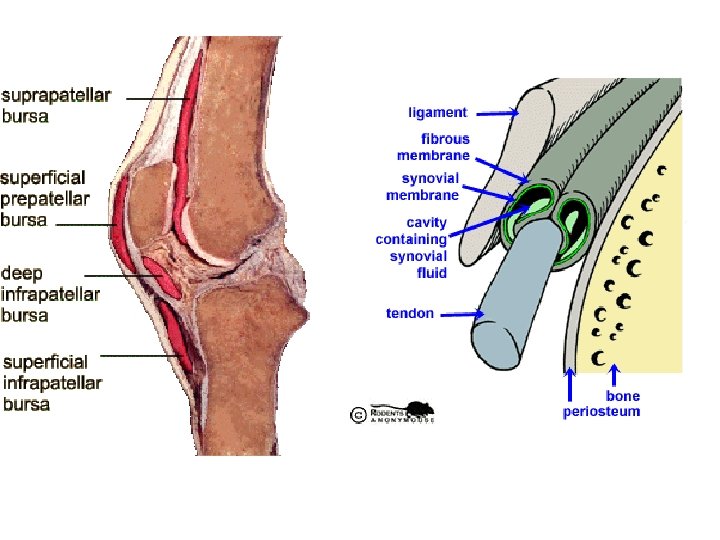
1. Connective: Synovial membranes • Soft areolar tissue • No epithelial cells • Line fibrous capsules surrounding joints (joint is where bones align next to each other) • Line bursae (purse) and tendon sheaths (both cushion organs that move against each other during muscle contractions)

2. Epithelial Membranes • Coverings and linings • Types: – A. Mucous – B. Serous – C. Cutaneous (skin) • All have epithelial tissue on a layer of connective tissue
 • Tissues: Epithelial tissue resting on loose connective tissue")
Epithelial: a. Mucous Membranes (mucosa) • Tissues: Epithelial tissue resting on loose connective tissue called lamina propria (areolar) • Lines all body cavities that open to the exterior (lungs, nasal, mouth, esophogus, anus/rectum, reproductive openings, urethra)

Epithelial: a. Mucous Membranes • Different types of epithelial tissues: • Ex: lungs have simple squamous, intestines have simple columnar and mouth has stratified squamous. • All mucosa membranes are WET (continuously bathed in mucous or with urine)
 • Line body cavities that are closed to the")
Epithelial: b. Serous Membranes (serosa) • Line body cavities that are closed to the exterior • Not the dorsal cavity or joint cavities (so basically ventral cavities) • Tissues: simple squamous on a thin layer of areolar connective tissue

• Serous membranes come in layered pairs • Parietal: lines the wall of the ventral body cavity • Visceral: lines the outside of the organ • Both layers secrete serous fluid: thin, clear fluid that separates the layers • Function of serous fluid: allows organs to slide past each other (ex: digestion, heart pumping)
 • Membranes named according to where they are located:")
Epithelial: b. Serous Membranes (serosa) • Membranes named according to where they are located: • Three names: – Peritoneum: abdominal cavity – Pleura: lungs – Pericardium: heart
 • Tissues: Stratified squamous on a layer of dense")
Epithelial: c. Cutaneous Membrane (skin) • Tissues: Stratified squamous on a layer of dense connective tissue • Squamous is keratinizing: produces keratin which makes skin • Unlike other epithelial membranes, cutaneous is exposed to air and is dry!
 • The skin and its appendages (hair, nails, oil")
Epithelial: c. Cutaneous Membrane (skin) • The skin and its appendages (hair, nails, oil and sweat glands) make up the Integumentary System

Review Membranes!

Integument means covering: 1. Protective covering 2. Keeps water and other substances out of the body (dirt, gases, microbes) 3. Insulates and protects deeper body organs 4. Protects body from mechanical damage, chemical damage, thermal damage, UV damage 5. Keeps water in Skin: Basic Functions
 7. Mini-excretion system:")
Skin: Basic Functions 6. Regulates heat loss (capillaries and sweat glands) 7. Mini-excretion system: sweat contains water, salt and urea 8. Manufactures vitamin D 9. Sensory receptors allow us to understand our environment (pain, temperature, pressure, tissuedamaging factors)

• Two layers: – Epidermis: epithelial tissue • Stratified squamous that produces keratin: allows the skin to be hard and tough – Dermis: dense connective tissue • Epidermis and dermis are tightly connected. – Friction can lead to blister • Underlying subcutaneous layer: adipose tissue – Cushions – Insulates Skin: Structure

Skin: Structure • Epidermis contains up to five layers called strata • Avascular • Most cells are keratinocytes: produce keratin (allows skin to be tough and waterproof) • Deepest layer: (1) Stratum basale – Closest to dermis, most nourished – Constantly undergoing mitosis (millions of new cells daily)
 Stratum spinosum and (3) Stratum granulosum – Become flatter, increasingly")
Skin: Structure • (2) Stratum spinosum and (3) Stratum granulosum – Become flatter, increasingly full of keratin (allows skin to be tough and waterproof) and finally die forming… • (4) Stratum lucidum – Not seen in all skin region – Only where skin is hairless and extra thick…soles of feet and palms
 Stratum corneum – Thickest layer of 2030 cells in thickness –")
• (5) Stratum corneum – Thickest layer of 2030 cells in thickness – Dead cells are like shingles on a roof – Completely filled with keratin (allows skin to be tough and waterproof) – Slough off steadily and is replaced by the Stratum basale (the deepest layer…and so it comes full circle ) Skin: Structure

• Your hide! • Tissue: dense connective • Varies in thickness – Palms of hands and soles of feet, very thick – Eyelids, quite thin • Two layers: – 1. Papillary; called dermal papillae – 2. Reticular Skin Structure: Dermis

Skin Structure: Dermis • 1. Papillary: indented into epidermis • Have capillaries: supply nutrients to epidermis (stratum basale) • Pain receptors (free nerve endings) • Touch receptors: Meissner’s corpuscles (light touch)

Skin Structure: Dermis • Make finger, toe, palm, foot prints by forming looped/whorled ridges on epidermis • Prints are result of sweat begin left behind

• 2. Reticular Layer • Deepest layer • Contains blood vessels, sweat and oil glands • Deep pressure receptors: Pacinian corpuscles • Phagocytes eat bacteria that has managed to break into skin Structure: Dermis

Skin Structure: Dermis • Reticular layer has many collagen and elastic fibers • Collagen: toughness, bind water to keep skin hydrated • Elastic: stretch • Age: number of fibers decreases

• Based on three factors: • 1. Amount and kind of melanin (yellow, reddish brown, black) • 2. Amount of carotene (orange-yellow pigment) in the corneum layer and subcutaneous tissue • 3. Blood Supply to dermis Skin: Color


• Stratum basale contains melanocytes: cells that produce melanin • Roughly same concentration of melanocytes regardless of skin color. • Amount of melanin determined genetically and environmentally. • Sunlight stimulates melanocytes to produce more melanin…hence, we tan • Melanin absorbs UV light and protects against DNA damage • Freckles and moles are concentrated with melanin Skin: Color

The Skin and UV Light • A small amount of UV radiation is important! It stimulates the synthesis of Vitamin D. • Melanocytes concentrate around the nuclear membrane and absorb UV light before it can damage the DNA.

Making Vitamin D 3 • Cells in epidermis convert a cholesterol related steroid into Vitamin D 3 • Vitamin D 3 is absorbed, modified, and released by the liver. It travels to the kidney and is converted into calcitrol (hormone essential for the absorption of calcium by the small intestine).

The Subcutaneous Layer • Not part of skin! • Binds skin to underlying tissue. Made of Loose CT and Fat cells

Appendages of the Skin • Includes: – 1. Cutaneous glands • A. Sebaceous glands • B. Sweat glands – 2. Hair – 3. Hair follicles – 4. Nails • Each arises (forms in) the epidermis

Appendages of the Skin • 1. Cutaneous glands – A. Sebaceous – B. Sweat (sudoriferous) • Exocrine glands (have ducts) • Release secretions on the skin • Made in epidermis but reside in dermis
: • Found everywhere except")
Appendages of the Skin • A. Sebaceous glands (oil glands): • Found everywhere except palms and soles • Most associated with hair • Sebum (product) – Mixture of fats, Cholesterol, proteins, Organic salts

Appendages of the Skin • Sebum functions: • Lubricant – Skin stays moist and soft – Hair not brittle • Chemicals that kill bacteria • During adolescence, very active – Male sex hormones trigger increased production • Whiteheads can lead to blackheads • Acne • Cradle cap in babies (seborrhea)
 • 2. 5 million/person •")
Appendages of the Skin • B. Sweat glands (sudoriferous) • 2. 5 million/person • Two types: – 1. merocrine – 2. apocrine

Appendages of the Skin • Sweat glands: Merocrine • Found all over body • Produce sweat: clear secretion mostly water, salts, vit C, lactic acid and urea • Sweat is acidic: p. H 4 -6 – Inhibits bacterial growth on skin • “pores” on face are not sweat pores but hair follices • Huge role in cooling body

Sudoriferous Glands aka Sweat Glands • Merocrine Glands – Most sweat glands are this type – Can make 600 m. L/day!!! – Found almost everywhere –palms, soles – Secretion is 99% water – cool body and lower body temp.

• Sweat glands: Apocrine • Found in axillary and genital regions • Secretions contains fatty acids and proteins in addition to water, salt, lactic acid, urea, vit C • Bacteria live on this and cause body odor • Start functioning during puberty when androgens are released in body • Not a huge role in cooling body Appendages of the Skin

Sudoriferous Glands aka Sweat Glands • Apocrine – Associated with hair follicles – Secretion is sticky, cloudy and odorous • Where does odor come from? ? ?

Appendages of the Skin • Hair • Parts: root, follicle and shaft • Formed from epithelial tissue in skin (epidermis) and extend into dermis/hypodermis • Dead cells filled with keratin and melanin • Arrector pilli muscle causes hair to stand straight from skin

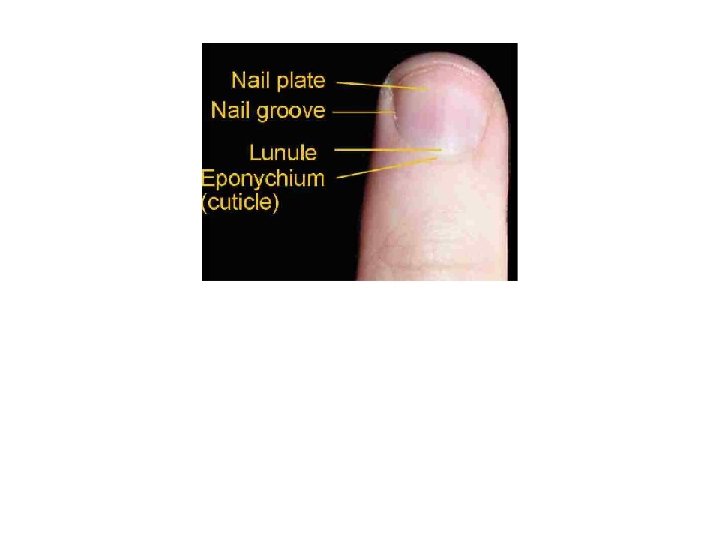
Appendages of the Skin • Nails • Parts: Nail root, nail bed and free nail • Formed from epithelial tissue (epidermis) • Dead cells filled with keratin • Modified scale or hoof

Nails • Like hair dead • Hard keratinized stratified squamous ET found over the dorsal surface of the terminal portions of the digits! Function: To help grasp small objects To prevent trauma to ends of digits To Scratch


Skin Structure: Dermis

Skin Structure: Dermis • Well supplied with blood • Ulcers • Bed sores

• Emotions – Redness/erythema: embarrassment, fever, allergy, inflammation, hypertension – Pallor/blanching: emotional stress (anger, fear), anemia, low blood pressure, impaired blood flow • Jaundice – Yellow tone from excess bile pigments in blood – Indicates liver damage • Bruises – Hematomas: blood clots Skin: Color
- Slides: 46