The Integumentary System Skin Hair Glands Nails The


- Slides: 30
The Integumentary System Skin, Hair, Glands, & Nails
The Skin � Cutaneous membrane ◦ Waterproof, stretchable, and invisibly repairs cuts, rips, or burns � Functions ◦ ◦ ◦ ◦ Protection Maintains body temperature Prevents excessive loss of inorganic & organic materials Receives stimuli Stores chemical compounds Synthesizes vitamin D Excretes water, salts, & several organic compounds
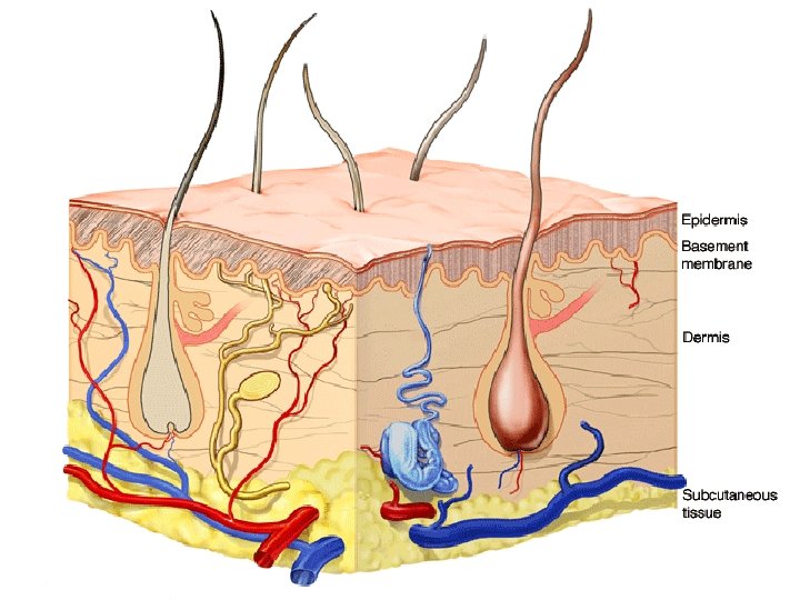
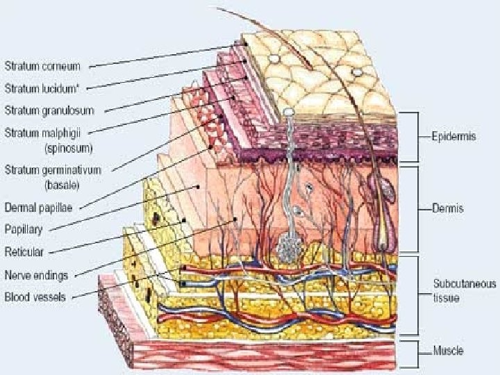
Structure of the Skin �Composed of two layers of tissue ◦ Epidermis �Outer layer composed of squamous epithelium ◦ Dermis �Underlying layer composed of dense connective tissue �The epidermis & dermis are fully cemented together �Skin is anchored to the underlying organs by the subcutaneous layer (Adipose Tissue) ◦ Acts as a shock absorber & insulation
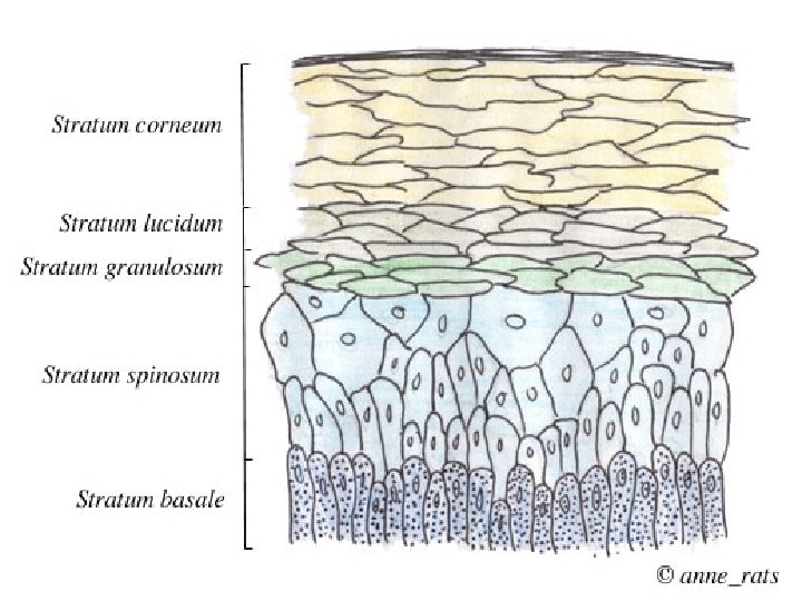
The Epidermis �Epidermis ◦ ◦ ◦ Layers from deepest to the surface Stratum Basale (Germinativum) Stratum Spinosum Stratum Granulosum Stratum Lucidum Stratum Corneum � The germinativum & spinosum undergo continual cell division & produce all the other layers � As cells move toward the surface, they become flatter, increasingly full of keratin (harden) & finally die ◦ Can no longer get adequate nutrients & oxygen from the dermis
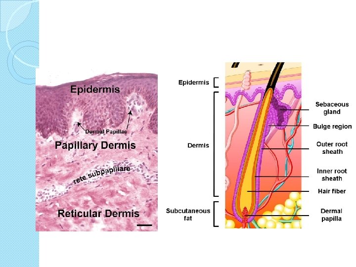
The Dermis �Two major regions: ◦ Papillary Area �Upper dermal region �Contains finger-like projections (dermal papillae) that indent the epidermis above �Dermal papillae bring nutrients to the epidermis ◦ Reticular Area – deepest skin layer �Contains blood vessels, sweat & oil glands, & deep pressure sensors �Contains collagen & elastic fibers �Varies in thickness depending on the locations on the body
Skin Color �Determined by 3 pigments ◦ Melanin �Brown & black pigment �Found in the epidermis �Produced by melanocytes �Acts as protection from UV rays ◦ Carotene �Orange-yellow pigment ◦ Hemoglobin �Blood in the capillaries of the dermis �Pigment in red blood cells – “rosy” skin tone
Alteration of Skin Color �Blue Color ◦ Indicates cyanosis: poor oxygenation ◦ Heart failure & severe breathing �Redness or Erythema ◦ Indicates embarrassment, fever, hypertension, inflammation, or allergy �Pallor or blanching ◦ Indicates fear, anger, or emotional stress ◦ Also anemia, low blood pressure, or impaired blood flow �Jaundice or a yellow cast ◦ Signifies a liver disorder due to excess bile
Skin Appendages These appendages come from the epidermis and help maintain the body’s homeostasis. � Cutaneous (relating to skin) glands ◦ Sebaceous glands ◦ Sweat glands � Hair follicles � Nails
Appendages of the Skin �Sebaceous glands ◦ Produce oil � Lubricant for skin which keeps skin soft and moist � Prevents brittle hair � Kills bacteria (slightly acidic) ◦ Most have ducts that empty into hair follicles; others open directly onto skin surface ◦ Glands are activated at puberty and this is what causes teenage acne
Appendages of the Skin
Facial Blemishes �Whitehead – sebaceous gland is blocked usually from the oily substance made by the gland. �Blackhead – when whitehead dries, it darkens forming blackhead �Acne – an active infection of the sebaceous glands caused by bacteria
Appendages of the Skin �Sweat glands ◦ Produce sweat ◦ Widely distributed in skin �(2. 5 million person) ◦ helps cool the body ◦ Two types � Eccrine � Open via duct to pore on skin surface � Most numerous on the body � Apocrine � Ducts empty into hair follicles � Found mostly in armpits and genital areas � Precise function is unknown but are they are activated during pain, stress and during sexual foreplay.
Appendages of the Skin
Sweat and Its Function �Composition ◦ Mostly water ◦ Salts and vitamin C ◦ Some metabolic waste (urea and uric acid) ◦ Fatty acids and proteins (apocrine only) �Function ◦ Helps rid body of excess heat ◦ Excretes waste products ◦ Acidic nature inhibits bacteria growth �Odor is from associated bacteria
Appendages of the Skin �Hair ◦ Produced by hair follicle which are made of hard keratinized epithelial cells ◦ Melanocytes provide pigment for hair color
Structure of Hair Follicle
Hair Anatomy �Hair anatomy ◦ Central medulla ◦ Cortex surrounds medulla ◦ Cuticle on outside of cortex � Most heavily keratinized
Hair Structures �Associated hair structures ◦ Hair follicle � Dermal and epidermal sheath surround hair root ◦ Arrector pili muscle � Smooth muscle � Pulls hairs upright when cold or frightened ◦ Sebaceous gland
Nails � Nails ◦ Scale-like modifications of the epidermis �Heavily keratinized ◦ Stratum basale extends beneath the nail bed �Responsible for growth ◦ Lack of pigment makes them colorless
Nail Anatomy �Nail structures ◦ Free edge ◦ Body is the visible attached portion ◦ Root of nail embedded in skin ◦ Cuticle is the proximal nail fold that projects onto the nail body
Severity of Burns �First-degree burns ◦ Only epidermis is damaged ◦ Skin is red and swollen �Second-degree burns ◦ Epidermis and upper dermis are damaged ◦ Skin is red with blisters �Third-degree burns (worst) ◦ Destroys entire skin layer ◦ Burn is gray-white or black
Severity of Burns
Rule of Nines �Way to determine the extent of burns �Body is divided into 11 areas for quick estimation �Each area represents about 9% of total body surface area
Skin Cancer Types �Basal cell carcinoma ◦ Malignant tumor of the basal cell layer of the epidermis �Squamous cell carcinoma ◦ Metastasizes to lymph nodes if not removed ◦ Early removal allows a good chance of cure ◦ Believed to be sun-induced ◦ Arises from stratum spinosum
Skin Cancer Types
Skin Cancer Types �Malignant melanoma ◦ Cancerous growth composed of melanocytes