CH 5 Integumentary System SKIN The integumentary system


 can pass through the skin,")
 –")
")


– ex. Warts, calluses, moles")


 Glands – Each sweat gland consists of a coiled tube (duct) –")
 • Adipose (fat) tissue helps conserve body heat • Contains")


 are small gray bugs. Symptoms: itchy scalp, inflammation, bacterial infection, swollen lymph")


 Varicella is a disease caused by the varicella-zoster virus (ZVZ). Symptoms: blistery,")


- Slides: 84
CH 5 Integumentary System
SKIN • The integumentary system includes the skin and its accessory organs • Another term for the skin is the “cutaneous membrane”.
Facts about the SKIN… • Skin is waterproof • The outer surface of the skin is made of dead cells. House dust is mainly skin flakes! • If you laid out all your skin on a flat surface, it would have an area of about 2 square meters (22 square feet). Skin weighs about 2. 5 kilograms (1215% of body weight) - the largest organ in the body. • What hurts if you pull it, but doesn't hurt if you cut it? – Your hair, of course! • Skin is elastic - it springs back into shape when stretched.
Skin Facts Continued…. • Some medicines (eg: oestrogen, nicotine) can pass through the skin, but others cannot (eg: insulin). Why is that? Because only fat-soluble substances can enter the skin, not water-soluble ones. • Your hair stands on end and you develop goose bumps because there are tiny muscles attached to the hair follicles and they contract when you are frightened or cold.
The Skin and its Tissues • Skin functions: • Protective covering (primary function) – barrier to the entry of microbes, viruses, and UV exposure • Aids in regulating body temperature • Slows water loss • Houses sensory receptors • Synthesizes various chemicals (vitamin D and melanin) • Excretes waste • The skin is composed of an epidermis, and subcutaneous layer (hypodermis).
THE SKIN Epidermis Dermis Subcutaneous (Hypodermis)
The EPIDERMIS • 4 -5 layers of 4 different types of cells. • The deepest layer of the epidermis contains cells undergoing cell division (mitosis) to produce new cells. • About 90% of the epidermis are keratinocytes. • Epidermal cells become keratinocytes when they undergo keratinization. During this process, they mature and are pushed toward the surface while producing keratin (takes about 2 -4 weeks). What does keratin do? – Gives tissue a waterproof quality • Melanocytes produce the skin pigment melanin. Melanin is a brown/black pigment that covers the nucleus to protect the DNA from UV radiation- which can cause mutation. The more that cells are exposed to UV radiation, the more melanin is produced. A sun tan is a sign of injury!!
Epidermis Continued…. • Langerhans cells- Arise from red bone marrow and move to the epidermis where they participate in immune responses. • Merkel cells- Found in the deepest layer of the epidermis where they contact the flattened part of a sensory nerve cell and function in the sensation of touch.
Layers of the Epidermis- from the bottom to top 1. Stratum Basale- Here is where cell division takes place, producing new skin cells and pushing older cells toward the surface. This layer contains prekeratinocytes, melanocytes, Langerhans cells and Merkel cells. This layer is attached to a layer of fibers called the basement membrane. 2. Stratum Spinosum- Contains spiny keratinocytes that are not yet mature enough to produce keratin. 3. Stratum Granulosum- Contains keratinocytes that produce keratin. 4. Stratum Lucidum- Clear, flat, dead cells found only in the thick skin of palms and soles. 5. Stratum Corneum- Thickest layer- 30 layers of flat, dead keratinocytes.
LAYERS OF EPIDERMIS
Quick Review 1. Another name for skin is the _______ membrane. 2. What are 5 functions of skin? 3. What are the 3 layers of skin? 4. What are the 4 types of cells found in the epidermis? 5. What are the 5 layers of the epidermis? 6. Which layer contains functioning keratinocytes? 7. Which layer is attached to the basement membrane? 8. Which layer is the thickest?
Skin Cancer Facts • Most skin tumors are benign (non-cancerous)– ex. Warts, calluses, moles • Skin cancers metastasize, which means that they invade other tissues. If they enter the bloodstream and/or lymph system, they can travel anywhere and lodge themselves in other locations. • The cause of skin cancer is not known, but overexposure to ultraviolet (UV) radiation is the main risk factor. The DNA in a cell can become mutated, and consequently, it divides out of control.
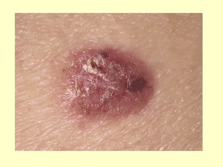
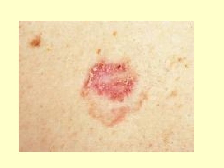
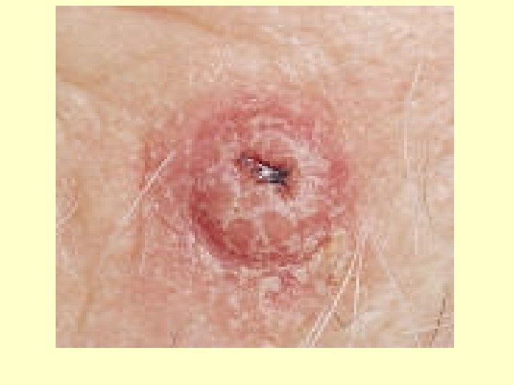
Basal Cell Carcinoma • Least malignant and most common skin cancer • Most often appears on the exposed areas of the face • Appears as a shiny dome-shaped nodule that later develops a central ulcer with a “pearly” beaded edge • Full cure in 99% that are removed surgically • Cells of the stratum basale no longer form keratin and do not honor the boundary between dermis and epidermis
Basal Cell Carcinoma
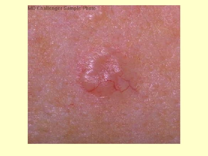
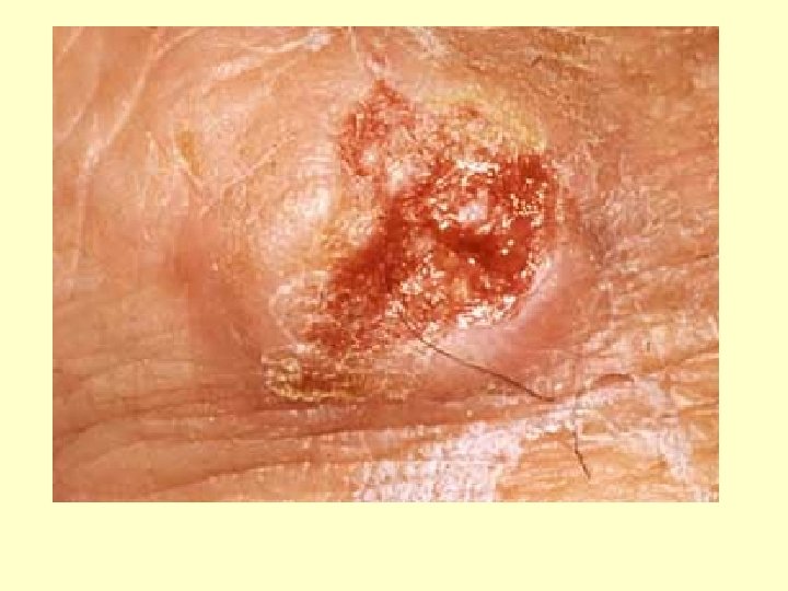
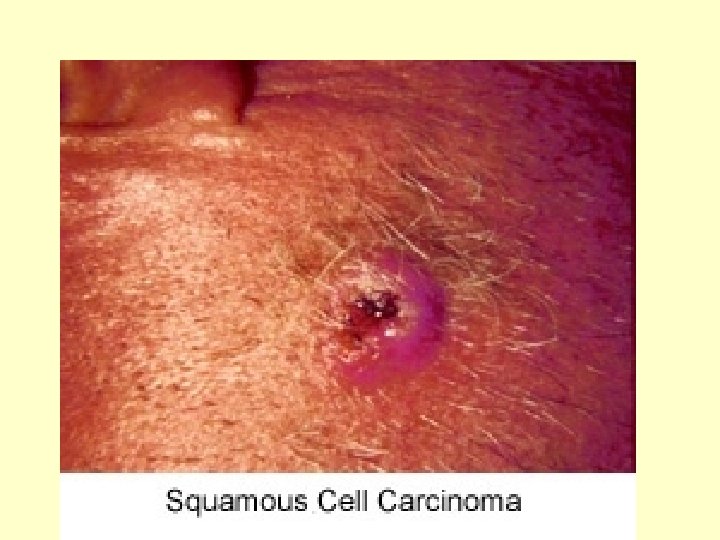
Squamous Cell Carcinoma • Arises from the cells of the stratum spinosum • Occurs most often on the scalp, ears, hands, and lower lip • Appears as a scaly, reddened elevation that gradually forms a shallow ulcer with a firm, raised border • It grows rapidly and metastasizes to adjacent lymph nodes if not removed • Chance for complete cure is good if caught early and removed surgically or by radiation therapy
Squamous Cell Carcinoma
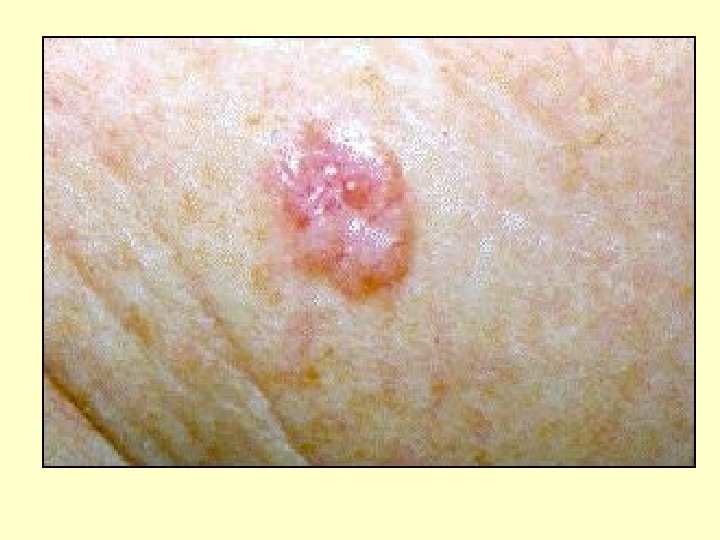
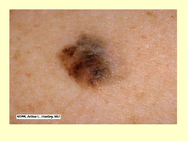
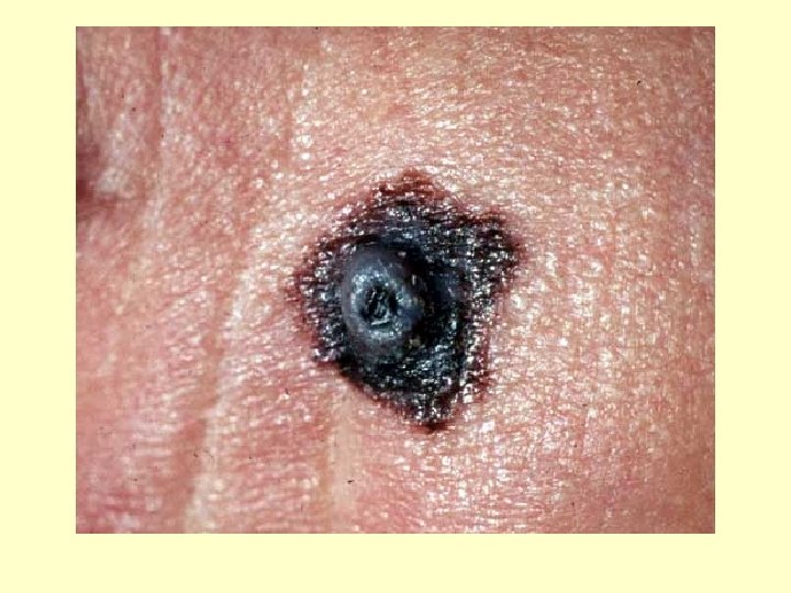
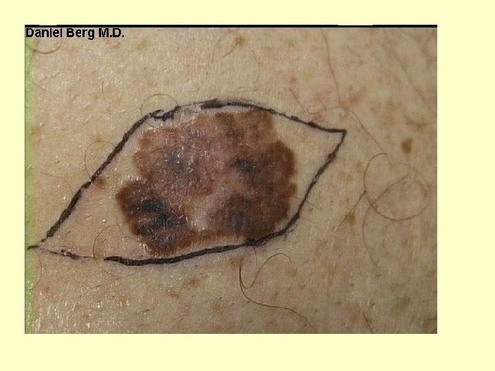
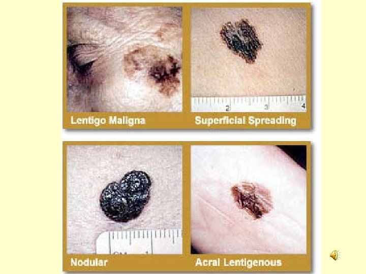
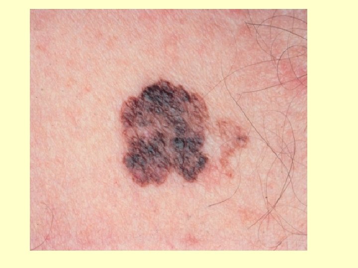
Malignant Melanoma • Cancer of the melanocytes • 5% of all skin cancers • Is often deadly and chance of survival is 50%. . . early detection helps. • Can begin anywhere there is pigmentation, some develop from pigmented moles. • Usually appears as a spreading brown to black patch that metastasizes rapidly to surrounding lymph nodes and blood vessels. • Use the ABCDE Rule to recognize it
Malignant Melanoma
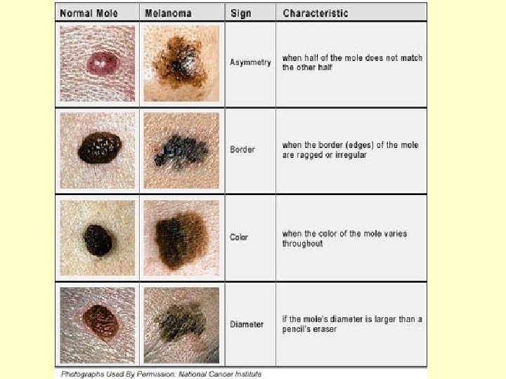
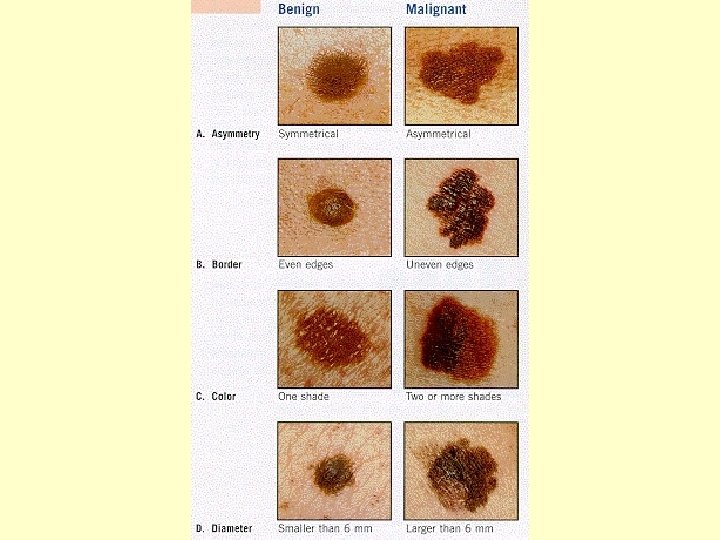
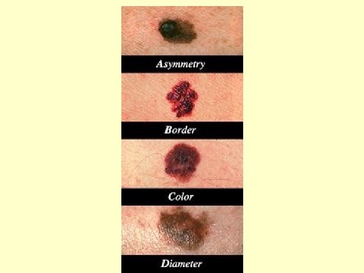
The ABCDE rule is a convenient guide to the usual signs of melanoma. • A is for ASYMMETRY: Onehalf of a mole or birthmark does not match the other. • B is for BORDER: The edges are irregular, ragged, notched, or blurred. • C is for COLOR The color is not the same all over, but may have differing shades of brown or black, sometimes with patches of red, white, or blue. • D is for DIAMETER: The area is larger than 6 millimeters (about ¼ inch -- the size of a pencil eraser) • E is for EVOLVING: If the growth changes at all
IMPORTANT NOTE: One blistering sunburn in childhood or adolescence more than doubles a person's chances of developing melanoma later in life. A person's risk for melanoma also doubles if he or she has had five or more sunburns at any age.
The DERMIS • Composed of 2 layers: papillary layer and reticular layer. • Papillary layer contains a thin arrangement of collagen fibers and supplies nutrients to select layers of the epidermis and regulates temperature (by increasing and reducing blood supply to the epidermis). The fingerlike projections of this layer are called dermal papillae. These cause ridges in the epidermis and are what produce fingerprints. • Inside the dermal papillae are blood vessels and nerve endings. The nerve endings are called corpuscles of touch or Meissner corpuscles. These are sensitive to touch. What type of epidermal cell is closely associated with these? – Merkel Cells!
Dermis continued…. . • Reticular layer- The reticular layer is contains thicker collagen fibers than the papillary dermis. It strengthens the skin, provides structure and provides elasticity. It also supports other components of the skin, such as hair follicles, nerves, oil glands, muscles, and sweat glands.
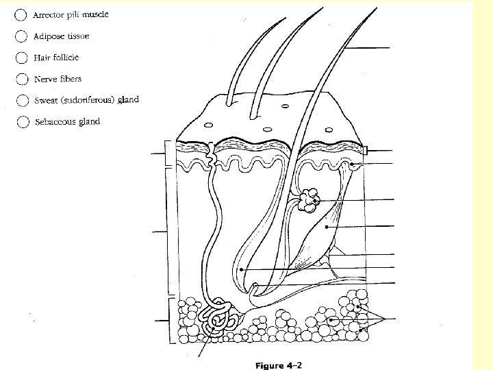
HAIR • • Also called pili- Each strand is dead, keratinized cells that consist of a shaft (above the skin) and a root (below the skin). Surrounding the root is a hair follicle which includes an external root sheath, internal root sheath, and connective tissue sheath The base of the follicle is the matrix- where new hair cells are formed from cell division Surrounding the follicle base is/are: • blood vessels- provide nourishment to the hair • arrector pili muscle-contracts and causes the hair to stand up (goose bumps) • hair root plexus (nerve endings)
HAIR
Sebaceous Glands • Sebaceous glands usually are associated with hair follicles • Secrete sebum, which keeps hair from drying out • If plugged and infected…a pimple develops
Sweat (Sudoriferous) Glands – Each sweat gland consists of a coiled tube (duct) – 2 Types (Apocrine and Eccrine) – Apocrine glands respond to emotional stress – larger and occur in armpits (axillary regions) and groin areas…these produce a solution that bacteria act on to produce “body odor” – Eccrine glands respond to elevated body temperature – Sweat is primarily water, but also contains salts and wastes
The SUBCUTANEOUS Layer (Hypodermis) • Adipose (fat) tissue helps conserve body heat • Contains blood vessels that branch into the dermis • The layer where you receive shots and vaccinations…
Nails • Nails are produced by epidermal cells originating at the nail matrix that undergo keratinization HOW SWEET ARE THOSE NAILS? ?
Burns • • Serious threat to skin 2 Life Threatening Problems associated with Burns: 1. Body Fluid Loss-. Dehydration and electrolyte imbalance follow and can lead to shutdown of kidneys and circulatory shock 2. Infection- The leading cause of death in burn victims. Burned skin is sterile for about 24 hrs. The bacteria and fungi easily invade areas where the skin is destroyed and feed off of the dead tissues. The patient’s immune system becomes overwhelmed and suppressed after severe burn injury.
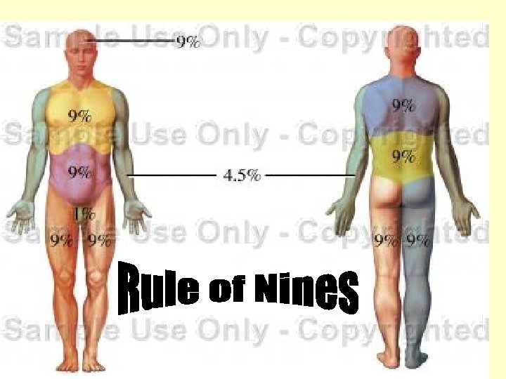
Rule of Nines • The severity of a burn can be estimated by determining how much of a body’s surface is burned using the RULE OF NINES. • This divides the body into regions and states the surface area % of the body for each region. See next slide.
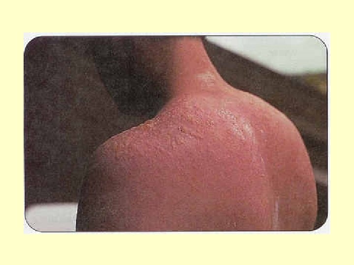
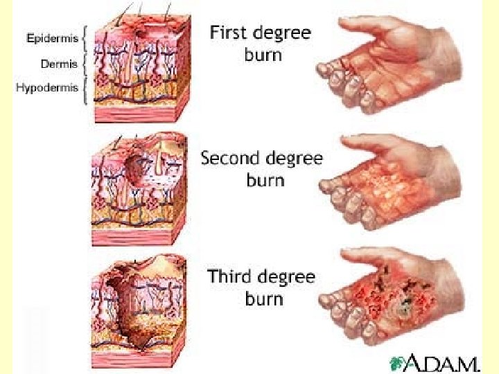
Severity of Burns • First Degree: Only the epidermis is damaged. Not usually serious and can heal in 2 -3 days. – Ex. Mild sunburn
First Degree Burns • First-degree burns involve minimal tissue damage and they involve upper layers of the epidermis (skin surface). These burns cause pain, redness and swelling.
First – Degree Burns
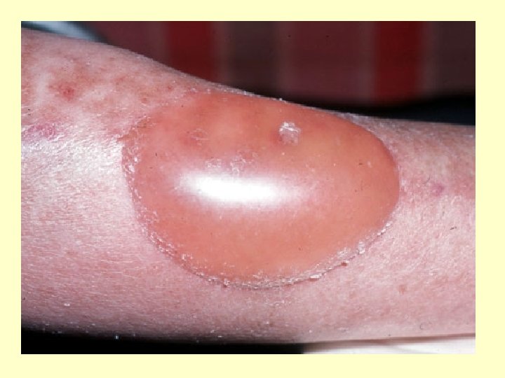
Second-Degree Burns • Epidermis and upper region of dermis is damaged. • Usually no permanent scars.
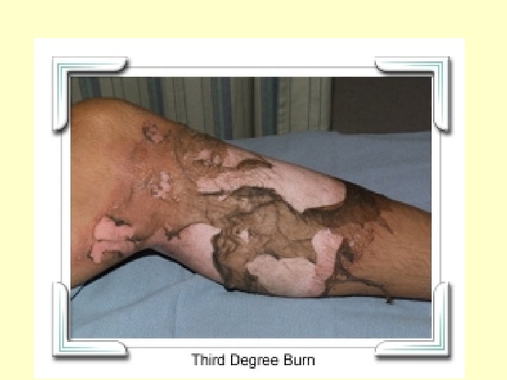
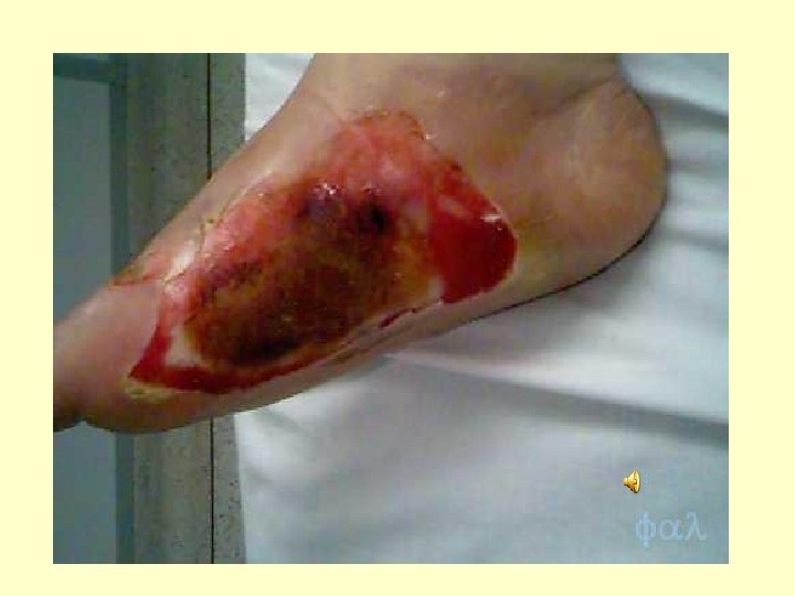
Third-Degree Burns • Third-degree burns affect the epidermis, dermis and hypodermis, causing charring of skin or a translucent white color, with coagulated vessels visible just below the skin surface. These burn areas may be numb because nerves are destroyed. Regeneration is not possible and skin grafting must be performed.
SEVERITY OF BURNS 1. Minor burns treated at home= First or seconddegree burns covering less than 15% of an adult's body or less than 10% of a child's body, or a third-degree burn on less than 2% BSA. . 2. Moderate burns treated at a hospital= These are defined as First or second-degree burns covering 15%-25% of an adult's body or 10%20% of a child's body, or a third-degree burn on 2%-10% BSA.
• Critical, or major, burns should be treated in a specialized burn unit of a hospital. These are defined as First or seconddegree burns covering more than 25% of an adult's body or more than 20% of a child's body, or a third-degree burn on more than 10% BSA. In addition, burns involving the hands, feet, face, eyes, ears, or genitals are considered critical.
For the following scenarios, indicate the body surface area % that is burned and rate the burn as minor, moderate or critical. 1. A child is burnt at a family bonfire. His anterior right arm and entire left leg are affected. 2. An adult woman was burnt using a chicken fryer. Her left foot and posterior left leg were affected.
Regulation of Body Temperature • Regulation of body temperature is vital because heat affects the rates of metabolic reactions • Normal body temperature of deeper body parts is about 37° C (98. 6° F) – Heat Production and loss • When body temperature rises above normal, dermal blood vessels dilate and sweat glands secrete sweat • In hot weather, 4 liters per hour can be lost…so drink your fluids!!! • If body temperature drops below normal, dermal blood vessels constrict and sweat glands become inactive • During excessive heat loss, skeletal muscles are stimulated to contract involuntarily (shivering)
Some actions involved in Body Temperature Regulation: Body Temp. Rises Above Normal Nervous System Signals dermal Blood vessels to dilate and sweat glands to secrete Body Heat is lost to surroundings Body Temp returns to normal Normal Body Temperature 37 C or 98. 6 F Body Temp rises towards normal Body heat is generated by muscle contractions Nervous System signals dermal blood vessels to constrict and sweat glands remain inactive Body Temp drops below normal If body temp continues to drop, Nervous system signals muscles to Contract involuntarily (shivering)
ACNE Acne is a common skin condition in which the sebaceous glands become clogged. This causes a pimple and inflamed infected abscesses, or collections of pus. The symptoms of this condition may include blackheads, whiteheads, pimples, pustules, and cysts. Acne is caused by a hormonal change in the body. It is usually inherited.
Allergic Contact Dermatitis – A bumpy patch of itchy, flaky, red skin. It occurs when someone has a reaction after coming into contact with something that irritates his/her skin. Symptoms are: redness, itchy skin, crackling skin, blisters, spreading patch, and oozing. Causes are: repeated contact, soaps, fabric softeners, perfumes, cosmetics, nickel, deodorants, plants such as poison ivy, oak, or sumac.
Alopecia is a medical name for baldness or hair loss. Symptoms: hair loss. Causes: Male-Patterned Baldness, which is the most common, inflammation of the scalp, medications, chemotherapy, radiation treatment of the head, infections such as syphilis, trauma to the hair or scalp, low iron, low thyroid hormone levels, lupus, and cancer
Athlete’s Foot is a fungal infection that affects the top layer of skin on the foot. Symptoms: Itching, itchy red rash between toes or underneath the arch of the foot, small blisters, which contain pus, skin may look inflamed, dry, and scaly. Causes: fungus
Boil/ Carbuncle Boil/Carbuncle is an abscess which is a collection of pus in soft tissues of the skin caused by bacterial infections and usually involves a hair follicle. Symptoms: tender red swollen skin , with an overlying pus head or a very tender soft lump in which the pus may not be visible.
Cold Sores are blisters inside the mouth or on the lips. Causes: a virus called Herpes Simplex Virus Type I. Symptoms: tiny blisters, swelling and redness, fever, not feeling well, feeling tired, hard to eat, and sore throat. Spread from person to person easily, able to spread to other parts of the body, may become infected with bacteria
Warts are non-cancerous skin growths caused by a viral infection in the top layer of the skin. Viruses that cause warts are called human papillomavirus (HPV). Warts are usually skin-colored and feel rough to the touch, but they can be dark, flat and smooth. The appearance of a wart depends on where it is growing.
Eczema is a non-contagious skin condition that causes patches of dry, scaly, extremely itchy skin. Symptoms: dry patches of skin, red extremely itchy skin, rash, blisters, itching, constant dry, scaly skin. Causes: related to history hypersensitivity or reaction in the body, people who have asthma or hay fever are more likely to get eczema. Stress, dry climate, high temperature, soaps, chlorine, and other irritating substances. Foods such as peanut butter, milk, or eggs.
Fungal Nail Infection A fungal nail infection is a condition in which a fungus or yeast causes a nail to become misshapen, discolored, and thick. Symptoms: unattractive nails, itching, peeling, skin dryness, and small blisters. Causes: fungal infection, bacterial buildup
Impetigo is a contagious bacterial infection on the surface of the skin. Symptoms: honey colored crusts, mild sores, pus filled blisters, blisters containing clear yellow or slightly cloudy fluid. Causes: scratches, cuts, or prior existing skin diseases such as eczema put a person at risk.
Keloids are patches of excessive scar tissue that may form following a skin injury. Symptoms: thick, smooth humped-up pink scar tissue larger that the original site of injury, occasional itching or tenderness. Causes: cosmetics, piercings
Pediculosis (lice) are small gray bugs. Symptoms: itchy scalp, inflammation, bacterial infection, swollen lymph nodes, nits. Causes: sharing hats, combs or headphones, wearing dirty clothing for weeks.
Psoriasis is an inherited disease that causes an increase in skin cells on the outer layer of the skin. Cause: unknown, but allergies are thought to play a role. Symptoms: red bumps or patches under dead skin, reddened areas that itch or are tender, itching, scaling and red patches in the scalp, crumbling or abnormal fingernails or toenails, thickening, cracking, and blistering of the palms or soles of the feet.
Rosacea is a chronic inflammation that occurs on the face. Symptoms: redness of the face, overproduction of sebum, inflamed acne like bumps, mild swelling of the skin on the cheeks and nose, thickening of the oil glands in skin of the nose, swelling of tiny blood vessels on the face. Cause: unknown
Scabies is a skin infestation caused by a scabies mite. Symptoms: itchy bumps in characteristic locations such as between fingers, or on the wrists, or on the genitals. Other symptoms include generalized itching and tiny burrow lines
Varicella (Chickenpox) Varicella is a disease caused by the varicella-zoster virus (ZVZ). Symptoms: blistery, itchy rash, usually on all body surfaces, fever, difficulty waking, trouble walking, stiff neck, breathing difficulty, vomiting, red tender skin, a child who looks or acts sick, scabs that become soft and drain a yellow pus (Chickenpox). We have a vaccine now that prevents this disease.
Shingles is a painful rash of blisters that develops due to the virus that causes chickenpox. Symptoms: sensations, pain, numbness, tingling, itching, groups of blisters. Causes: reactivation of the chickenpox virus, weakness of the immune system
What is the skin disorder? 1. 2. 3. 4. 5. 6. 7. skin infestation caused by a mite increase in skin cells on the outer layer of the skin baldness or hair loss small gray bugs caused by HPV- Human Papilloma Virus reactivation of the chicken pox virus redness of the face, overproduction of sebum, inflamed acne like bumps 8. caused by Herpes Simplex Virus Type 1 9. fungal infection that affects the top layer of skin 10. patches of excessive scar tissue