
Chapter 32 Skin Integrity and Wound Care Copyright




- Slides: 52
Chapter 32 Skin Integrity and Wound Care Copyright © 2011 Wolters Kluwer Health | Lippincott Williams & Wilkins
Functions of the Skin • Protection • Body temperature regulation • Psychosocial • Sensation • Vitamin D production • Immunological • Absorption • Elimination Copyright © 2011 Wolters Kluwer Health | Lippincott Williams & Wilkins
Question Tell whether the following statement is true or false. Blood vessels in the skin dilate to dissipate heat. A. True B. False Copyright © 2011 Wolters Kluwer Health | Lippincott Williams & Wilkins
Answer: A. True Blood vessels in the skin dilate to dissipate heat. Copyright © 2011 Wolters Kluwer Health | Lippincott Williams & Wilkins
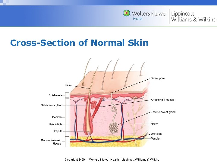
Cross-Section of Normal Skin Copyright © 2011 Wolters Kluwer Health | Lippincott Williams & Wilkins
Factors Affecting the Skin • Unbroken and healthy skin and mucous membranes defend against harmful agents. • Resistance to injury is affected by age, amount of underlying tissues, and illness. • Adequately nourished and hydrated body cells are resistant to injury. • Adequate circulation is necessary to maintain cell life. Copyright © 2011 Wolters Kluwer Health | Lippincott Williams & Wilkins
Developmental Considerations • Infant’s skin and mucous membranes are easily injured and subject to infection. • A child’s skin becomes increasingly resistant to injury and infection. • The structure of the skin changes as a person ages. • The maturation of epidermal cells is prolonged, leading to thin, easily damaged skin. Copyright © 2011 Wolters Kluwer Health | Lippincott Williams & Wilkins
Causes of Skin Alterations • Very thin and very obese people are more susceptible to skin injury. – Fluid loss during illness causes dehydration. – Skin appears loose and flabby. • Excessive perspiration during illness predisposes skin to breakdown. • Jaundice causes yellowish, itchy skin. • Diseases of the skin cause lesions that require care. Copyright © 2011 Wolters Kluwer Health | Lippincott Williams & Wilkins
Types of Wounds • Intentional or unintentional • Open or closed • Acute or chronic • Partial thickness, full thickness, complex Copyright © 2011 Wolters Kluwer Health | Lippincott Williams & Wilkins
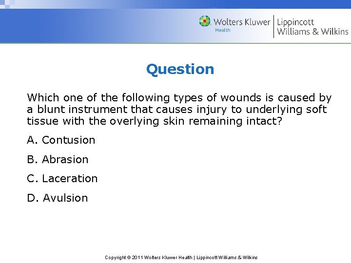
Question Which one of the following types of wounds is caused by a blunt instrument that causes injury to underlying soft tissue with the overlying skin remaining intact? A. Contusion B. Abrasion C. Laceration D. Avulsion Copyright © 2011 Wolters Kluwer Health | Lippincott Williams & Wilkins
Answer: A. Contusion Rationale: A contusion is caused by a blunt instrument and may result in bruising or hematoma. An abrasion is the rubbing or scraping of epidermal layers of skin. A laceration is the tearing of skin and tissue with a blunt or irregular instrument. Avulsion is the tearing of a structure from normal anatomic position. Copyright © 2011 Wolters Kluwer Health | Lippincott Williams & Wilkins
Principles of Wound Healing • Intact skin is the first line of defense against microorganisms. • Surgical asepsis is used in caring for a wound. • The body responds systematically to trauma of any of its parts. • An adequate blood supply is essential for normal body response to injury. • Normal healing is promoted when wound is free of foreign material. Copyright © 2011 Wolters Kluwer Health | Lippincott Williams & Wilkins
Principles of Wound Healing (cont. ) • The extent of damage and the person’s state of health affect wound healing. • Response to wound is more effective if proper nutrition is maintained. Copyright © 2011 Wolters Kluwer Health | Lippincott Williams & Wilkins
Phases of Wound Healing • Hemostasis • Inflammatory • Proliferation • Maturation Copyright © 2011 Wolters Kluwer Health | Lippincott Williams & Wilkins
Question In which one of the following phases of wound healing is new tissue built to fill the wound space, primarily through the action of fibroblasts? A. Hemostasis B. Inflammatory phase C. Proliferation phase D. Maturation phase Copyright © 2011 Wolters Kluwer Health | Lippincott Williams & Wilkins
Answer: C. Proliferation phase Rationale: In the proliferation phase, granulation tissue is formed to fill the wound. In hemostasis, involved blood vessels constrict and blood clotting begins. In the inflammatory phase, white blood cells move to the wound. In the maturation phase, collagen is remodeled forming a scar. Copyright © 2011 Wolters Kluwer Health | Lippincott Williams & Wilkins
Inflammatory Phase • Begins at time of injury • Prepares wound for healing – Hemostasis (blood clotting) occurs – Vascular and cellular phase of inflammation Copyright © 2011 Wolters Kluwer Health | Lippincott Williams & Wilkins
Hemostasis • Occurs immediately after initial injury • Involved blood vessels constrict and blood clotting begins • Exudate is formed causing swelling and pain • Increased perfusion results in heat and redness • Platelets stimulate other cells to migrate to the injury to participate in other phases of healing Copyright © 2011 Wolters Kluwer Health | Lippincott Williams & Wilkins
Inflammatory Phase • Follows hemostasis and lasts about 4 to 6 days. • WBCs move to the wound. • Macrophages enter wound area and remain for extended period. • They ingest debris and release growth factors that attract fibroblasts to fill in wound. • Patient has generalized body response. Copyright © 2011 Wolters Kluwer Health | Lippincott Williams & Wilkins
Proliferation Phase • Phase begins within 2 to 3 days of injury and may last up to 2 to 3 weeks. • New tissue is built to fill wound space through action of fibroblasts. • Capillaries grow across wound. • A thin layer of epithelial cells forms across wound • Granulation tissue forms a foundation for scar tissue development. Copyright © 2011 Wolters Kluwer Health | Lippincott Williams & Wilkins
Maturation Phase • Final stage of healing begins about 3 weeks to 6 months after injury. • Collagen is remodeled. • New collagen tissue is deposited. • Scar becomes a flat, thin, white line. Copyright © 2011 Wolters Kluwer Health | Lippincott Williams & Wilkins
Factors Affecting Wound Healing • Age—children and healthy adults heal more rapidly • Circulation and oxygenation—adequate blood flow is essential • Nutritional status—healing requires adequate nutrition • Wound condition—specific condition of wound affects healing • Health status—corticosteroid drugs and postoperative radiation therapy delay healing Copyright © 2011 Wolters Kluwer Health | Lippincott Williams & Wilkins
Wound Complications • Infection • Hemorrhage • Dehiscence and evisceration • Fistula formation Copyright © 2011 Wolters Kluwer Health | Lippincott Williams & Wilkins
Question Which one of the following wound complications is caused by overhydration related to urinary and fecal incontinence? A. Necrosis B. Edema C. Desiccation D. Maceration Copyright © 2011 Wolters Kluwer Health | Lippincott Williams & Wilkins
Answer: D. Maceration Rationale: Maceration is caused by overhydration related to incontinence that causes impaired skin integrity. Necrosis is dead tissue present in the wound that delays healing. Edema is swelling at a wound site that interferes with blood supply to the area. Desiccation is the process in which the cells dehydrate and die. Copyright © 2011 Wolters Kluwer Health | Lippincott Williams & Wilkins
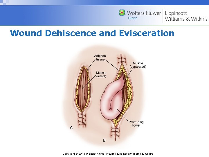
Wound Dehiscence and Evisceration Copyright © 2011 Wolters Kluwer Health | Lippincott Williams & Wilkins
Psychological Effects of Wounds • Pain • Anxiety • Fear • Change in body image Copyright © 2011 Wolters Kluwer Health | Lippincott Williams & Wilkins
Factors Affecting Pressure Ulcer Development • Aging skin • Chronic illnesses • Immobility • Malnutrition • Fecal and urinary incontinence • Altered level of consciousness • Spinal cord and brain injuries • Neuromuscular disorders Copyright © 2011 Wolters Kluwer Health | Lippincott Williams & Wilkins
Mechanisms in Pressure Ulcer Development • External pressure compressing blood vessels • Friction or shearing forces tearing or injuring blood vessels Copyright © 2011 Wolters Kluwer Health | Lippincott Williams & Wilkins
Stages of Pressure Ulcers • Stage I—nonblanchable erythema of intact skin • Stage II—partial-thickness skin loss • Stage III—full-thickness skin loss; not involving underlying fascia • Stage IV—full-thickness skin loss with extensive destruction • Unstageable—base of ulcer covered by slough and or eschar in wound bed Copyright © 2011 Wolters Kluwer Health | Lippincott Williams & Wilkins
Measurement of a Pressure Ulcer • Size of wound • Depth of wound • Presence of undermining, tunneling, or sinus tract Copyright © 2011 Wolters Kluwer Health | Lippincott Williams & Wilkins
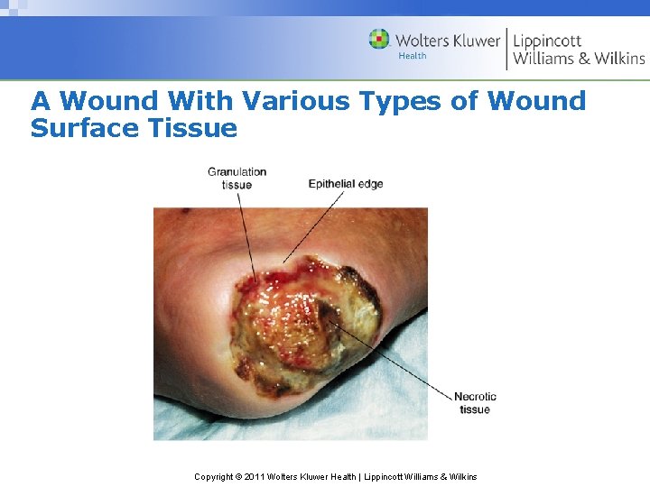
A Wound With Various Types of Wound Surface Tissue Copyright © 2011 Wolters Kluwer Health | Lippincott Williams & Wilkins
Question Tell whether the following statement is true or false. A Stage III pressure ulcer requires debridement through wet-to-dry dressings, surgical intervention, or proteolytic enzymes. A. True B. False Copyright © 2011 Wolters Kluwer Health | Lippincott Williams & Wilkins
Answer: A. True A Stage III pressure ulcer requires debridement through wet-to-dry dressings, surgical intervention, or proteolytic enzymes. Copyright © 2011 Wolters Kluwer Health | Lippincott Williams & Wilkins
Cleaning a Pressure Ulcer • Clean with each dressing change. • Use careful, gentle motions to minimize trauma. • Use 0. 9% normal saline solution to irrigate and clean the ulcer. • Report any drainage or necrotic tissue. Copyright © 2011 Wolters Kluwer Health | Lippincott Williams & Wilkins
Wound Assessment • Inspection for sight and smell • Palpation for appearance, drainage, and pain • Sutures, drains or tube, and manifestation of complications Copyright © 2011 Wolters Kluwer Health | Lippincott Williams & Wilkins
Presence of Infection • Wound is swollen. • Wound is deep red in color. • Wound feels hot on palpation. • Drainage is increased and possibly purulent. • Foul odor may be noted. • Wound edges may be separated with dehiscence present. Copyright © 2011 Wolters Kluwer Health | Lippincott Williams & Wilkins
Assessment of Wound Drainage • Serous • Sanguineous • Serpsamgiomepis • Purulent Copyright © 2011 Wolters Kluwer Health | Lippincott Williams & Wilkins
Purposes of Wound Dressings • Provide physical, psychological, and aesthetic comfort. • Remove necrotic tissue. • Prevent, eliminate, or control infection. • Absorb drainage. • Maintain a moist wound environment. • Protect wound from further injury. • Protect skin surrounding wound. Copyright © 2011 Wolters Kluwer Health | Lippincott Williams & Wilkins
Types of Wound Dressings • Telfa • Gauze dressings • Transparent dressings Copyright © 2011 Wolters Kluwer Health | Lippincott Williams & Wilkins
Types of Bandages • Roller bandages • Circular turn • Spiral turn • Figure-of-eight turn • Recurrent-stump bandage Copyright © 2011 Wolters Kluwer Health | Lippincott Williams & Wilkins
Types of Binders • Straight—used for chest and abdomen • T-binder—used for rectum, perineum, and groin area • Sling—used to support an arm Copyright © 2011 Wolters Kluwer Health | Lippincott Williams & Wilkins
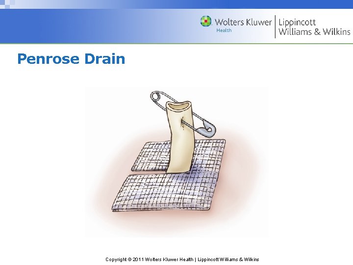
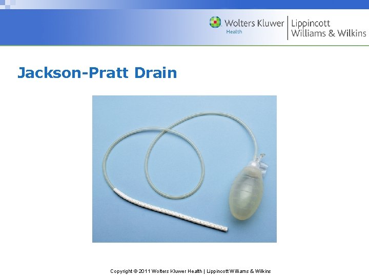
Type of Drainage Systems • Open systems – Penrose drain • Closed systems – Jackson-Pratt drain – Hemovac drain Copyright © 2011 Wolters Kluwer Health | Lippincott Williams & Wilkins
Penrose Drain Copyright © 2011 Wolters Kluwer Health | Lippincott Williams & Wilkins
Jackson-Pratt Drain Copyright © 2011 Wolters Kluwer Health | Lippincott Williams & Wilkins
Color Classification of Open Wounds • R = red—protect • Y = yellow—cleanse • B = black—debride • Mixed wound—contains components of RY&B wounds Copyright © 2011 Wolters Kluwer Health | Lippincott Williams & Wilkins
Topics for Home Care Teaching • Supplies • Infection prevention • Wound healing • Appearance of the skin/recent changes • Activity/mobility • Nutrition • Pain • Elimination Copyright © 2011 Wolters Kluwer Health | Lippincott Williams & Wilkins
Factors Affecting the Response to Hot and Cold Treatments • Method and duration of application • Degree of heat and cold applied • Patient’s age and physical condition • Amount of body surface covered by the application Copyright © 2011 Wolters Kluwer Health | Lippincott Williams & Wilkins
Effects of Applying Heat • Dilates peripheral blood vessels • Increases tissue metabolism • Reduces blood viscosity and increases capillary permeability • Reduces muscle tension • Helps relieve pain Copyright © 2011 Wolters Kluwer Health | Lippincott Williams & Wilkins
Effects of Applying Cold • Constructs peripheral blood vessels • Reduces muscle spasms • Promotes comfort Copyright © 2011 Wolters Kluwer Health | Lippincott Williams & Wilkins
Devices to Apply Heat • Hot water bags or bottles • Electric heating pads • Aquathermia pads • Hot packs • Moist heat • Sitz baths • Warm soaks Copyright © 2011 Wolters Kluwer Health | Lippincott Williams & Wilkins
Devices to Apply Cold • Ice bags • Cold packs • Hypothermia blankets • Moist cold Copyright © 2011 Wolters Kluwer Health | Lippincott Williams & Wilkins