The Cellular Level of Organization Chapter 1 Section


")


 �# of")


 are isotonic so RBCs will not")


")


 Na+ in cytosol bind to pump 2. that triggers")


 �lipoproteins membrane (proteins + lipids) �packaged")

 & steroids �inactivates (detoxifies) drugs &")


 �ends with 2 daughter")


 �eliminates potentially harmful")


 and do a")


- Slides: 170
The Cellular Level of Organization Chapter 1 Section 1: Parts of a Cell
Typical Structures Found in a Cell
Plasma Membrane �forms a cell’s flexible outer surface �separates inside from outside �acts as a selective barrier that regulates what gets in or out of cell �plays key role in communication with other cells
Plasma Membrane Fluid Mosaic Model �resembles an ever-moving sea of fluid lipids that has large proteins bobbing along throughout the lipids
Fluid Mosaic Model of Plasma Membrane �membrane proteins either float around like icebergs or are anchored at specific locations
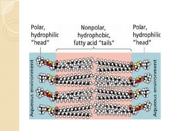
The Lipid Bilayer �basic structure of plasma membrane �made of 2 back-to-back layers using 3 types of lipids �asymmetric
Lipid Bilayer 1 st Lipid Phospholipids 1. ◦ ◦ 75% of lipid membrane lipids with phosphate groups
Lipid Bilayer 2 nd Lipid 2. Cholesterol � 20% of lipid membrane �a steroid �makes bilayer less fluid �weakly amphipathic (-OH group on 1 end can form Hbonds)
Lipid Bilayer 3 rd Lipid �Glycolipids �~5% of membrane lipids �lipid with attached carbohydrate group �stick out into extracellular space
Phospholipids are Amphipathic
Phospholipid Bilayer
“likes like” �hydro: water �philic: loving �phobic: hating �Polar molecules hydrophilic �Nonpolar molecules hydrophobic
Arrangement of Membrane Proteins �Integral Membrane Proteins �embedded in membrane �most are transmembrane: protrude both into cytosol & ECF �amphipathic �Peripheral Protein �loosely ass’c with polar heads of membrane lipids or with integral proteins �inner or outer surface of membrane
Membrane Proteins
Glycoproteins �proteins with carbohydrate groups attached to the ends that protrude into ECF �these carbohydrate groups are oligosaccharides (few-sugars): straight or branched chains of 2 – 60 monosaccharides
Glycoproteins
Glycocalyx �carbohydrate portions of glycolipids + glycoproteins = glycocalyx �acts like name tag for cells to recognize each other
Glycocalyx �enables cells to adhere to each other �protects some cells from being digested by enzymes in ECF
Types & Functions of Membrane Proteins: Integral Proteins ION CHANNELS � are holes or pores thru which specific ions flow in or out of cell � most are very selective � Examples: K+ ion channels 1.
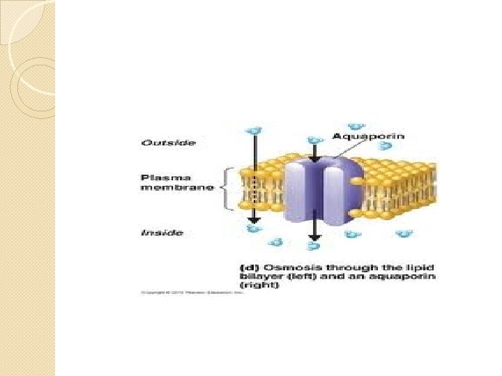
Types of Integral Membrane Proteins 2. TRANSPORTERS �selectively move a polar substance (like water) across the plasma membrane �water transporters called aquaporins �Example: aquaporin
Types of Integral Membrane Proteins RECEPTORS � serve as cellular recognition sites � recognizes & binds specific type of molecule � the specific molecule that binds to a receptor protein is called a ligand 3.
Receptor Proteins
Types of Integral Membrane Proteins 4. ENZYMES �catalyze specific chemical reactions at inside or outside cell
Types of Integral Membrane Proteins 5. LINKERS �integral proteins anchor proteins in the plasma membranes of neighboring cells or to protein filaments inside & outside the cell
Cell-Identity Markers �glycoproteins or glycolipids �Example: ABO blood type markers
Selective Permeability �some substances can easily pass thru, others not at all �lipid-bilayer nonpolar so ◦ permeable to small nonpolar substances ◦ impermeable to polar substances ◦ Water is an exception ◦ is polar ◦ small amts cross plasma membrane
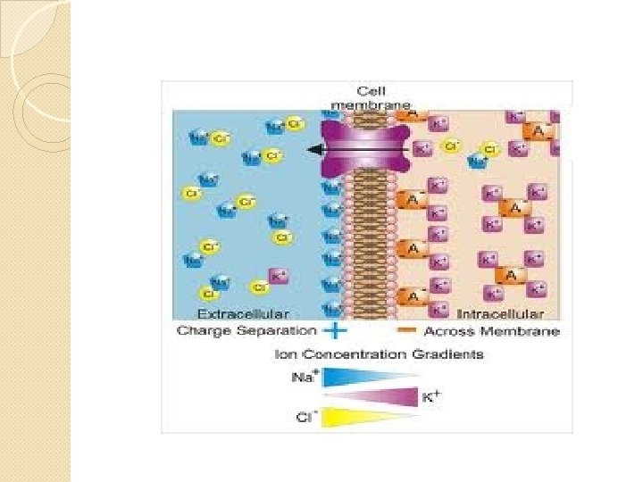
Gradients Across the Plasma Membrane �selective permeability across plasma membrane maintains different concentrations of certain substances on either side of plasma membrane �concentration gradient: a difference in the concentration of a chemical from 1 place to another
Gradients across the Plasma Membrane �because several of the substances kept in different concentrations on either side of membrane have (+) or (-) charges there is an electrical gradient across the membrane �typically, inside surface of membrane is slightly (-) & outside surface of membrane is slightly (+)
Gradients across the Plasma Membrane �a difference in charge across membrane is called: membrane potential
�just like a battery where there is separation of charge used as source of energy so too a membrane potential can be thought of as potential energy for cell to move substances in or out of cell
�The combined influence of the concentration gradient & membrane potential on movement of a particular ion is referred to as its electrochemical gradient
Chapter 3 Section 2 Transport Across the Plasma Membrane
Essential Question �What is the main difference between primary & secondary active transport mechanisms?
Kinetic Energy Transport �diffusion is a passive process in which the random mixing of particles in a solution occurs because of the particle’s kinetic energy �solute: dissolved substances �solvent: liquid that does dissolving
Diffusion �solutes move down their concentration gradient until they are evenly distributed
Diffusion �solute particles continue to move due to their kinetic energy �http: //highered. mcgraw- hill. com/sites/0072495855/student_view 0/chapter 2/animation__how_diffusion_ works. html
Factors that Influence Rate of Diffusion across Plasma Membrane Steepness of Concentration Gradient � greater the difference between 2 sides, higher rate of diffusion � steepness of electrochemical gradient determines rate of diffusion 1.
Factors that Influence the Rate of Diffusion across Plasma Memebrane 2. Temperature �higher temperature higher KE of particles faster it diffuses �all diffusion processes go faster when person has a fever
Factors that Influence Rate of Diffusion across Plasma Membrane 3. Mass of the Solute Diffusing �larger masses diffuse slower than smaller masses
Factors that Influence Rate of Diffusion across Plasma Membrane 4. Surface Area � larger membrane surface area, faster the diffusion rate
Factors that Influence Rate of Diffusion across Plasma Memebrane 5. Diffusion Distance �greater distance over which diffusion must occur, the longer it takes �plasma membranes very thin so diffusion is rapid, processes that thicken that space (autoimmune disease) slow down rate
What Diffuses Across Plasma Membrane �nonpolar, hydrophobic, small molecules: �O 2 & CO 2 gases �Lipids (fatty acids, steroids) �fat-soluble vitamins: ◦ A, D, E, K
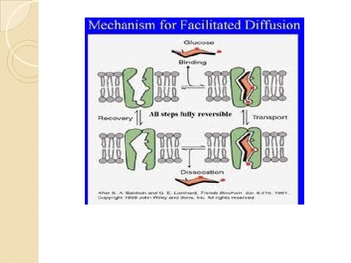
Facilitated Diffusion �diffusion through an integral membrane protein from side with higher concentration to side with lower concentration
Facilitated Diffusion
Facilitated Diffusion �transporters very specific �passive process (no nrg output by cell) �# of transporters limited so there is an upper limit to the amount that can cross membrane: called a transport maximum or Tmax �once all transporters occupied the diffusion rate cannot increase
Facilitated Diffusion �http: //programs. northlandcollege. edu/ biology/Biology 1111/animations/passi ve 3. swf
Gated Channels when part of the channel protein acts as a “plug” or “gate” changing shape to open pore & in another shape to close it
Gated Channels �some randomly alternate between open & closed �some regulated by chemical or electrical changes inside & outside cell
Voltage Gated Channels
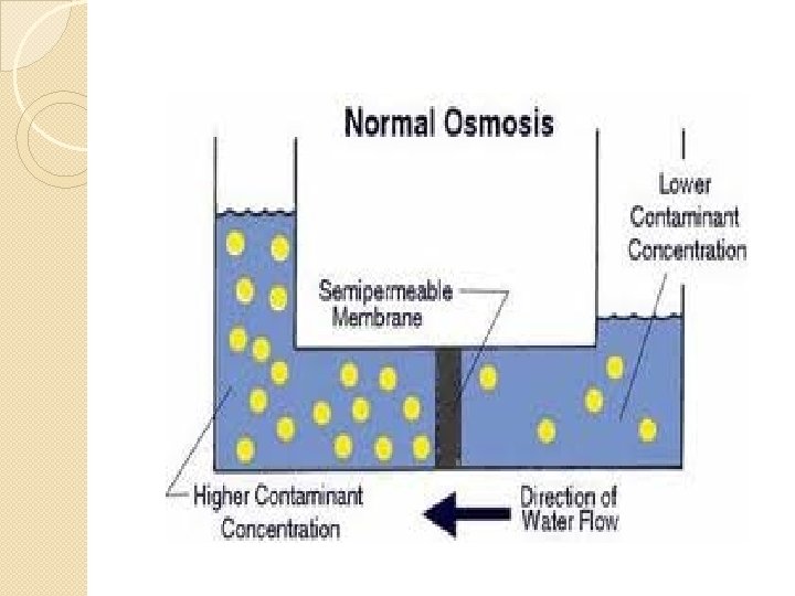
Osmosis �net movement of water thru selectively permeable membrane ◦ water can move across membrane but solute cannot �water moves down water’s concentration gradient
Osmosis �during osmosis water molecules pass through a plasma membrane in 2 ways: 1. moving thru lipid bilayer 2. moving thru aquaporins
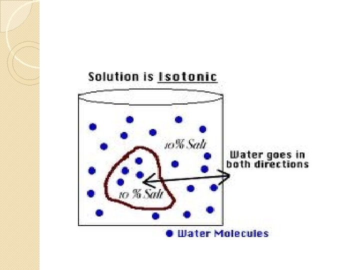
Isotonic Solutions �concentration of solutes that cannot cross membrane same on both sides of membrane �in human body isotonic solution is 0. 9% Na. Cl (normal saline) �allows cells to maintain normal shape & volume
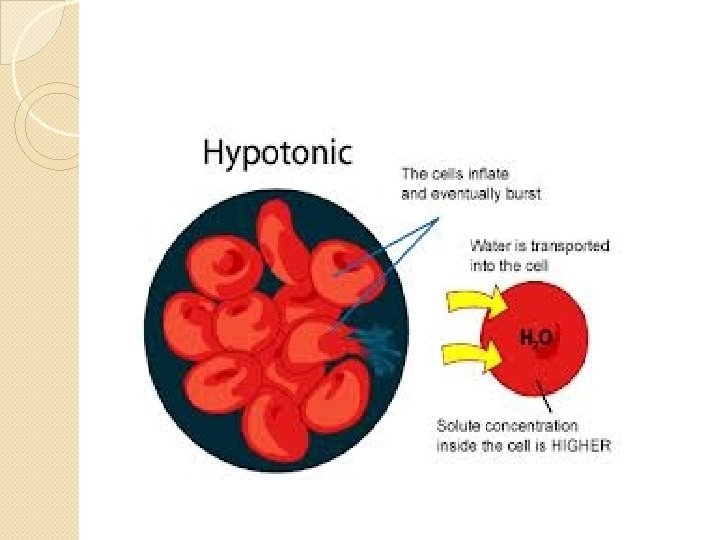
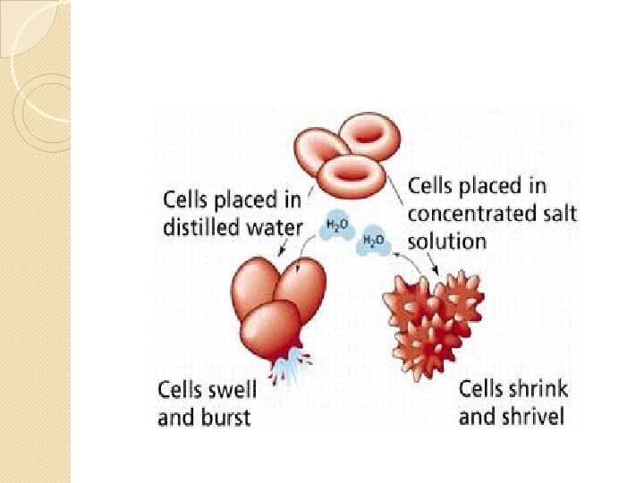
Hypotonic Solution �solution has lower concentration of solutes than the cytosol (higher concentration of water outside cell) �water molecules enter cell faster than they leave cell volume enlarges cell bursts (if RBC called hemolysis)
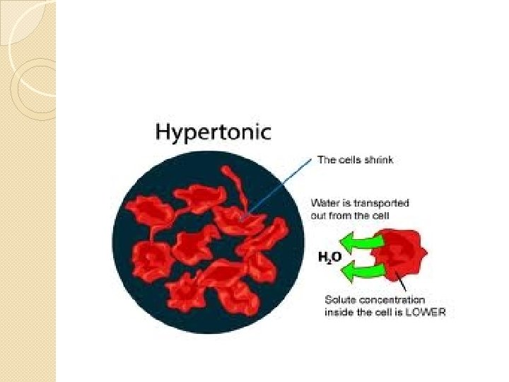
Hypertonic Solution �solution has a higher concentration of solute than cytosol �water molecules will move out of cell into ECF �RBCs will shrink, called crenation
Crenation
Medical Uses of Isotonic Solutions �IV solutions (intravenous) are isotonic so RBCs will not change shape or volume �example : 0. 9% Na. Cl
Medical Uses for Hypotonic Solutions �given orally or IV used to treat dehydration �water & sports drinks hypotonic
Medical Uses for Hypertonic Solutions �used to treat cerebral edema (increased interstitial fluid around the brain) �example: mannitol ◦ causes excess water in interstitial fluid into blood kidney for excretion in urine
Active Transport �used for polar solutes that must enter or leave cells need to move “uphill” against their concentration gradients �requires cell to spend some energy moving things against concentration gradients
Primary Active Transport �nrg required comes from hydrolysis of ATP (adenosine triphosphate)
ATP Pumps
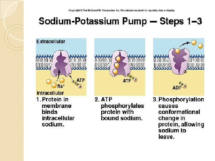
�a typical body expends 40% of its ATP on primary active transport �most prevalent primary active transport mechanism is the sodium-potassium pump (Na/K pump) part of the pump acts as enzyme ATPase (hydrolyzes ATP) so pump also called: sodium-potassium-ATPase pump
Steps in Na+/K+/ATPase Pump (3) Na+ in cytosol bind to pump 2. that triggers hydrolysis of 1 ATP; phosphate removed attached to pump which changes shape of the pump causing all 3 Na+ to be expelled out into the ECF. This new shape also favors the binding of 2 K+ from ECF onto pump 1.
Steps in Na+/K+/ATPase Pump
Steps in Na+/K+/ATPase Pump 3. binding K+ triggers release of that phosphate group that attached itself in Step 1, releasing it causes change in shape of protein pump 4. when shape changes in Step 3, the 2 K+ are released into the cytosol. Pump now ready to attach 3 Na+ and start over
Na+/K+/ATPase Pump �http: //www. brookscole. com/chemistry_ d/templates/student_resources/shared _resources/animations/ion_pump/ionp ump. html
Why is the Na+/K+/ATPase Pump so Important? �must maintain the different concentrations of Na+ and K+ in order for cells to maintain normal volume & for ability of some cells to generate electrical signals (action potentials in neurons)
Secondary Active Transport �potential electricalchemical nrg stored in concentration gradient of Na+ or H+ is used to push substances across membrane “uphill” (against their concentration gradient) �these concentration gradients established by primary active transport so the 2’ active transport indirectly uses ATP
Secondary Active Transport
Symporters �transporters that move 2 substances in same direction
Antiporters �transporters that move 2 substances in opposite directions across the plasma membrane
Transport in Vesicles �vesicle: small spherical sac formed by membrane
Endocytosis �materials move into cell in a vesicle formed by plasma membrane � 3 Types: 1. Receptor-mediated endocytosis 2. Phagocytosis 3. Pinocytosis
Receptor-Mediated Endocytosis
Receptor-Mediated Endocytosis
Phagocytosis
Macrophage Phagocytosing Bacteria
Pinocytosis �means cell-drinking �tiny droplets of ECF taken into cell �no receptor proteins involved
Exocytosis �releases material from cell �all cells do it but is most important in: 1. Secretory cells that release digestive enzymes, hormones, mucus 2. end of axons on neuron that release neurotransmitters
Exocytosis
�segments of plasma membrane lost in endocytosis and gained in exocytosis �usually evens out �if necessary ER makes more
Chapter 3 Section 3 Cytoplasm & Organelles �Essential Question: �How would you describe the structure & function of the cytoplasm and main organelles of the cell?
Cytosol �same as intracellular fluid �fluid portion of cytoplasm � 55% of cell volume �composition varies from cell to cell: mostly it is 75 -90% water, rest is dissolved & suspended components ◦ ions , glucose, a. a. , fatty acids, lipids, proteins, ATP, waste products
Cytosol �site of many chemical reactions ◦ glycolysis
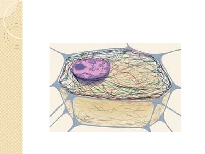
Cytoskeleton �a network of protein filaments that extends throughout the cytosol
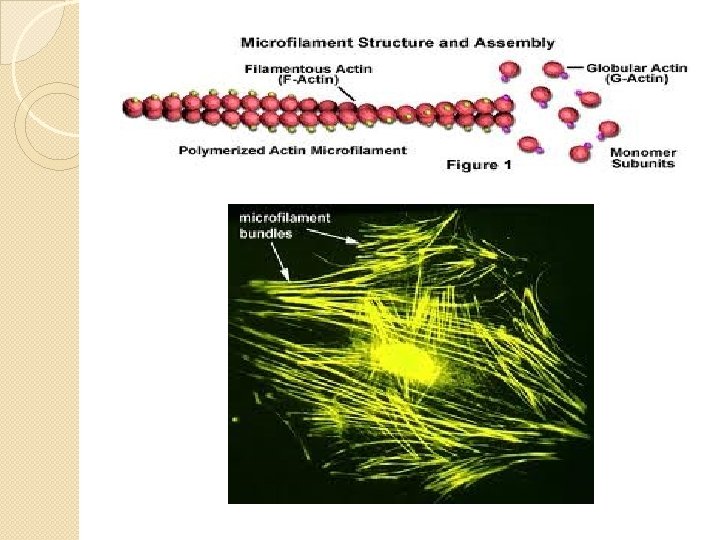
3 Types of Cytoskeleton 1. Microfilaments thinnest of the 3 made of protein actin most prevalent at periphery of cell 2 functions: 1. movement 2. support
Microfilaments �also provide mechanical support for microvilli : fingerlike projections of plasma membrane; function is to greatly increase surface area �abundant in cells of small intestine that absorb
Cytoskeleton 2. Intermediate Filaments medium-sized exceptionally strong (used in parts of cell under mechanical stress) also function to anchor nucleus & attach cells to each other
Intermediate Filaments
Microtubules �largest of cytoskeleton components �Long, unbranched hollow tubes �made mostly of protein tubulin �made in centrosomes an organelle near nucleus �function: cell shape, movement of organelles, chromosomes, flagella, & cilia
Microtubules
Cilia
Flagella
Ribosomes �sites of protein synthesis �made of r. RNA + >50 proteins ◦ 2 subunits: made separately in nucleolus ◦ apart until making protein
Ribosomes �found: in cytoplasm 1. ◦ ◦ called “free” ribosomes make proteins used in cytosol attached to membrane of RER 2. ◦ make proteins to be used by specific organelles or to be exported from cell
Ribosomes
Endoplasmic Reticulum ER �network of membranes in form of flattened sacs or tubules �extend from its connection to nuclear envelope to throughout the cytoplasm � >1/2 membranous surfaces w/in most cells �types: rough or smooth
Rough ER = RER �continuous with nuclear membrane �folded in series of flattened sacs �outer surface RER studded with ribosomes which make proteins that then travel thru spaces of ER for processing & sorting
Proteins made in RER �glycoproteins (proteins + carbohydrate) �lipoproteins membrane (proteins + lipids) �packaged inside vesicles Golgi or directly to export
RER
Smooth ER = SER �makes fatty acids (fa) & steroids �inactivates (detoxifies) drugs & other harmful substances �stores & releases Ca++ that trigger muscle contractions
SER & Drug Tolerance � 1 of functions of liver is to detoxify& it does this because liver cells, hepatocytes, have a lot of SER �prolonged use of a medication or illegal drug causes hepatocytes to increase the amount of SER to accommodate detoxifying extra drug & so person has to take more & more drug to get same affect
SER
Hepatocytes
SER or RER?
Golgi Complex Functions �modifies, sorts, packages, & transports proteins received from RER �forms secretory vesicles that discharge processed proteins via exocytosis into ECF; vesicle membrane then added to plasma membrane
Golgi Complex
Lysosomes Function �digest substances that enter cell via endocytosis & transport final products of digestion into cytosol �autophagy (digestion of worn out organelles) �autolysis (digest entire cell) �extracellular digestion
Lysosomes
Mitochondria �generate ATP thru reactions of aerobic cellular respiration �have a double membrane ◦ both similar to plasma membrane �mitochondrial DNA (maternal) ◦ replicate when demand for ATP increases
Mitochondria
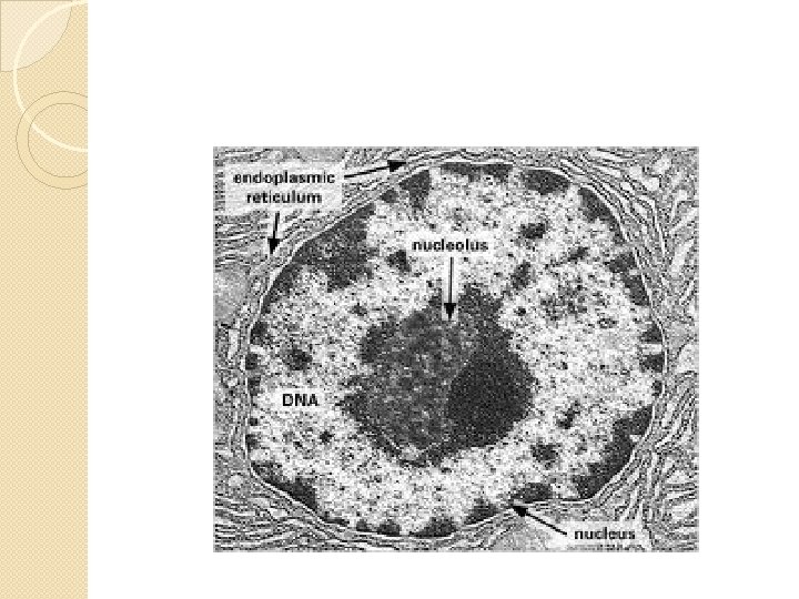
NUCLEUS �Functions: contains genetic information 2. controls cellular structure 3. controls cell activities 4. produces ribosomes in nucleolus 1.
NUCLEUS �usually most prominent structure in cell �most human cells have 1 ◦ mature RBC have 0 ◦ mature WBC have multiple �dbl membrane surrounds it, called nuclear envelope ◦ holes in it called nuclear pores
NUCLEOLUS �prominent, dark, spherical structure in nucleus �site of manufacture of subunits of ribosomes �disperse & disappear during cell division
Name & Function Please 1. 2.
3. 4.
5. 6.
7. 8.
9. 10.
Chapter 3 Section 4 Cell Division �Essential Question: How would you explain the stages and significance of somatic & reproductive cell division?
Definitions �somatic cell: any cell other than ova or sperm �mitosis: nuclear division of somatic cell �cytokinesis: division of cytoplasm �gametes: sex cells, ova or sperm �meiosis: nuclear division of gametes
Definitions �homologous chromosomes: 2 copies of same chromosomes: 1 maternal, 1 paternal �haploid: having only 1 set of chromosomes as found in gametes, designated n �diploid: having 2 sets of chromosomes as found in somatic cells, designated 2 n
Cell Cycle
Cell Cycle �G 0 when cells are not going to go thru cell division again or not for very long periods of time �typically: ◦ G 1 ◦S ◦ G 2 8 – 10 hours 8 hours 4 – 6 hours
Definitions �centromere: constricted portion of a duplicated chromosome; serves as point of attachment for microtubules that pull chromatids apart during anaphase �kinetochore: protein complex attached to outside of centromere
Definitions �mitotic spindle: collective term for a football-shaped assembly of microtubules that is responsible for movement of chromosomes during mitosis �sister chromatids: pair of identical DNA strands that are joined at the centromere and separate during mitosis becoming a chromosome of 1 of 2 daughter cells
Chromosomes
Mitosis
Mitotis Phases: Prophase 1. ◦ ◦ Early Prophase chromatin condenses & shorten into chromosomes visible under light microscope made up of sister chromatids with centromere
Late Prophase �mitotic spindle begins to form extending to poles of cell
Metaphase �microtubules align the centromeres at exact center of mitotic spindle �called metaphase plate
Anaphase �centromeres split & sister chromatids move toward opposite poles �once separated chromatids called chromosomes
Telophase �identical sets of chromosomes now at opposite poles of cell �chromosomes uncoil chromatin �nuclear envelope re-forms �cleavage furrow starts to form
Cytokinesis �division of cytoplasm completed �cleavage furrow usually midway between poles (actin microfilaments form a contractile ring) �organelles on each side
Mitosis �start with parent cell with diploid # chromosomes (46) �ends with 2 daughter cells each with a diploid # of chromosomes (46) genetically the same as parent cell
Name That Stage of Mitosis 1. 2.
3. 4.
5. 6.
Control of the Cell Cycle �Cells have 3 choices: 1. Keep going thru Cell Cycle 2. enter G 0 3. Die Homeostasis is maintained when there is a balance between cell proliferation & cell death
Cell Cycle Regulation �cyclins are proteins that are responsible for activating enzymes called Cdks (cyclin-dependent protein kinases) �Cdks transfer a phosphate group from ATP to a protein which activates the protein; other enzymes remove the phosphate group thus de-activating it
Cell Cycle Regulation �activation/deactivation of Cdks is crucial to DNA replication, mitosis & cytokinesis �activation of specific cyclin-Cdk complexes responsible for a cell going from G 1 S G 2 mitosis
Cell Death �apoptosis: programmed cell death �trigger from outside or inside cell causes “cell-suicide” genes to produce enzymes that damage the cell in several ways �phagocytes (cells that engulf debris, bacteria; “clean-up crew”) ingest the dying cell
Apoptosis �removes unneeded cells during fetal development (like webbing between digits) �eliminates potentially harmful cells like cancer cells
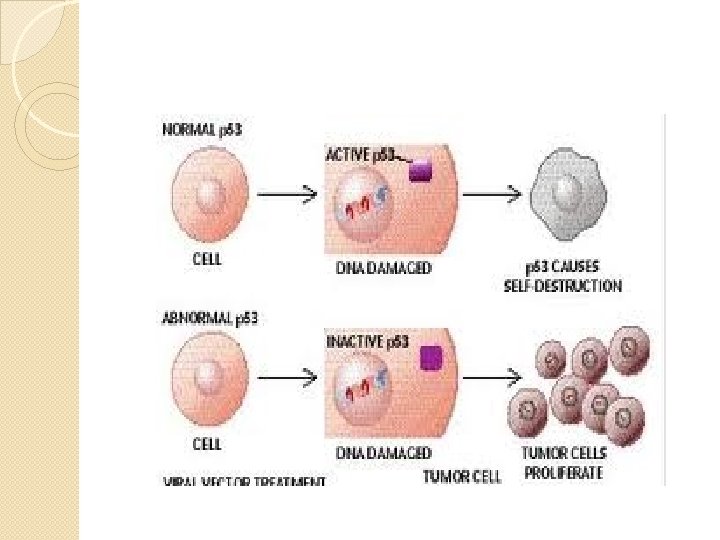
Tumor-Suppressor Genes �produce proteins that inhibit cell division �damage to these genes causes some types of cancer �example: p 53 is a tumor-suppressor gene on chromosome 17 which normally arrests cells in G 1 & assists in repair of DNA… if repair not successful p 53 induces apoptosis of that cell ◦ abnl in this gene have led to different cancers (breast, colon)
Meiosis �for reproduction of sex cells �must have haploid # so when ova & sperm unite offspring will have normal diploid # of chromosomes �starts with diploid cell meiosis 4 daughter cells each with a haploid # of chromosomes (23) that are genetically different than original cell
Meiosis
Meiosis I: Prophase I � homologous chromosomes matched up (called synapse) and do a little trading of alleles (crossing over): results in genetic variation: the 4 daughter cells will be genetically different
Crossing Over
Meiosis I Metaphase I �homologous chromosomes line up on metaphase plate Anaphase I �homologous chromosomes move to opposite poles
Meiosis I �Telophase I �nuclear envelopes re -form �each nucleus has 23 duplicated chromosomes �Cytokinesis I � 2 daughter cells each with n chromosomes (in duplicated state)
Meiosis II Prophase II Metaphase II
Meiosis II Anaphase II Telophase II
Meiosis II
Homeostatic Imbalances Cancer �Large group of diseases characterized by uncontrolled or abnormal cell proliferation �Abnl cells also called: ◦ Tumor ◦ Malignancy ◦ Neoplasm Metastasis: cells spread from organ of origin
Causes of Cancer �Carcinogen: chemical or radiation that causes cancer �Carcinogens cause DNA mutations (permanent changes in DNA) �Oncogenes: cancer-causing genes �Oncogenic Viruses: stimulate abnl proliferation of cells (HPV, Hepatitis. C)