INFLAMMATION INFLAMMATION Defn Local response of living mammalian


 § Tissue necrosis from")


 § A hallmark of acute inflammation is increased")

 Vascular dilation and increased blood flow")


 Recognition and attachment of the particle")

 for which the phagocytes express")


, which,")
 to generate the highly reactive")


- Slides: 35
INFLAMMATION
INFLAMMATION Defn: § Local response of living mammalian tissues to injury due to any agent. Signs of inflammation § Rubor (Redness) § Tumor (Swelling) § Callor (Heat) § Dollar (Pain) § Functeo leasa (Loss of function)
Types of inflammation § Acute inflammation § Chronic inflammation
Acute inflammation § Rapid host response that serves to deliver leukocytes and plasma proteins, such as antibodies, to sites of infection or tissue injury.
Stimuli for acute inflammation § Infections (bacterial, viral, fungal, parasitic) § Tissue necrosis from any cause, including ischemia trauma, physical and chemical injury § Foreign bodies (splinters, dirt, sutures) § Immune reactions (hypersensitivity reactions)
Events in acute inflammation § Vascular events § Cellular events
Vascular events 1. Changes in Vascular Flow and Caliber 2. Altered vascular permeability
1. Changes in Vascular Flow and Caliber § Vasodilation, sometimes it follows a transient constriction of arterioles, lasting a few seconds. § Vasodilation first involves the arterioles and then leads to opening of new capillary beds in the area. § The result is increased blood flow, which is the cause of heat and redness (erythema).
1. Changes in Vascular Flow and Caliber § Vasodilation is followed by increased permeability of the microvasculature. § The loss of fluid and increased vessel diameter will slow the blood flow resulting stasis § Blood leukocytes, principally neutrophils, accumulate along the vascular endothelium.
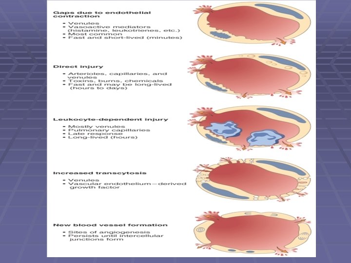
2. Increased Vascular Permeability (Vascular Leakage) § A hallmark of acute inflammation is increased vascular permeability leading to the escape of a protein-rich exudate into the extravascular tissue, causing edema.
Mechanisms responsible for increased vascular permeability § Contraction of endothelial cells resulting in increased interendothelial spaces is elicited by histamine and other chemical mediators § Endothelial injury, resulting in endothelial cell necrosis and detachment. § Transcytosis Increased transport of fluids and proteins, through the endothelial cell.
The major local manifestations of acute inflammation (1) Vascular dilation and increased blood flow (causing erythema and warmth) (2) Extravasation and extravascular deposition of plasma fluid and proteins (edema) (3) Leukocyte emigration and accumulation in the site of injury.
The major local manifestations of acute inflammation
Cellular events A. Exudation of leucocytes B. Phagocytosis
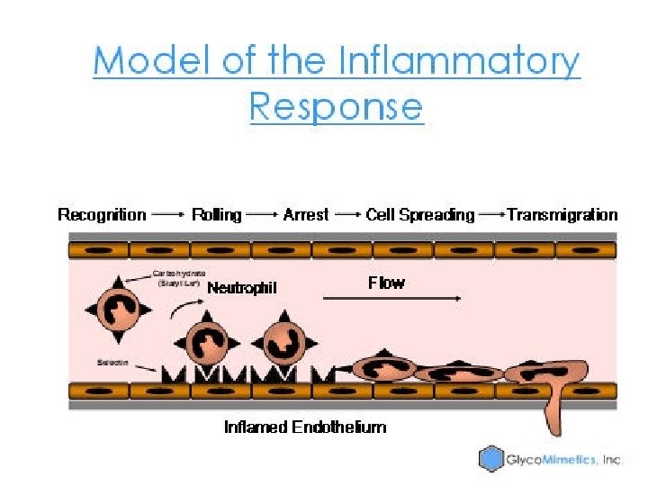
A. Exudation of leucocytes 1. Cellular margination 2. Pavementing 3. Rolling and adhesion 4. Emigration and chemotaxis
1. Exudation of leucocytes § Margination : white cells assume a peripheral position along the endothelial surface. § Rolling : Individual and then rows of leukocytes adhere transiently to the endothelium. § Pavementing : Endothelium lined by white cells. § Adhesion : The cells adhere firmly to endothelial cells mediated by adhesion molecules.
Adhesion molecules § The initial rolling interactions are mediated by a family of proteins called selectins. There are three types of selectins: § one expressed on leukocytes (L-selectin), § one on endothelium (E-selectin), § one in platelets and on endothelium (Pselectin).
Adhesion molecules Firm adhesion is mediated by a family of heterodimeric leukocyte surface proteins called integrins. § Vascular cell adhesion molecule 1 (VCAM-1) § Intercellular adhesion molecule-1 (ICAM-1).
Endothelial-Leukocyte Adhesion Molecules § Endothelial Molecule /Leukocyte Molecule / Role § P-selectin / Sialyl-Lewis X–modified proteins /Rolling § E-selectin / Sialyl-Lewis X–modified proteins /Rolling § § § and adhesion Gly. Cam-1, CD 34 / L-selectin / Rolling ICAM-1 / CD 11/CD 18 (β 2) integrins / Adhesion, arrest, transmigration VCAM-1 / VLA-4 (β 1) integrin / Adhesion
2. Transmigration § Migration of the leukocytes through the § § § endothelium. Chemokines stimulate the cells to migrate through interendothelial spaces towards the site of injury. Adhesion molecules PECAM-1 (platelet endothelial cell adhesion molecule) present in the intercellular junctions between endothelial cells are involved in the migration of leukocytes. Leukocytes pierce the basement membrane by secreting collagenases, and enter the extravascular tissue.
3. Chemotaxis § Locomotion oriented along a chemical gradient. § Exogenous and endogenous substances act as § § § chemoattractants. Exogenous agents are bacterial products, Endogenous chemoattractants (1) cytokines, (e. g. , IL-8); (2) components of the complement system, particularly C 5 a (3) arachidonic acid (AA) metabolites, mainly leukotriene B 4 (LTB 4).
Chemotaxis § The leukocyte moves by extending filopodia that pull the back of the cell in the direction of extension.
MULTISTEP PROCESS OF LEUCOCYTE MIGRATION
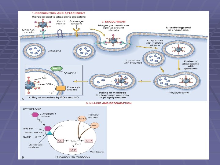
4. Phagocytosis § Three sequential steps : (1) Recognition and attachment of the particle to be ingested by the leukocyte (2) Engulfment (3) Killing or degradation of the ingested material.
1. Recognition and attachment § Mannose receptors, scavenger receptors, and receptors for various opsonins all function to bind and ingest microbes.
Opsonins § Microbes are opsonized by specific proteins (opsonins) for which the phagocytes express receptors. The major opsonins § Ig. G antibodies § C 3 b breakdown product of complement § Certain plasma lectins.
2. Engulfment. § Extensions of the cytoplasm pseudopods flow around it § Plasma membrane pinches off to form a vesicle phagosome that encloses the particle. § The phagosome then fuses with a lysosomal granule, resulting in phagolysosome. § During this process the phagocyte may also release granule contents into the extracellular space.
3. Killing and Degradation. § The final step is killing and degradation within neutrophils and macrophages. § Microbial killing is accomplished largely by reactive oxygen species and reactive nitrogen species, mainly derived from NO.
Reactive oxygen species § Azurophilic granules of neutrophils contain the enzyme myeloperoxidase (MPO), which, in the presence of a halide such as Cl-, converts H 2 O 2 to hypochlorite (OCl • ). § (OCl • ) destroys microbes by halogenation or by oxidation of proteins and lipids § The H 2 O 2 -MPO-halide system is the most efficient bactericidal system of neutrophils. § H 2 O 2 is also converted to hydroxyl radical ( • OH), another powerful destructive agent.
NO § NO reacts with superoxide ( o- ) to generate the highly reactive free radical peroxynitrite (ONOO • ). § These oxygen- and nitrogen-derived free radicals attack and damage the lipids, proteins, and nucleic acids of microbes.
Microbial killing by the action substances in leukocyte granules. § Neutrophil granules contain many enzymes (elastase), § § defensins, cationic arginine-rich granule peptides, Cathelicidins, antimicrobial proteins found in neutrophils and other cells Lysozyme, found in the glycopeptide coat of all bacteria Lactoferrin, an iron-binding protein is cytotoxic to many parasites; Bactericidal/permeability increasing protein, important in defense against some gram-negative bacteria
§THANK YOU