Inflammation lab Dr Bushra AlTarawneh Inflammation The response

Inflammation lab Dr. Bushra Al-Tarawneh

Inflammation The response of living tissue to injury. 1 - Acute inflammation 2 - Chronic inflammation: A- Nonspecific chronic inflammation B- Granulomatous inflammation.
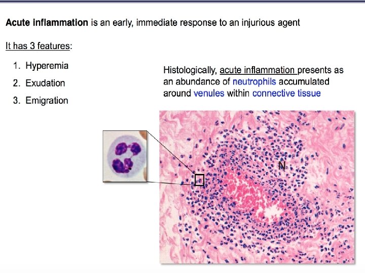

Microscopic changes of acute inflammation �Dilatation of vessels. �Fluid leaks into interstitium. �Increased permeability of vessels: (not to water but to protein). �Cells move into interstitium.

Acute inflammatory response show vasodilation with margination of neutrophils and exudation of fluid with edema.

Tissue oedema Tissue edema Neutrophil margination …. And emigration Neutrophil margination …. emigration

Myocardial infarction- neutrophil infiltration Dead myocytes Neutrophils

Acute inflammation in myocardium

Lobar pneumonia

Lobar pneumonia ls wal r a l o e v Al Alveoli should contain AIR… Not EXUDATE!

Acute bronchopneumonia

A variety of inflammatory cell types may be present, though one may predominate mainly neutrophils , but also plasma cells, lymphocytes and macrophages.

ABSCESS Localized area of pus formation surrounded by abscess membrane.

Abscess
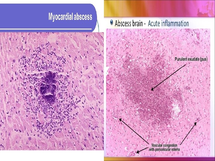

Liver abscess

Chronic inflammation 1. Long duration: 2. Tissue destruction greater than in acute inflam. 3. The inflammatory infiltrate: Macrophages, lymphocytes, plasma cells 4. Productive: Production of fibrous tissue through formation of granulation T.

Chronic endometritis: Lymphocytes and plasma cells in the endometrial stroma.

Chronic inflammation in the lung: 1 - Chronic inflammatory cells infiltration 2 - Destruction of the normal tissue (normal alveoli are replaced by spaces lined by cuboidal cells (arrow heads) 3 -Replacement by fibrosis (arrows)

Chronic cholecystitis

Rheumatoid arthritis This deformity of the hand is due to rheumatoid arthritis. This autoimmune disease leads to synovial proliferation and joint destruction, involving small joints of hands and feet, followed by wrists, ankles, elbows, and knees.

Rheumatoid arthritis: Chronic inflammation in the synovium with collections of dark blue lymphocytes.

Chronic inflammation in skin
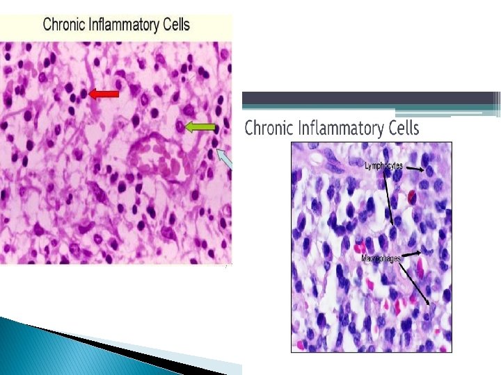

Plasma cells

Eosinophils

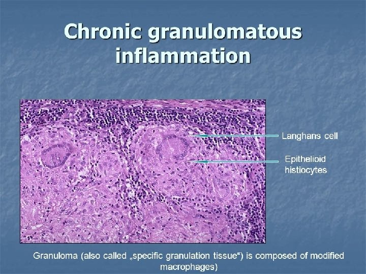
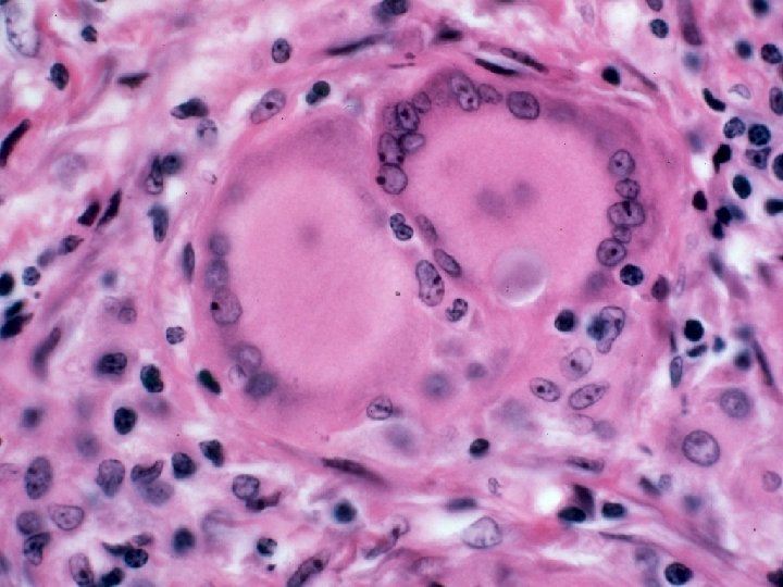
Granulomatous Inflammation �Distinctive • • pattern of chronic inflammation characterized by aggregation of : Activated macrophages (epithelioid). Multinucleated giant cells.

Diagram of typical TB granuloma Multinucleated GC Epithelioid cells Caseation Lymphocytes fibroblasts

T. B granuloma

Caseating granuloma. Epithelioid cells surround a central area of necrosis that appears irregular, amorphous, and pink.

Lung, granulomatous infl. with caseation Grossly, areas of caseation appear cheese-like. Yellowish mottling of lung tissue. Small, yellow nodules (caseating TB granulomas). Some have fused together to form larger areas of yellow caseation (arrow)

TB granulomas lung Low power: Two, adjacent, well- defined, rounded granulomas with multinucleated giant cells. High power: Amorphous, pinkish central caseation, surrounded by a rim of epithelioid cells. caseation Ep ith eli oid ce lls

Thank you
- Slides: 36