Chapter 7 1 Life Is Cellular What is


 n Anton n 1674 Van Leeuwenhoek Lensmaker n Observed single-celled organisms")


 create enlarged image as light")


")

")


 · Allow exchange of materials (into & out of")
 · Clumps up to form")

 Boundary between inside and outside of cell n")


 · Makes and Transports materials: Rough ER =")
 · Fixes, stores, and sends proteins and")

 Copyright © 2003 Pearson Education,")

 plasma membrane · Makes up boundary between inside and outside")


 Organelle where photosynthesis takes place n Uses sunlight")

 n Protects the plant cell and is")


 phospholipid layers.")


 •")


 1. Is osmosis passive or active transport? 2. What specific")


 DO NOT WRITE ON")


 – the process of expelling material out of the")

 & export (move out) large materials")

 - You. Tube PASSIVE ACTIVE")


 n 1.")


- Slides: 110
Chapter 7. 1 Life Is Cellular
What is a cell? The basic unit of structure and function in living things – All carry out life processes – Take in food – Break it down – Produce energy
All Organisms are Made of Cells n All organisms are made of cells n Organisms are either Unicellular (single cell) or Multicellular (many cells) n Most cells can not be seen without magnification
Scientists n Robert Hooke n 1665 n English Scientist n Observed cork cells (dead plant cells) n Saw “compartments” and named them “cells”
Scientists (Cont. ) n Anton n 1674 Van Leeuwenhoek Lensmaker n Observed single-celled organisms in pond water
n Additional Scientists: – Theodor Schwann (Stated that the cell was the basic unit of structure in animals) - 1839 – Rudolph Virchow (Said that new cells come from existing cells) - 1855 – Ernst Ruska (invented the electron microscope) - 1931 – Matthias Schleiden & Robert Brown (Both stated that the cell was the basic unit of structure in plants) - 1838 All the observations and discoveries by the different scientists were tested over and over again. As a result, the Cell Theory was developed!
The Cell Theory States. . . 1. All living things are made up of one or more cells 2. Cells are the basic units of structure in living things, and cells carry on all life processes 3. Cells come only from other living cells
Looking Forward! What do you Already Know? Directions: On a piece of lined paper, follow the following prompts and answer the following questions. 1. Complete a QUICK sketch of a microscope and label as many parts as you can. 2. Who might use microscopes and why are they important? 3. If you could look at one thing under a microscope, what would it be and WHY?
Warm – Up n Be ready to describe three components of the Cell Theory. n Also, be ready to identify the parts of a microscope!
Light Microscope Eyepiece – lens to look through Adjustment Knobs - focusing Arm Objectives – rotating lenses Stage clips Stage – supports the slide Diaphragm – controls amount of light Base – support & carrying Light Source
WARM-UP Number 1 -6 in your warm-up book and match the correct scientist with his discovery/contribution. 1. ______ Leeuwenhoek microscope 2. ______ Shleiden A. Invented the first electron B. Discovered that all animals are made of of cells 3. ______ Schwann C. Discovered that all plants are composed of cells 4. ______ Ruska D. Observed and named “cells” in cork 5. ______ Hooke E. Discovered that all cells come from other cells 6. ______ Virchow F. Was the first person to observe living cells in pond water under the microscope
Objectives n Identify Parts of a Microscope n Compare/Contrast 3 different types of microscopes based on: 1. 2. 3. Total Magnification Advantages & Disadvantages Purpose & Micrograph Images
Key Words n Lens – A piece of curved glass or other clear material – Light bends as it passes through to enlarge images n Micrograph – a photograph of the view through a microscope n Total Magnification – Degree of image/specimen enlargement
3 Types of Microscopes Compound Light Microscope 2. Transmission Electron Microscope 3. Scanning Electron Microscope 1.
Compound Light Microscope n 2 lenses (eyepiece & objective) create enlarged image as light shines through specimen n Total 800 x Magnification = Plant Cell
Transmission Electron Microscope n Electrons pass through specimens to study the INSIDE of things (instead of light) n Total magnification = 1, 000 x Inside Bacteria
Scanning Electron Microscope n Bounces electrons off specimens to study the SURFACE of specimens n Total Magnification = 1, 000 x
Short Video Clip n Microscopes Types n For each type of microscope pick out: – Advantages – Disadvantages
Practice C A B
Scanning Electron Microscope Staple in a piece of paper
Light Microscope Plant Cells (with chloroplasts)
Scanning Electron Microscope Embryo embedded in a Uterus
Scanning Electron Microscope Grain of Salt (Na. Cl)
Transmission Electron Microscope Red Blood Cell
Just because science is fun… Eye of a Fruit Fly Velcro Mascara Brush
Wrap - Up n For each Scenario, choose which type of microscope (LM, SEM, or TEM) would be most useful. 1. Check a CD for scratches because the music “skips” 2. Examine the internal structure of a cell taken from an autopsy 3. Study the swimming movements of plankton from an area in the ocean near an oil spill
Prokaryotes vs Eukaryotes n Two Main Kinds of cells 1. Prokaryotes – Cells that do not have a nucleus. The DNA is not separated from the rest of the cell. 2. Eukaryotes – Cells that enclose their DNA in a nucleus
Prokaryotes 1. Smaller and simpler than eukaryotes. 2. DNA floats freely in the cell. 3. Prokaryotes grow, reproduce and respond to their environment. 4. Some prokaryotes can glide along surfaces and swim through liquids. 5. An example of a prokaryote is bacteria.
Eukaryotes 1. Larger and more complex than prokaryotes. 2. Most eukaryotes have dozens of structures and membranes inside them. 3. The nucleus separates the DNA from the rest of the cell. 4. Protists live as single cells. 5. Others make up large organisms with many cells such as plants, animals, and fungi.
Prokaryotes vs Eukaryotes
Warm-Up 1. _____ are the basic units of life. 2. According to the _______, all living things are made of cells. 3. Cells whose DNA is held in a nucleus are _______.
Chapter 7. 2 Cell Structure
THINK ABOUT IT – At first glance, a factory is a puzzling place, and the sheer diversity of activity can be confusing. – However, if you take your time and watch carefully, what might at first seem like chaos begins to make sense. – The same is true for the living cell.
Cells are different by. . ·Size ·Shape ·Function ·Location ·Parts • A cell’s form fits function Copyright © 2003 Pearson Education, Inc. publishing as Benjamin Cummings Slide 3. 2
What are some similarities that cells share? · All eukaryotic cells (plant & animal cells) have the same 3 general structures: 1) Nucleus 2) Cytoplasm If they are Eukaryotic 3) Cell membrane Figure 3. 1 a Copyright © 2003 Pearson Education, Inc. publishing as Benjamin Cummings Slide 3. 2
1. Nucleus = control center! DNA · Has three main parts: 1. Nucleolus 2. Nuclear envelope 3. DNA (“chromatin”) Figure 3. 1 b Copyright © 2003 Pearson Education, Inc. publishing as Benjamin Cummings Slide 3. 3
Nuclear envelope (Membrane around nucleus) · Allow exchange of materials (into & out of nucleus) · Made up of a phospholipid bilayer Copyright © 2003 Pearson Education, Inc. publishing as Benjamin Cummings Slide 3. 4
Chromatin of nucleus · DNA (and protein mixed in) · Clumps up to form chromosomes during cell division. Copyright © 2003 Pearson Education, Inc. publishing as Benjamin Cummings Slide 3. 6
2. Cytoplasm · Gel-filled space that organelles float around in. ·Organelles- Mini “organs” - Perform metabolic functions in cytoplasm. · (ex. Mitochondria) Copyright © 2003 Pearson Education, Inc. publishing as Benjamin Cummings Slide 3. 9
Cell Membrane ( aka plasma Membrane) Boundary between inside and outside of cell n Made up of phospholipid bilayer n
Warm-Up n What are the 2 major parts of cellular membranes? n Label the structures: (phospholipid bilayer, phosphate head, hydrophobic fatty acid tails, protein)
ORGANELLES n. A part/piece inside of a cell that has a specific job n Organelle n There means “mini-organ” are many different types of organelles in cells
2. Organelles in cytoplasm Figure 3. 4 Copyright © 2003 Pearson Education, Inc. publishing as Benjamin Cummings Slide 3. 10
2. Organelles · Mitochondria · Location for Cellular respiration process that makes ATP molecules (usable energy) Copyright © 2003 Pearson Education, Inc. publishing as Benjamin Cummings Slide 3. 15
2. Organelles · Ribosomes • Site for Protein synthesis • (process by which proteins are made) Copyright © 2003 Pearson Education, Inc. publishing as Benjamin Cummings Slide 3. 11
2. Organelles · Endoplasmic reticulum (ER) · Makes and Transports materials: Rough ER = w/ ribosomes Smooth ER = no ribosomes Copyright © 2003 Pearson Education, Inc. publishing as Benjamin Cummings Slide 3. 12
2. Organelles · Golgi apparatus (Golgi body) · Fixes, stores, and sends proteins and other chemical products around cell Copyright © 2003 Pearson Education, Inc. publishing as Benjamin Cummings Slide 3. 13 a
2. Organelles · Lysosomes · Contain digestive enzymes that breakdown macromolecules (proteins, nucleic acids, lipids) or harmful bacteria Copyright © 2003 Pearson Education, Inc. publishing as Benjamin Cummings Slide 3. 14
2. Organelles · Vacuoles · Storage!!! (water, food, waste) Copyright © 2003 Pearson Education, Inc. publishing as Benjamin Cummings Slide 3. 17
On a piece of scrap paper number 1 -7 and write the organelle that matches the appropriate function. WITHOUT LOOKING AT YOUR NOTES! 1. 2. 3. 4. 5. 6. 7. Stores water, food, and waste Produces energy for the cell Breaks down and recycles waste Makes proteins Controls all the cell processes Packages and distributes materials throughout the cell Network of tubes for materials to travel through the cell Golgi Bodies, Nucleus, Ribosome, Endoplasmic Reticulum, Lysosome, Vacuole, Mitochondria
Phosp holipid 3. (Cell) plasma membrane · Makes up boundary between inside and outside of cell · Made of phospholipid bilayer cell membrane Copyright © 2003 Pearson Education, Inc. publishing as Benjamin Cummings Slide 3. 7 a
3. Plasma Membrane Copyright © 2003 Pearson Education, Inc. publishing as Benjamin Cummings Figure 3. 2 Slide 3. 7 b
Warm-Up n What are the 2 major parts of cellular membranes? n Label the structures: (phospholipid bilayer, phosphate head, hydrophobic fatty acid tails, protein)
Special structures of some plasma membranes 1. Cilia moves materials across the cell surface 2. Flagellum helps the cell “swim” (pushes it through water like a tail) cilia flagella Copyright © 2003 Pearson Education, Inc. publishing as Benjamin Cummings Slide 3. 18
Review Warm-Up With a partner, list the functions of each of these organelles on scrap paper. • Nucleus • ER • Golgi • Ribosome • Lysosome • Mitochondria • Cell/plasma Membrane
Chloroplasts (found only in plant Cells) Organelle where photosynthesis takes place n Uses sunlight (Energy) to make glucose (food)
What is a Chloroplast? n Chloroplasts! Chloroplasts are like “solar power packs”. n Trap light energy and turn it to chemical energy for the cell via PHOTOSYNTHESIS!! n
Cell Wall (only found in plant cells) n Protects the plant cell and is strong to keeps the cell’s box-like shape n Outer-most layer of plant cells
Cell Wall The cell wall protects the plant cell’s insides and gives it shape. Cool fact: Many cell walls stacked on top of each other are responsible for holding up the weight of a tall tree against the force of gravity.
What are some differences between Plant Cells and Animal Cells? • Cell wall • Cell membrane • Large central vacuole (H 2 O) • Chloroplasts • Box-like shape • Various organelles • Only cell membrane • Many small vacuoles • Rounded shape • various organelles
Plant Animal
Chapter 7. 3 Cell TRANSPORT!!!
Cell Membranes Function of Cell Membranes n Form a boundary/barrier between inside and outside the cell n Controls transport of substances that pass into and out of the cell Structure of Cell Membranes n Parts include mainly: 1. 2. Phospholipids Membrane-bound proteins (squeezed in)
Phospholipid Bilayer n Made up of 2 (tail-to-tail) phospholipid layers.
Phospholipid Bilayer n Proteins are squeezed in among the phospholipid bilayer with many functions.
Protein Functions n The proteins in the phospholipid bilayer carry out many functions: 1. Enzymes – (catylists) speed up chemical reactions 2. Transport proteins – carry materials into and out of cell 3. Communication- with other cells (“cell to cell recognition”) 4. Cell signaling as “chemical messengers”
“Fluid Mosaic” Model n “Fluid Mosaic Model” – phospholipids movement is “fluid” meaning they drift/float freely – Proteins float like “icebergs” in a “sea of phospholipids”
“Semipermeable Membrane” n “Semi-Permeable” – allow some materials to pass through and restricts/stops others!
What do you think… Make 2 sketches to diagram your answer: 1. How would phospholipids orient/situate themselves if they were placed on the SURFACE of water? 2. How would phospholipids orient/situate themselves if they were IMMERSED in water? n
Transport n Cellular membranes control the traffic of molecules into and out of the cell. n Objectives: – Compare/Contrast Passive & Active Transport – Compare/Contrast Diffusion and Osmosis – Explain how large molecules move across membranes (to get into or out of cells)
n Think of a time when you were in a large group of people and it was so crowded that you could hardly move. (AKA…they were totally in your personal bubble!) n Write down 3 words describing how you felt in that situation.
TRANSPORT KEY CONCEPT Materials move across cell membranes because of concentration differences Kool Aid
Passive Transport n Types of transport in which it is easy for molecules to move through cell membranes n NO ENERGY IS REQUIRED for the molecules to move because particles naturally move down a concentration gradient – (high low concentration) n There are 3 types of Passive Transport: – Simple Diffusion (of molecules) – Facilitated Diffusion (of molecules) – Osmosis (of water molecules)
Simple Diffusion n Movement of the particles of a substance from where they are more concentrated to where they are less concentrated (molecules move from high low concentration) – Examples: perfume spreading etc. n Doesn’t stop until equilibrium is established
Example Dynamic Equilibrium
Diffusion Demos • Molecules diffuse down a concentration “gradient” (high to low concentration) • Particles eventually reach a dynamic equilibrium. • What do you think it will look like when equilibrium is reached? ? ?
Review Simple Diffusion 1. What type of transport is simple diffusion? (passive or active? ) _______ 2. Molecules move from areas of ____ concentration to ____ concentration. 3. It is ____(easy or hard? ) for molecules to squeeze through phospholipids of cell membranes. 4. Molecules won’t stop diffusing until ______ is reached.
Balloon Demo n What do you notice? n What does the balloon represent? n What does the extract represent? n What characteristic of the balloon allows us to smell the extract?
Starch & Iodine Molecules Starch WHAT IS THE MAJOR DIFFRENCE BETWEEN THESE 2 MOLECULES? ? ? Iodine HOW YOU THINK IT WILL AFFECT HOW THEY MOVE THROUGH SEMIPERMEABLE MEMBRANES?
DEMO n Which molecule will be able to move across the semipermeable membrane? n FYI…When iodine and starch mix, it turns a blue/black color! Iodine water (yellow) Starch water (white)
Facilitated Diffusion • Some molecules cannot easily diffuse across the cell membrane! (boo hoo ) • They can only diffuse across membranes using transport proteins. • Facilitated diffusion is diffusion through transport proteins. Molecules move from high low concentration
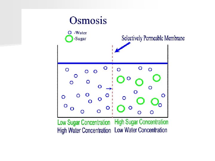
Osmosis • Type of diffusion where water molecules move across a semipermeable membrane. • Movement occurs from high to low concentration • Water molecules don’t mix well with lipid bilayers. Cells have channel proteins called aquaporins in their membranes. Review: Semipermeable membranes allow some materials to cross but not others = solute (ex. dissolved salt) = water molecule
3 Types of Solutions Note: solutions are always named by comparing them to an object (usually a cell) n Hypertonic – LOWER concentration of water molecules (than the cell) n Hypotonic – – solution with: – HIGHER concentration of water molecules (than the cell) n Isotonic – solution with equal concentration of water & solutes
WARM-UP (review of osmosis) 1. Is osmosis passive or active transport? 2. What specific molecule moves by osmosis? 3. Does osmosis require energy? 4. In what direction are the molecules moving? _____
Scenarios for Osmosis 1. 2. 3. A cell in an Isotonic solution (equal amount of water) A cell in a Hypertonic solution (less H 20 than cell) A cell in a Hypotonic solution (more H 20 than cell)
WARM UP Draw these situations in your notes. Which way will water flow for each of these cells in solution (draw arrows)? Are the solutions Hyper, Hypo, or isotonic? ? 90 % H 20 70% H 20 90% H 20 10 % Salt/Na. Cl 60 % H 20 90% Salt/Na. Cl
DI Osmosis Problems Activity 1. Obtain: 1. Scenario Card (BLUE) DO NOT WRITE ON IT!! 2. Yellow piece of paper to write your answers on. 2. Write out and Answer these questions on the yellow paper: 1. The cell is in a ________ solution. (hypotonic, hypertonic, isotonic) 2. Water will move _________ the cell. (into OR out of) 3. The cell will _________. (Shrink, Burst, or Stay the Same)
Plasmolysis n Definition: Loss of water by a plant cell n Causes plants to wilt!
Video Clip n Diffusion and Osmosis
WARM UP Answer the following questions about this osmosis problem: 1. What type of solution is the cell sitting in? 70% H 2 O 30% salt (hypertonic, hypotonic, isotonic) 2. Which move? 3. What cell? way will water (into/out of the cell) will happen to the 10% H 2 O 90% salt
Active Transport KEY CONCEPT Cells use energy to transport large materials that cannot diffuse across a membrane OR need to be pumped against the normal concentration gradient. Sometimes cells must use active transport to stay alive!!!!!
Active Transport n Allows a cell to move a substance against its normal concentration gradient. so now……low high concentration n Requires energy from the cell in the form of ATP molecules.
3 Types of Active Transport 1. Protein pumps (proteins push molecules across membranes from low high concentration)
2. Exocytosis (“sounds like exit”) – the process of expelling material out of the cell Exocytosis
3. Endocytosis –The process of taking large materials into the cell Endocytosis
Endocytosis and Exocytosis Cells can import (bring in) & export (move out) large materials using Endocytosis and exocytosis. Cells use energy to transport material in vesicles/compartments. • Endocytosis is the process of taking material into the cell. • Phagocytosis (solid materials) • Pinocytosis (liquid materials)
Active vs. Passive Transport Passive High Low No Energy Needed! Active Low High Against Conc. Gradient Requires Energy (ATP)!!
Passive vs. active transport n Membrane Transport Animation (LEGENDADO) - You. Tube PASSIVE ACTIVE
Crash Course n In Da Club - Membranes & Transport: Crash Course Biology #5 - You. Tube
Organize your thoughts… Using the graphic organizer provided, write in each shape: Protein Pumps 1. a brief summary/description of the process or topic a basic picture to illustrate it 2. **Proteins pumps use ATP energy to pump molecules from low high concentration.
Chapter 7. 4 Homeostasis and cells
Key Questions n 1. How do individual cells maintain homeostasis? n 2. How do the cells of multicellular organisms work together to maintain homeostasis?
n Homeostasis – Relatively constant internal physical and chemical conditions. n Single-celled organisms 1. grow 2. respond to their environment 3. change food or sunlight into useful energy 4. Reproduce
Unicellular Organisms n Prokaryotes are very adaptable. They live everywhere (Ex: Bacteria) n 1. Soil, on leaves, in the ocean, in the air and within the human body.
Multicellular Organisms n Human cells and other multicellular organisms do not live on their own. n They need other cells to survive. n They become specialized for certain jobs. n They communicate with one another to maintain homeostasis.
Cell Specialization 1. Some cells are specialized for movement. 2. Some must react to their environment. 3. Some must take in substances that the organism needs. Ex: air passages have cilia to sweep dust away to keep lungs clean
Levels of Organization n Specialized cells are organized into tissues. n Tissues are organized into organs. n Organs are organized into organ systems.
Cellular Communication n 1. Cells in large organisms use chemical signals to communicate. n 2. They can speed up or slow down activities of the cells that receive them. 3. Cells have cellular junctions to help communicate with other cells. n Receptors to receive communication n