CHAPTER 5 KNEE GENU VALGUM The alignment pattern

 The alignment pattern of genu valgum commonly demonstrates: 1. Excessive Internal")


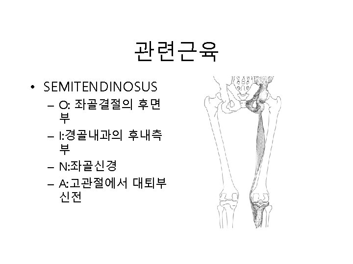
 – I: medial condyle of tibia")
 – I: lateral margin of patella(")


 The alignment pattern of genu varum commonly demonstrates: 1. Excessive External")


 – I: lateral margin of patella(")
 – I: medial condyle of tibia")


. MOD: Internally rotate, traction")


 2. Biceps Femoris (short head) 3. Tensor Fascia")


 – I: medial condyle of tibia")


 – I: lateral margin of patella(")


 2. Biceps Femoris (short head) 3. peroneus Longus")


 2. Biceps Femoris (short head) 3. Peroneus Longus")


 2. Biceps Femoris (short head) 3. Tibialis Anterior")


 2. Biceps Femoris (short head) 3. Tibialis Anterior")


 2. Biceps Femoris (short head) 3. Peroneus Longus")


- Slides: 148
CHAPTER 5 KNEE
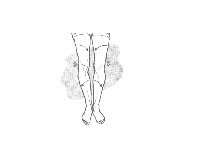
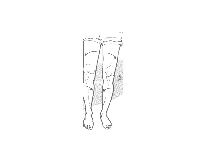
GENU VALGUM (외반슬) The alignment pattern of genu valgum commonly demonstrates: 1. Excessive Internal Rotation of the femur (external rotation restriction) 2. Excessive External Rotation of the tibia (Internal Rotation restriction) 3. Medial Patella deviation due to femur rotation
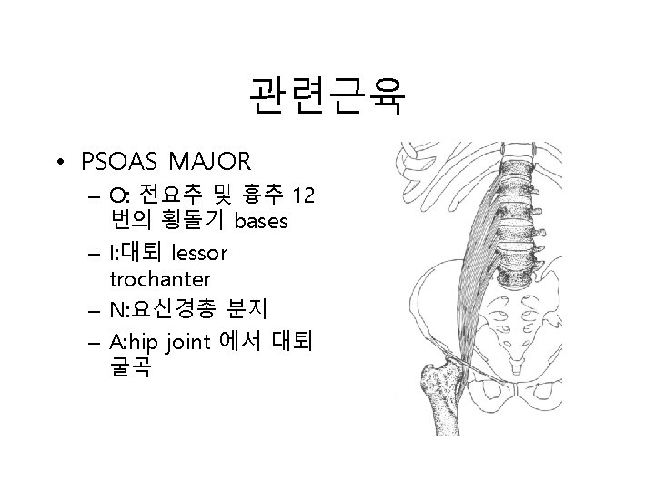
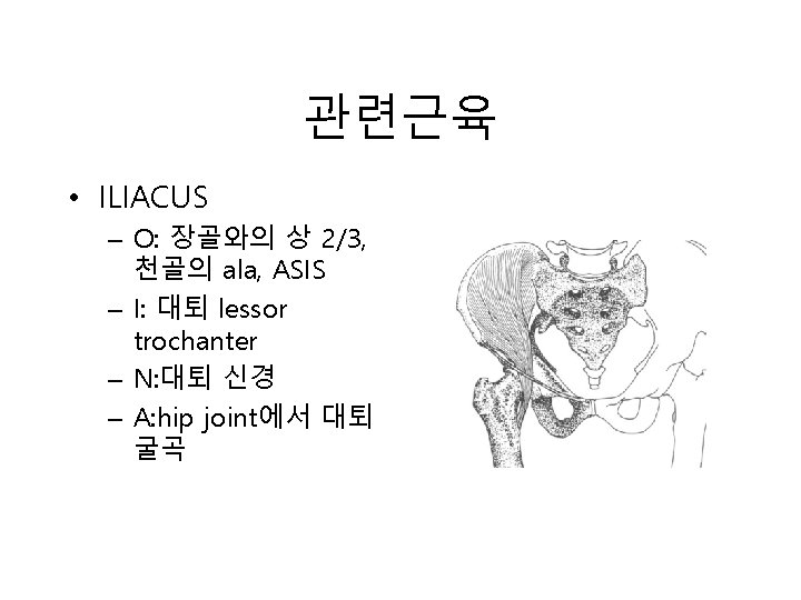
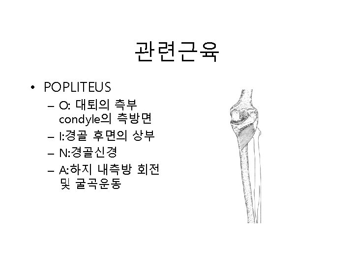
DYSKINETIC MUSCLES Knee: 1. Gracilis 2. Semitendinosus 3. Sartorius Hip 1. Psoas 2. lliacus 3. Piriformis 4. Semimembranosus 5. Popliteus 6. Vastus Medialis and Lateralis 4. Gluteus Maximus & Medius 5. Obturator Externus & Internus 6. Quadratus Femoris
관련근육 • VASTUS MEDIALIS – O: intertrochanteric line(medial) – I: medial condyle of tibia – N: 대퇴 신경 – A: Extends leg at knee joint
관련근육 • VASTUS LATERALIS – O: intertrochanteric line(lateral) – I: lateral margin of patella( 경골조면 ) – N: 대퇴신경 – A: Extends leg at knee joint
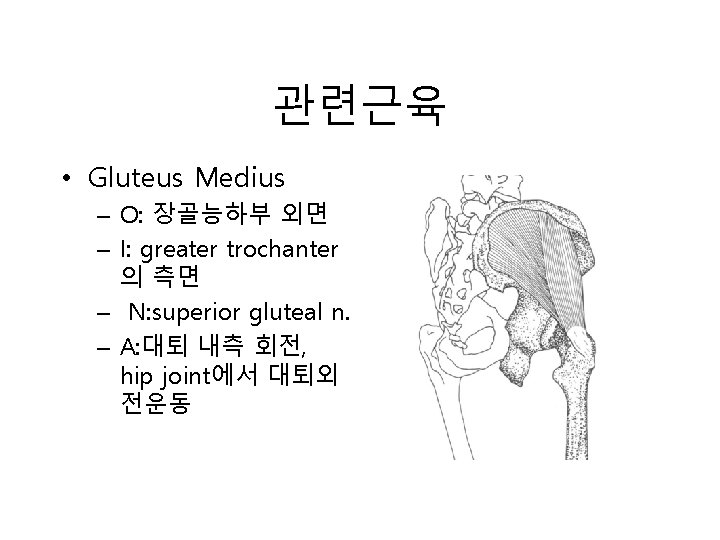
관련근육 • PIRIFORMIS – O: internal surface of sacrum, sacrotuberous ligament – I: greater trochanter 의 상연 – N: S 1, S 2 – A: 대퇴외회전, 외전운 동(abd. )
관련근육 • GLUTEUS MAXIMUS – O: 천골후방, sacrotuberous lig. – I: 대퇴근막장근, 대퇴의 gluteal tuberosity – N: inf. Gluteal n. – A: 대퇴 abd. 외회전운동. 몸체 신전
관련근육 • Obturator externus – O: pubic bone, 폐쇄 공 주변 좌골부 – I: trochanteric fossa of femur – N: Obturator nerve – A: 대퇴 외회전
관련근육 • Obturator internus – O: pelvic surface of obturator membrane – I: greater trochanter 의 내측면 – N: 천신경총 분지 – A: hip joint에서 대퇴 외회전
관련근육 • Quadratus femoris – O: ischial tuberosity 의 lateral border – I: below intertrochanteric crest – N: 천신경총 분지 – A: hip joint에서 대퇴 외회전
GENU VARUM (내반슬) The alignment pattern of genu varum commonly demonstrates: 1. Excessive External Rotation of the femur (Internal rotation restriction) 2. Excessive Internal Rotation of the tibia (External Rotation restriction) 3. Lateral Patella deviation due to femur rotation
DYSKINETIC MUSCLES Knee: 1. Biceps Femoris 2. Tensor Fascia Lata Hip 1. Tensor Fascia Lata 2. Gluteus Minimus 4. Vastus Lateralis 5. Vastus Medialis 4. Adductor Brevis 5. Adductor longus
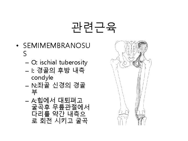
관련근육 • Biceps femoris – O: 장두-ischial tuberosity , 단두linea aspera – I: 비골두 측면, 경골측 방condyle. – N: 좌골신경분지 – A: hip joint에서 대퇴 신전, knee joint에서 leg 굴곡
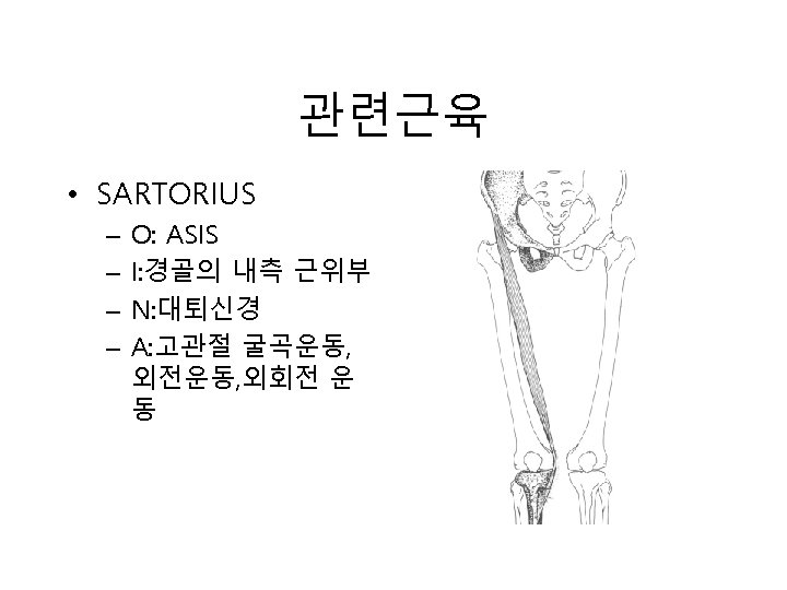
관련근육 • Tensor fascia lata – O: 장골능외측 – I: 대퇴상부의 iliotibial tract – N: superior gluteal n. – A: 대퇴 내회전, 굴곡
관련근육 • VASTUS LATERALIS – O: intertrochanteric line(lateral) – I: lateral margin of patella( 경골조면 ) – N: 대퇴신경 – A: Extends leg at knee joint
관련근육 • VASTUS MEDIALIS – O: intertrochanteric line(medial) – I: medial condyle of tibia – N: 대퇴 신경 – A: Extends leg at knee joint
관련근육 • Gluteus minimus – O: middle and inferior gluteal line – I: greater trochanter 의 anterior surface – N: superior gluteal n. – A: hip joint에서 대퇴 외전, 대퇴 내측회전
관련근육 • Adductor brevis – O: pubis의 inf. Ramus. – I: linea aspera의 근위 부 – N: Obturator n. – A: hip joint에서 대퇴 내전
관련근육 • Adductor longus – O: pubic bone의 ant. – I: linea aspera의 중간 부위 – N: Obturator n. – A: Adduct thigh, 내회 전보조
EXTERNALLY ROTATED TIBIA ① IND: Medial capsular ligament pain, genu valgum, prominent medial tibial condyle and plateau, tightness of the pes anserine tendons, chondromalacia patellae, restricted internal tibia rotation. ② PP: Prone with involved knee flexed 50˚ to 70˚. ③ DP: End of table with involved knee resting on the Dr. s shoulder. ④ CH: Thenar contact behind tibial condyles. ⑤ IH: Interlocked with CH.
LOC: Internally rotate with caudad thrust (toward the Dr. ). MOD: Internally rotate, traction and thrust. DM: 1. Semitendinosus 2. Gracilis 3. Sartorius 4. Semimembranosus 5. Popliteus
교정법 • EXTERNALLY ROTATED TIBIA
교정법 2 • EXTERNALLY ROTATED TIBIA
교정법 3 • EXTERNALLY ROTATED TIBIA
교정법 4 -1 • EXTERNALLY ROTATED TIBIA
교정법 4 -2 • EXTERNALLY ROTATED TIBIA
INTERNALLY ROTATED TIBIA ① IND: Lateral capsular ligament pain, genu varum, prominent lateral tibial condyle and plateau, tightness of the ilio-tibial and lateral hamstring endons, chondromalacia patellae, restricted external tibia rotation. ② PP: Prone with involved knee flexed 50˚ to 70˚. ③ DP: End of table with involved knee resting on the Dr. 's shoulder. ④ CH: Thenar contact behind tibial condyles. ⑤ IH: Interlocked with CH. ⑥ LOC: Externally rotate with caudad thrust (toward the Dr. ). ⑦ MOD: Externally rotate, traction and thrust.
DM: 1. Biceps Femoreis (long head) 2. Biceps Femoris (short head) 3. Tensor Fascia Lata
교정법 • INTERNALLY ROTATED TIBIA
교정법 2 • INTERNALLY ROTATED TIBIA
교정법 3 • EXTERNALLY ROTATED TIBIA
관련근육 • Biceps femoris – O: 장두-ischial tuberosity , 단두linea aspera – I: 비골두 측면, 경골측 방condyle. – N: 좌골신경분지 – A: hip joint에서 대퇴 신전, knee joint에서 leg 굴곡
관련근육 • Tensor fascia lata – O: 장골능외측 – I: 대퇴상부의 iliotibial tract – N: superior gluteal n. – A: 대퇴 내회전, 굴곡
POSTERIOR TIBIA ① IND: Popliteal fossa tenderness, anterior tivial trauma, posterior cruciate ligament damage, posterior drawer sign, patellar tendon hypotonicity and depression, restricted anterior tibia motion. ② PP: Prone, involved knee flexed 70˚. ③ DP: End of table with involved tibial condyles. ④ POC: Both hands interlock on the posterior aspect of the tibial condyles. ⑤ LOC: Toward the Dr. (posterior to anterior).
MOD: Traction and thrust toward the Dr. DM: 1. Vastus Intermedius 2. Rectus Femoris 3. Hamstrings
교정법 • POSTERIOR TIBIA
관련근육 • Rectus femoris – O: ASIS, ilium above acetabulum – I: patella tendon을 통 한 경골조면 – N: femoral n. – A: knee joint에서 leg 신전, hip joint에서 대퇴굴곡
관련근육 • Hamstring muscles – – – – 1. 2. 3. 4. 5. 6. 7. Sciatic nerve. Quadratus femoris Biceps femoris Semimebranosus Semitendinosus Tibial n. Common peroneal n.
관련근육 • Quadratus femoris – O: ischial tuberosity 의 lateral border – I: below intertrochanteric crest – N: 천신경총 분지 – A: hip joint에서 대퇴 외회전
관련근육 • Biceps femoris – O: 장두-ischial tuberosity , 단두linea aspera – I: 비골두 측면, 경골측 방condyle. – N: 좌골신경분지 – A: hip joint에서 대퇴 신전, knee joint에서 leg 굴곡
ANTERIOR TIBIA ① IND: Patellar tendon tenderness, Anterior Drawer Sign, Anterior Cruciate Ligament damage, patellar tendon hypertonicity or tendonitis, restricted posterior tibia motion. ② PP: Supine with involved leg extended. ③ DP: Side of involvement. ④ CH: Proximal anterior tibia. ⑤ IH: Supporting distal tibia and ankle. ⑥ LOC: Anterior to posterior. ⑦ MOD: Dr. traction leg distally and thrusts anterior to posterior.
DM: 1. Semimembranosus 2. Semitendinosus 3. Biceps Femoris 4. Quadriceps
교정법 • ANTERIOR TIBIA
관련근육 • Quadratus femoris – O: ischial tuberosity 의 lateral border – I: below intertrochanteric crest – N: 천신경총 분지 – A: hip joint에서 대퇴 외회전
관련근육 • Biceps femoris – O: 장두-ischial tuberosity , 단두linea aspera – I: 비골두 측면, 경골측 방condyle. – N: 좌골신경분지 – A: hip joint에서 대퇴 신전, knee joint에서 leg 굴곡
MEDIAL TIBIA ① IND: Trauma to lateral tibia, valgus trauma, medial collateral ligament damage, restricted lateral motion. ② PP: Supine, involved hip flexed approximately 45˚. The knee is slightly flexed. ③ DP: Side of involvement standing laterally with leg in Dr. 's axilla. ④ CH: Pisiform at proximal medial tibial condyle. ⑤ IH: Wrist of the CH. ⑥ LOC: Medial to lateral. ⑦ MOD: Traction toward the Dr. then thrust with the pisiform contact at the medial tibial condyle.
DM: 1. Tensor Fascia Lata 2. Biceps Femoris 3. Gracilis 4. Semitendinosus 5. Sartorius 6. Semimembranosus
교정법 • MEDIAL TIBIA
관련근육 • Tensor fascia lata – O: 장골능외측 – I: 대퇴상부의 iliotibial tract – N: superior gluteal n. – A: 대퇴 내회전, 굴곡
관련근육 • Biceps femoris – O: 장두-ischial tuberosity , 단두linea aspera – I: 비골두 측면, 경골측 방condyle. – N: 좌골신경분지 – A: hip joint에서 대퇴 신전, knee joint에서 leg 굴곡
LATERAL TIBIA ① IND: Trauma to medial tibia, valgus trauma, medial collateral ligament damage, restricted lateral motion. ② PP: Supine, involved hip flexed approximately 45˚. The knee is slightly flexed. ③ DP: Side of involvement standing laterally with leg in Dr. 's axilla. ④ CH: Pisiform at proximal lateral tibial condyle. ⑤ IH: Wrist of the CH. ⑥ LOC: Lateral to medial. ⑦ MOD: Traction toward the Dr. then thrust with the pisiform contact at the lateral tibial condyle.
DM: 1. Gracilis 2. Semitendinosus 3. Sartorius 4. Semimembranosus 5. Tensor Fascia Lata 6. Biceps Femoris
교정법 • LATERAL TIBIA
관련근육 • Tensor fascia lata – O: 장골능외측 – I: 대퇴상부의 iliotibial tract – N: superior gluteal n. – A: 대퇴 내회전, 굴곡
관련근육 • Biceps femoris – O: 장두-ischial tuberosity , 단두linea aspera – I: 비골두 측면, 경골측 방condyle. – N: 좌골신경분지 – A: hip joint에서 대퇴 신전, knee joint에서 leg 굴곡
SUPERIOR PATELLA ① IND: Patellar tendonitis, quadricep spasm, chondromalcia patellae, restricted inferior patella motion. ② PP: Supine, leg extended. ③ DP: Side of involvement. ④ CH: Superior hand takes a web contact over superior aspect of the patella. ⑤ IH: Stabilizes tibia below the knee. ⑥ LOC: Superior to inferior. ⑦ MOD: Traction inferior and thrust.
DM: 1. Vastus Intetmedius 2. Rectus Femoris 3. Hamstrings
교정법 • Superior Patella
관련근육 • Rectus femoris – O: ASIS, ilium above acetabulum – I: patella tendon을 통 한 경골조면 – N: femoral n. – A: knee joint에서 leg 신전, hip joint에서 대퇴굴곡
관련근육 • Quadratus femoris – O: ischial tuberosity 의 lateral border – I: below intertrochanteric crest – N: 천신경총 분지 – A: hip joint에서 대퇴 외회전
SUPERIOR-LATERAL PATELLA ① IND: Patellar tendonitis, quadricep spasm, chrondromalacia patellae, genu valgum, restricted inferior-medial motion. ② PP: Supine, leg extended. ③ DP: Side of involvement. ④ CH: Superior hand takes a web contact over superior-lateral aspect of the patella. ⑤ IH: Stabilizes tibia below the knee. ⑥ LOC: Superior to inferior and lateral to medial. ⑦ MOD: Traction according to the LOC and thrust.
DM: 1. Vastus Medialls 2. Vastus Intermedijus 3. Rectus Femoris 4. Hamstrings.
교정법 • Superior Lateral Patella
관련근육 • VASTUS MEDIALIS – O: intertrochanteric line(medial) – I: medial condyle of tibia – N: 대퇴 신경 – A: Extends leg at knee joint
관련근육 • Rectus femoris – O: ASIS, ilium above acetabulum – I: patella tendon을 통 한 경골조면 – N: femoral n. – A: knee joint에서 leg 신전, hip joint에서 대퇴굴곡
SUPERIOR-MEDIAL PATELLA ① IND: Patellar tendonitis, quadricep spasm, chrondromalacia patellae, genu valgum, restricted inferior-lateral patella motion. ② PP: Supine, leg extended. ③ DP: Side of involvement. ④ CH: Superior hand takes a web contact over superior-medial aspect of the patella. ⑤ IH: Stabilizes tibia below the knee. ⑥ LOC: Superior to inferior and lateral to medial. ⑦ MOD: Traction according to the LOC and thrust.
DM: 1. Vastus Lateralis 2. Vastus Intermedius 3. Rectus Femoris 4. Hamstrings.
교정법 • Superior Medial Patella
관련근육 • VASTUS LATERALIS – O: intertrochanteric line(lateral) – I: lateral margin of patella( 경골조면 ) – N: 대퇴신경 – A: Extends leg at knee joint
관련근육 • Rectus femoris – O: ASIS, ilium above acetabulum – I: patella tendon을 통 한 경골조면 – N: femoral n. – A: knee joint에서 leg 신전, hip joint에서 대퇴굴곡
INFERIOR FIBULA ① IND: Inversion sprain, tender fibular collateral ligament due to injury, restricted superior fibula motion. ② PP: Lateral recumbant position. ③ DP: End of table in plane with distal fibula malleolus. ④ CH: Lateral malleolus between thenar pads. ⑤ IH: Placed on wrist of CH. ⑥ LOC: Inferior to superior. ⑦ MOD: Traction cephalad, thrust is made with the CH at the lateral malleolus.
DM: 1. Biceps Femoris (long head) 2. Biceps Femoris (short head) 3. peroneus Longus 4. Peroneus Brevis 5. Peroneus Tertius 6. Extensor Hallucis Longus 7. Extensor Digitorum Longus
교정법 • Inferior fibula
관련근육 • Biceps femoris – O: 장두-ischial tuberosity , 단두linea aspera – I: 비골두 측면, 경골측 방condyle. – N: 좌골신경분지 – A: hip joint에서 대퇴 신전, knee joint에서 leg 굴곡
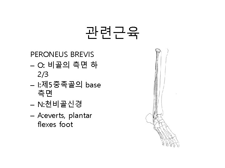
관련근육 • Peroneus longus – O: 비골의 측면 상 2/3 – I: medial cuneiform bone의 측면, 제 1중족 골의 base – N: 천비골신경 – A: plantar FLEXES, EVERTS FOOT.
관련근육 • Peroneus tertius – O: 비골전방 하 1/3, interosseous membrane – I: 제 5중족골 base의 dorsal surface – N: 심비골신경 – A: dorsiflexes and everts foot.
관련근육 Extensor hallucis longus – O: 비골의 전면 중간 부 – I: great toe 의 base – N: 심비골신경 – A: extends great toe, dorsiflexes and inverts foot.
관련근육 Extensor digitorum longus – O: 비골의 전면 상 2/3 – I: 4개의 측방 toe 발 등부위 – N: 심비골신경 – A: extends toes, dorsiflexes and inverts foot.
POSTERIOR INFERIOR FIBULA ① IND: Pain at fibular head, inversion sproin, lateral collateral ligament pain at the ankle, lateral hamstring complaints, restricted anterior-superior fibula motion. ② PP: Supine, knee flexed. ③ DP: Stand lateral to involved leg with superior hand placed in popliteal fossa. ④ CH: In popliteal fossa with thenar pad contacting the fibular head. ⑤ IH: Distal tibia and fiblua. ⑥ LOC: Inferior to superior and posterior to anterior. ⑦ MOD: Dr. flexes leg with inferior hand contacting at the ankle area and thrusting in a lifting motion with superior hand at the head of the fibula.
DM: 1. Biceps Femoris (long head) 2. Biceps Femoris (short head) 3. Peroneus Longus 4. Peroneus Brevis 5. Peroneus Tertius 6. Extensor Hallucis Longus 7. Extensor Digitorum Longus
교정법 1 -1 • Posterior Inferior fibula
교정법 1 -2 • Posterior Inferior fibula
관련근육 • Biceps femoris – O: 장두-ischial tuberosity , 단두linea aspera – I: 비골두 측면, 경골측 방condyle. – N: 좌골신경분지 – A: hip joint에서 대퇴 신전, knee joint에서 leg 굴곡
관련근육 • Peroneus longus – O: 비골의 측면 상 2/3 – I: medial cuneiform bone의 측면, 제 1중족 골의 base – N: 천비골신경 – A: plantar FLEXES, EVERTS FOOT.
관련근육 • Peroneus tertius – O: 비골전방 하 1/3, interosseous membrane – I: 제 5중족골 base의 dorsal surface – N: 심비골신경 – A: dorsiflexes and everts foot.
관련근육 Extensor hallucis longus – O: 비골의 전면 중간 부 – I: great toe 의 base – N: 심비골신경 – A: extends great toe, dorsiflexes and inverts foot.
관련근육 Extensor digitorum longus – O: 비골의 전면 상 2/3 – I: 4개의 측방 toe 발 등부위 – N: 심비골신경 – A: extends toes, dorsiflexes and inverts foot.
SUPERIOR FIBULA ① IND: Eversion sprain, tender fibular collateral ligament due to jamming injury, may result in a slight foot drop, restricted inferior fibula motion. ② PP: Supine, leg extended and hip flexed approximately 45˚. ③ DP: End of table with Pt. 's foot placed on anterior thigh. ④ CH: Proximal portion of lateral malleolus between thenar pads. ⑤ IH: Wrapped over CH to stabilize. ⑥ LOC: Superior to inferior. ⑦ MOD: Traction caudad, thrust is made with the CH at the lateral malleolus.
DM: 1. Biceps Femoris (long head) 2. Biceps Femoris (short head) 3. Tibialis Anterior 4. Peroneus Longus 5. Peroneus Brevis 6. Peroneus Tertius 7. Extensor Digitorum Longus
교정법 • SUPERIOR FIBULA
관련근육 • Biceps femoris – O: 장두-ischial tuberosity , 단두linea aspera – I: 비골두 측면, 경골측 방condyle. – N: 좌골신경분지 – A: hip joint에서 대퇴 신전, knee joint에서 leg 굴곡
관련근육 Tibialis anterior – O: 경골의 상 ½, 경골 의 lateral condyle – I: 제 1중족골 base , medial cuneiform bone 의 발바닥, 내 측 – N: 심비골신경 – A: dorsiflexes at ankle joint and inverts foot.
관련근육 • Peroneus longus – O: 비골의 측면 상 2/3 – I: medial cuneiform bone의 측면, 제 1중족 골의 base – N: 천비골신경 – A: plantar FLEXES, EVERTS FOOT.
관련근육 • Peroneus tertius – O: 비골전방 하 1/3, interosseous membrane – I: 제 5중족골 base의 dorsal surface – N: 심비골신경 – A: dorsiflexes and everts foot.
관련근육 Extensor digitorum longus – O: 비골의 전면 상 2/3 – I: 4개의 측방 toe 발 등부위 – N: 심비골신경 – A: extends toes, dorsiflexes and inverts foot.
POSTERIOR-MEDIAL FIBULA ① IND: Inversion sprain, hamstring pull, trauma to anterior-lateral knee, genu valgum. restricted anteriorlateral fibula motion. ② PP: Prone, leg flexed 70˚ to 90˚. ③ DP: End of table kneeling with invloved ankle resting on Dr. 's shoulder. ④ CH: Pisiform contact at the medial aspect of the fibular head. ⑤ IH: Interlaced with the CH. ⑥ LOC: Posterior to anterior and medial to lateral. ⑦ MOD: Traction in the direction of the LOC then thrust.
DM: 1. Biceps Femoris (long head) 2. Biceps Femoris (short head) 3. Tibialis Anterior 4. Peroneus Longus 5. Peroneus Brevis 6. Peroneus Tertius 7. Extensor Digitorum Longus
교정법 • POSTERIORMEDIAL FIBULA
관련근육 • Biceps femoris – O: 장두-ischial tuberosity , 단두linea aspera – I: 비골두 측면, 경골측 방condyle. – N: 좌골신경분지 – A: hip joint에서 대퇴 신전, knee joint에서 leg 굴곡
관련근육 Tibialis anterior – O: 경골의 상 ½, 경골 의 lateral condyle – I: 제 1중족골 base , medial cuneiform bone 의 발바닥, 내 측 – N: 심비골신경 – A: dorsiflexes at ankle joint and inverts foot.
관련근육 • Peroneus longus – O: 비골의 측면 상 2/3 – I: medial cuneiform bone의 측면, 제 1중족 골의 base – N: 천비골신경 – A: plantar FLEXES, EVERTS FOOT.
관련근육 • Peroneus tertius – O: 비골전방 하 1/3, interosseous membrane – I: 제 5중족골 base의 dorsal surface – N: 심비골신경 – A: dorsiflexes and everts foot.
관련근육 Extensor hallucis longus – O: 비골의 전면 중간 부 – I: great toe 의 base – N: 심비골신경 – A: extends great toe, dorsiflexes and inverts foot.
관련근육 Extensor digitorum longus – O: 비골의 전면 상 2/3 – I: 4개의 측방 toe 발 등부위 – N: 심비골신경 – A: extends toes, dorsiflexes and inverts foot.
ANTERIOR-LATERAL FIBULA ① IND: Lateral hamstring tendon pain, genu varum, hamstring pull, excessive pronation, eversion sprain, trauma to posterior-lateral knee, restricted posterior medial fibula motion. ② PP: Prone, leg fixed 70˚. ③ DP: End of table kneeling with involved ankle resting on Dr. 's shoulder. ④ CH: Pisiform contact at the anterior-lateral aspect of the fibula head. ⑤ IH: Interlaced with the CH. ⑥ LOC: Anterior to posterior and lateral to medial. ⑦ MOD: Traction in the direction of the LOC then thrust.
DM: 1. Biceps Femoris (long head) 2. Biceps Femoris (short head) 3. Peroneus Longus 4. Peroneus Brevis 5. Peroneus Tertius 6. Extensor Hallucis Longus 7. Extensor Digitorum Longus
교정법 • ANTERIORLATERAL FIBULA
관련근육 • Biceps femoris – O: 장두-ischial tuberosity , 단두linea aspera – I: 비골두 측면, 경골측 방condyle. – N: 좌골신경분지 – A: hip joint에서 대퇴 신전, knee joint에서 leg 굴곡
관련근육 • Peroneus longus – O: 비골의 측면 상 2/3 – I: medial cuneiform bone의 측면, 제 1중족 골의 base – N: 천비골신경 – A: plantar FLEXES, EVERTS FOOT.
관련근육 • Peroneus tertius – O: 비골전방 하 1/3, interosseous membrane – I: 제 5중족골 base의 dorsal surface – N: 심비골신경 – A: dorsiflexes and everts foot.
관련근육 Extensor hallucis longus – O: 비골의 전면 중간 부 – I: great toe 의 base – N: 심비골신경 – A: extends great toe, dorsiflexes and inverts foot.
관련근육 Extensor digitorum longus – O: 비골의 전면 상 2/3 – I: 4개의 측방 toe 발등부위 – N: 심비골신경 – A: extends toes, dorsiflexes and inverts foot.