MEDIAL AND LATERAL COLLATERAL LIGAMENT INJURIES IAN RICE


 • II -")

 with Grade III injuries can walk unaided and")


 • NSAIDs, rest, therapy • Therapy • Early weight-bearing • Emphasize")

 • More controversial • Trend toward nonop • Splint in full")


 • Arises from myotendinous jxn of popliteus •")


 function to resist: • Varus opening")


 JAAOS")

 • 13 patients isolated")


 JAAOS")


")

: La. Prade RF, Terry GC: Injuries to the posterolateral")

: Garrick JG (ed): Orthopaedic Knowledge Update: Sports Medicine 3.")

: La. Prade RF, Ly TV, Wentorf FA, et al:")
 tears")


- Slides: 90
MEDIAL AND LATERAL COLLATERAL LIGAMENT INJURIES IAN RICE MD
MEDIAL COLLATERAL LIGAMENT
MCL: ANATOMY • Tibial collateral ligament • 2 layers: superficial and deep
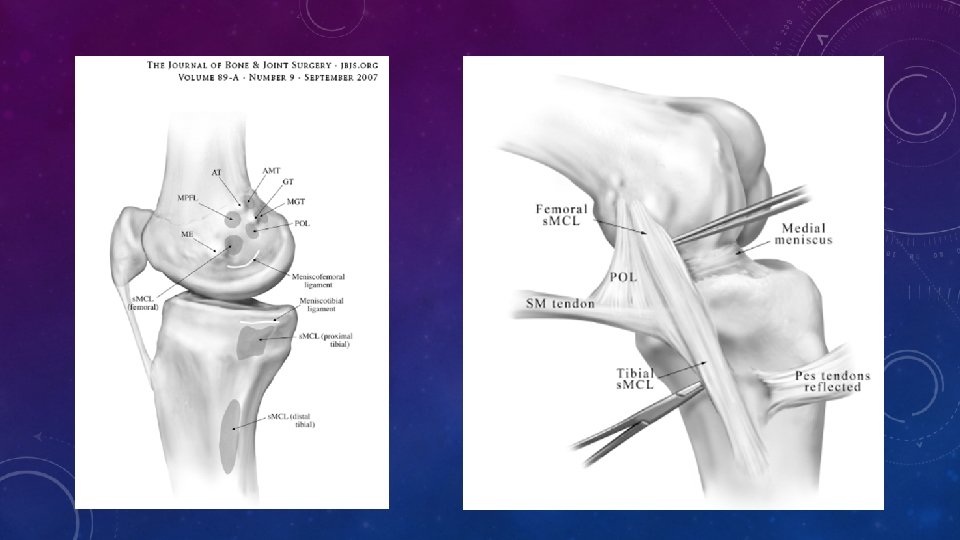
MCL: ANATOMY • Superficial MCL • Fibers • Anterior – parallel, distinct vertical margin • Posterior – become oblique • Femoral attachment • Circular, femoral epicondyle • 1 cm anterior and distal to adductor tubercle • Tibial attachment • 4. 5 cm distal to joint line on tibial metaphysis • Blends with tibial periosteum • Posterior and deep to pes anserinus • Bursa (of Voshell) • Primary valgus stabilizer
MCL: ANATOMY
MCL: ANATOMY • Deep MCL • AKA medial capsular ligament • Originates from distal femur, blends with superficial fibers • Divided into: • Meniscofemoral • Meniscotibial • Attached to medial meniscus by coronary ligaments • Secondary restraint to valgus stress (4 -8% restraint)
POSTERIOR OBLIQUE LIGAMENT: ANATOMY • Origin: medial femur distal to the adductor tubercle and posterior to the superficial MCL origin • Insertion: Posteromedial corner of tibia • Action: • Stabilize medial meniscus in conjunction with coronary ligaments • Posteromedial rotatory stability
SEMIMEMBRANOSUS • Tension on POL during knee flexion thought to retract posterior horn of medial meniscus, preventing impingement.
MCL: ANATOMY
MEDIAL KNEE STABILITY • Can be organized into static/dynamic forces • Static • Superficial MCL • Deep MCL • Posterior oblique ligament (POL) • Dynamic • Medial head of gastroc • Pes anserine tendons (Sartorius, Gracilis, semi. T) • Semimembranosus • Vastus medialis
MCL: ANATOMY • Medial knee capsuloligamentous structures can be divided into 3 layers
MCL: LAYER 1 • Sartorius and crural fascia • Covers both heads of gastroc and neurovascular structures • Blends with layer II anteriorly • Joins tibial periosteum near insertion of pes anserinus tendons
MCL: BETWEEN LAYERS 1 AND 2 • Semitendinosis • Gracicilis
MCL: LAYER 2 • Superficial MCL • Posterior oblique ligament
MCL: LAYER 3 • Capsule • Deep MCL • Coronary ligaments
MCL: BIOMECHANICS • Fan shaped, near instant center of rotation • Anterior fibers tense in flexion • Posterior fibers tense in extension • 1 -2 cm excursion during flexion • Tight in external rotation
MCL: RESEARCH • At 25 degrees of flexion, the superficial MCL provided 78% of the restraint to valgus stress, and at 5 degrees, the contribution was 57%. As the knee extends, the posterior capsule and POL tighten, providing increasing restraint to valgus stress. At 5 degrees, the POL provides 18% medial stability and at 25 degrees only 4%. Similarly, the deep ligament accounted for 8% at 5 degrees and 4% at 25 degrees. The cruciate ligaments provided the remaining stabilizing force with increasing amounts as the knee was extended. • “This study shows the increased awareness the physician should exhibit with increased laxity in extension owing to the possible rupture of the cruciates and posteromedial complex. ” Grood E. S. , Noyes F. R. , Butler D. L. , et al: Ligamentous and capsular restraints preventing straight medial and lateral laxity in an intact human cadaver knees. J Bone Joint Surg Am 1981; 63: 1257 -1269
MCL: RESEARCH • Sequentially sectioned the deep ligament, POL, and superficial ligament and found that the superficial ligament was the primary stabilizer against valgus and external rotation stresses. After the deep ligament and POL had been cut, sectioning the superficial MCL increased the medial joint opening 5 to 7 mm and increased external rotation 200% to 300%. Warren L. F. , Marshall J. L. , Girgis F. : The prime static stabilizer of the medial side of the knee. J Bone Joint Surg Am 1974; 56: 665 -674.
MCL: INJURY MECHANISM • Typical mechanism is force to the lateral aspect of the leg or lower thigh, ie. clipping injury in football • noncontact injury is from external rotation stress (cutting, pivoting, or twisting) • Knee dislocation • Skiers are more prone to medial-side injuries with 60% of all skiing knee injuries affecting the MCL
MCL: INJURY MECHANISM • Rupture typically occurs at femoral insertion • Tibial sided avulsions are rare
MCL: PHYSICAL EXAMINATION • Compare both sides • Point tenderness may identify location of injury 78% of time (Hughston, et al. ) • Localized swelling can identify tear in medial knee structures 64% of time • Valgus stress at 30 degrees is gold standard • Dropping leg over edge of exam table may make assessment easier
MCL: SEVERITY • Grade • I - <5 mm (Physiologic 2) • II - 5 -9 mm • III -10+ mm • Degree • I – tenderness, no instability • II – valgus laxity with firm end point • III – Mushy or absent end point
MCL: SLOCUM’S TEST • Rationale: disruption of the deep MCL allows the meniscus to move freely and allows the medial tibial plateau to rotate anteriorly • Modified anterior drawer • Valgus stress in 15 degrees external rotation and 80 degrees flexion • Prominence of medial tibial condyle is positive test
MCL: GAIT • Many patients (50%) with Grade III injuries can walk unaided and have minimal pain • Incomplete tears found to be more painful than complete tears • Vaulting type gait – Quad activation for medial stabilization • knee is hyperextended and locked at the end of the stance phase and the patient vaults over the extremity • Contrast with bent knee gait in ACL tear
MCL: IMAGING • Pellegrini – Stieda Lesion • Evidence of old, chronic MCL injury • Intra-substance calcification • Usually near proximal origin • Follows severe injuries • Can be treated with excision if symptomatic
MCL: IMAGING • MRI helpful for delineating of disruption of the collateral complex • Determine whether injury is proximal, mid substance, or distal • Determine whether the deep and/or superficial ligament is disrupted • Collateral ligament is best visualized on T 2 weight images: • high signal of edema and hemorrhage in the substance of the low signal ligament • Look for concomitant meniscal tear
MCL: MILLER’S TREATMENT ALGORITHM
Phisitkul P et al. “MCL Injuries of the Knee: Current Concepts Review. ” Iowa Orthop J. 2006; 26: 77– 90.
GRADE I (ISOLATED) • NSAIDs, rest, therapy • Therapy • Early weight-bearing • Emphasize early ROM • Strengthening • quad sets, SLRs, and hip adduction above the knee to begin immediately • cycling and progressive resistance exercises as tolerated • Functional ROM bracing to protect against further valgus insult • Return to play • Grade I returned to play at an average of 10. 6 days • Increased laxity and higher risk of reinjury, but not statistically significant
GRADE II • Treatment same as Grade I • Nonoperative • Emphasize early ROM, progressing to strengthening • Early weight-bearing • Functional ROM bracing to protect against further valgus insult • players with grade II injury returned to play at an average of 19. 5 days. • Again, higher incidence of reinjury than control knees, but not statistically significant
GRADE III (ISOLATED) • More controversial • Trend toward nonop • Splint in full extension (2 weeks) • Then ROM, WBAT • Return to play in 9. 2 weeks • Healing may continue for years • Strenghtening -> 80% then agility program • Brace for remainder of season to protect against valgus insult
• First to prospectively compare operative to nonoperative treatment of isolated third-degree ruptures. All patients underwent examination under anesthesia and arthroscopy to rule out other pathology • Objectively stable knees in 15 of 16 patients treated operatively and in 17 of 20 patients treated nonoperatively • Subjective scores were higher in the nonsurgical group • good to excellent results of 90% in the nonsurgical group • 88% in the surgically repaired group • No benefit to surgical intervention. J Bone Joint Surg Am. 1983 Mar; 65(3): 323 -9.
MCL REPAIR • Medial approach to the knee • Indicated for acute injuries • ligament avulsions: should be reattached with suture anchors in 30 degrees of flexion • interstitial disruption: anterior advancement of the MCL to femoral and tibial origins
MCL RECONSTRUCTION • Medial approach to the knee • Indications • chronic instability • insufficient tissue for repair • Graft type • Choice of semitendinosus autograft or hamstring, tibialis anterior or achilles tendon allograft
MCL: REHABILITATION PROTOCOL • Immobilization shown to have detrimental effects on the mechanical properties of the MCL, such as disorganization of collagen fibrils, decrease in the structural properties of the bone-ligament- bone complex, and resorption of bone at ligament insertion sites • Early controlled motion has become a part of standard nonoperative treatment protocols in most current series.
MCL: RETURN TO PLAY
MCL: ASSOCIATED INJURIES • Additional injury to surrounding structures increases with increasing force (Fetto and Marshall) • 20% of Grade I • 52% Grade II • 78% Grade III • 88% of surgically treated medial sided knee injuries had injury to more than one medial or posteromedial structure, most commonly the posterior oblique ligament (POL).
MCL: ASSOCIATED ACL INJURY • “The literature supports nonoperative treatment of the MCL tear with surgical reconstruction of the ACL. This is the trend that most surgeons are currently using. ” • Early versus late reconstruction continues to be a subject of debate, with studies supporting both sides
LATERAL COLLATERAL LIGAMENT
INTRODUCTION • Incidence of PLC injuries is not accurately known often undetected • Isolated injuries to PLC are rare • De. Lee & Rockwood– • 2 % of all ligamentous knee injuries • Usually combined with cruciate ligament injury (PCL > ACL) • MRI study of tibial plateau fxs showed PLC injuries in 68% of cases • Commonly being recognized when residual instability exists s/p ACL or PCL reconstruction. • Poor outcomes after ACL reconstruction? PLC injury?
LCL ANATOMY • Femur: proximal and posterior to lateral epicondyle • Fibula: 8. 2 mm posterior to anterior surface • Primary static restraint to varus opening
PLC ANATOMY • Popliteus • Originates posteromedial tibia • Intra-articular course through knee • Popliteal hiatus of coronary ligament • Inserts at popliteal saddle of lateral femoral condyle • Internally rotates • Popliteus Tendon Complex • Popliteus muscle/tendon unit • Connections from tendon to surrounding structures • Popliteofibular ligament • Popliteotibial ligament • Popliteomeniscal ligament • Restrict posterior tibial translation, ER of tibia, varus stress
PLC ANATOMY • Popliteofibular Ligament (PFL) • Arises from myotendinous jxn of popliteus • Inserts on fibular styloid • Present in 94 -100% of knees.
PLC ANATOMYOTHER SUPPORTS AND RESTRAINTS • • • Iliotibial Band Biceps femoris Oblique Popliteal Ligament of Wrisberg Fabellofibular ligament • Thickening of distal capsular edge of short head of biceps • Lateral capsular ligament (middle third) • Lateral meniscus • Posterior cruciate ligament
ANATOMY: LATERAL LAYERS • Anatomy Layers run from superficial to deep
ANATOMY – LAYER 1 • Iliotibial Band Biceps Femoris • Dynamic external rotators of the tibia. • Secondary restraint to varus stress. • Common peroneal nerve lies between layers 1 & 2
ANATOMY – LAYER 2 • Quadriceps Retinaculum • Lateral Patellofemoral Ligament
ANATOMY – LAYER 3 • Superficial • LCL • Fabellofibular ligament • Deep • Arcuate ligament • Coronary ligament • Popliteus tendon • Popliteofibular ligament • Capsule
ANATOMY – LAYER 3 • Arcuate ligament • Variable • Reinforces the posterolateral capsule and covers the popliteus • Y shaped with medial and lateral limbs • Fibular styloid to lateral femoral condyle
BIOMECHANICS • Structures of the PLC (including LCL) function to resist: • Varus opening • External tibial rotation • Posterior tibial translation • Act in combination with PCL for overall stability to lateral knee • PLC (excluding the LCL) • Primary stabilizer of ER at all flexion angles • Larger role at ~ 30° compared to 90 ° flexion • Resist posterior tibial translation • Isolated sectioning produced increased translation at all angles, greatest at early flexion • Primary restraint to posterior tibial translation at full knee extension
BIOMECHANICS • LCL • Tight in extension, lax in flexion • Greater ER resistance at full extension vs flexion • PCL • Resists posterior translation of tibia • Primary in greater knee flexion • Isloated sectioning had no effect on ER
BIOMECHANICS • Three most important structures to control varus and ER • LCL • Popliteus tendon • Popliteofibular ligament
CLINICAL EVALUATION • History • Common Mechanisms • Blow to anteromedial tibia • Hyperextension w/ ER (+/- contact) • Direct varus blow to flexed knee • Knee dislocation • Combined with ACL or PCL injury • More common than isolated PLC injury • Timing • Acute: 3 -4 weeks • Chronic: >6 weeks
PHYSICAL EXAMINATION • Complete knee examination • Remember association with other injuries • TTP & echymosis at posterolateral knee • Neurovascular exam • 12 – 29% with peroneal nerve palsy • Gait assessment • Varus or hyperextension/varus thrust can be seen • Numerous specific tests
PHYSICAL EXAMINATION • Adduction-Abduction stress test • Normal varus laxity 7° • Performed at 0° and 30° flexion. • Can get laxity at full extension with cruciate injury.
PHYSICAL EXAMINATION: DIAL TEST • May be done prone or supine • 10° difference in rotation • @ 30° = PLC • Reduced rotation at 90° = Isolated PLC • @ 90° = PLC and PCL • Veltri and Warren
PHYSICAL EXAMINATION • Reverse Pivot Shift – • Start – knee flexed 90° • Slowly extend knee with axial load and valgus stress. Foot in ER. • Tibia is subluxed > will “jump” or reduce at 20 -30° of flexion • IT band changes from flexor to extensor at this angle • La. Prade & Terry (1997) • + RPS associated with: • LCL injury • Mid-third lateral capsular ligment injury • Popliteal complex injury
PHYSICAL EXAMINATION • Posterolateral Drawer Test • Performed with the hip flexed 45°, knee flexed 80°, and foot is ER 15°. • A combined posterior drawer and external rotation force is then applied to the knee to assess for an increase in posterolateral translation (lateral tibia externally rotates relative to lateral femoral condyle) • Graded 1 -3 in 5 mm increments • 70 -75% sensitivity • Hughston & Norwood
PHYSICAL EXAMINATION • External Rotation Recurvatum • Positive when lower leg falls into external rotation and recurvatum when leg suspended by toes in supine patient
CLASSIFICATION • No universal classification system has been adoptied • Modification of Hughston system • Grade I = 0 -5 mm varus or 0 -5° rotation • Grade II = 6 -10 mm varus or 6 -10° rotation • Grade III = >10 mm varus and >10° rotation
IMAGING • X-Rays • AP/Lat weight-bearing, Patellofemoral views • Typically normal • BUT, fibular head fractures, avulsion of Gerdy’s, tibial plateau fractures, tibiofemoral dislocation, PCL avulsion fractures, Segond’s fracture, tibial spine fractures • Chronic injuries • May show arthrosis of lateral compartment • Full-extremity films to evaluate alignment • Stress views for laxity
IMAGING • MRI • Diagnostic study of choice • Standard sequences • Coronal oblique slices may improve accuracy & sensitivity of PLC specific injuries • May be especially useful in acute injuries • Difficult/Limited exam due to pain
TREATMENT OPTIONS Ranawat et al. (2008) JAAOS
TREATMENT • Nonoperative – • Isolated PLC injuries typically report little functional impairment initially • Brief period of protective weight-bearing followed by functional rehab can lead to good results. • Initial 2 -4 weeks immobilization (protected WB initial 2 weeks) • Hinged knee brace locked in extension • Increased ROM, WB, and strengthening • Return to full activity around 3 -4 months
TREATMENT • Nonoperative – • Baker et al (JBJS 1983) • 13 patients isolated PLC injury • All returned to full pre-injury activity. • Kannus (Am J Sports Med 1989) • Grade I & II injuries do well with nonoperative treatment (despite persistent laxity) • Grade III do poorly with non-op tx
SURGICAL TREATMENT • Indications • Grade III injuries • Persisent pain, instability, and/or functional limitations despite non-op management • Options • Direct repair +/- augmentation, or reconstruction • Acute disruptions • Primary repair with or without augmentation. • Chronic Injury • Occasionally possible to tighten existing structures • More commonly ligamentous reconstruction required
SURGICAL TREATMENT • PLC repair/reconstruction, ACL and/or PCL reconstruction, +/- HTO • Indications • In acute and chronic combined ligament injuries • Technique • PLC reconstruction should be performed at same time or prior to (as staged procedure) ACL or PCL to prevent early cruciate failure • Combined ACL and posterolateral corner reconstruction allows less anterior translation than isolated ACL reconstruction, but could not identify significant differences between the two groups in terms of functional outcomes (Kim et al JBJS 2012). • High Tibial Osteotomy • Indicated in patients with varus mechanical alignment • Failure to correct bony alignment jeopardizes ACL and PLC reconstruction success
SURGICAL TREATMENT • Early surgery is better! • Within 3 weeks • Better function and stability • More likely to restore native anatomy & biomechanics • Stannard (2005)– Early surgery • 37% failure with repair vs. 9% failure with reconstruction • Cruciate repairs were not done at initial surgery • Pearls: • • • 1: Diagnose and address all concomitant injuries 2: Treat avulsions w/ direct internal fixation or sutures 3: LCL should have midsubstance repair and graft reconstruction 4: Possible release of peroneal nerve 5: Fix all combined injuries at once if possible
LCL RECONSTRUCTION • There is no benchmark technique/procedure • Non-anatomic vs. Anatomic • Non-anatomic: more historical • Biceps tenodesis, arcuate copmlex/proximal bone block advancments, IT band sling, augments • Anatomic (nearly anatomic): more common recently • Fibular-based vs. tibial-fibular-based
BICEPS TENODESIS • Replaces LCL & PFL • Band of distal biceps femoris tendon is attached to lateral femoral epicondyle. • Attachment to fibular head kept • Variations on this theme whole tendon moved anterior, different modes of fixation, tendon can be looped. • Fixation on femur • 1 cm anterior to LCL attachment.
LCL RECONSTRUCTION • Fibular-based • Larson • Reconstruct both LCL and PFL • Two-tailed, figure of 8
LCL RECONSTRUCTION • Tib-fib-based • More closely resembles anatomy • No evidence of improved outcomes yet • More technically demanding • La. Prade technique • Reconstructs LCL, PFL, and popliteus tendon • 2 tendon grafts
LCL RECONSTRUCTION • Tib-Fib Based • Tendon graft is fixed to isometric point of the femoral epicondyle. • One branch is fixed to the fibular head with a bone tunnel and tranosseous sutures to reconstruct the LCL. • Second limb is brought through the posterior tibia to reconstuct the popliteofibular ligament
LCL RECONSTRUCTION
LCL RECONSTRUCTION Bone –PT-Bone Allograft. Achilles can be used as well.
TREATMENT OPTIONS Ranawat et al. (2008) JAAOS
REHABILITATION • Partial WB w/ Knee brace locked in extension for 6 weeks • Brace unlocked for ROM exercise early closed chain quad strengthening • Avoid active flexion exercises early • Quad sets • Return to full function at 10 -12 months
OITE QUESTIONS
• A soccer player sustains a knee injury. He is noted to have a significant increase in tibial passive external rotation both at 30 and 90 degrees. His MRI would most likely show which of the following? • 1. A fibular collateral ligament tear 2. A popliteus tendon tear 3. A posterior cruciate ligament tear 4. Answers 1 & 2 5. Answers 1, 2 & 3
PREFERRED RESPONSE: 5 • DISCUSSION: The fibular collateral ligament (also known as lateral collateral) and the popliteus tendon are both components of the postero-lateral corner (PLC). A positive dial test at 30 degrees indicates an isolated PLC injury, while a positive dial test at 30 and 90 degrees indicates PLC and concurrent posterior cruciate ligament (PCL) injury. The Fanelli article describes the evaluation and treatment of multi-ligament knee injuries including PCL and posterior-lateral corner tears. REFERENCES: • 1. OITE 07 #123 • 2. Fanelli GC, Orcutt DR, Edson CJ: The multiple-ligament injured knee: Evaluation, treatment, and results. Arthroscopy. 2005: 21: 471 -486. PMID: 15800529 (Link to Abstract)
A 25 -year-old man injured his knee in a motor vehicle collision. Abnormal examination findings include 10° increased external tibial rotation at 30° and 90° knee flexion. What additional examination finding is expected? • 1 - Increased opening to valgus stress at 30° of knee flexion • 2 - Increased varus opening at 0° of knee flexion • 3 - Positive apprehension sign with lateral patellar translation • 4 - Positive pivot shift test • 5 - Medial tibial plateau rests 10 mm anterior to the medial femoral condyle
Preferred Response: 2 Recommended Reading(s): La. Prade RF, Terry GC: Injuries to the posterolateral aspect of the knee: Association of anatomic injury patterns with clinical instability. Am J Sports Med 1997; 25: 433 -438. Gollehon DL, Torzilli PA, Warren RF: The role of posterolateral and cruciate ligaments in the stability of the human knee: A biomechanical study. J Bone Joint Surg Am 1987; 69: 233 -242.
A 25 -year-old competitive soccer player has chronic anterior knee pain and reports “sloppiness” since injuring it in a collision with another player 2 months ago. He missed several weeks of practice but has since attempted a return to play. Examination reveals no quadriceps atrophy, standing varus alignment of 8°, a posterior sag sign, 3+ posterior drawer, 2+ varus instability in extension, 3+ varus instability at 30°, and 20° increased prone external rotation at 30° and 90°. He walks with a varus thrust. What is the best treatment option? • 1 - High tibial osteotomy • 2 - Reconstruction of the posterior cruciate ligament (PCL) and repair of the posterolateral • corner (PLC) • 3 - Reconstruction of the PCL • 4 - Reconstruction of the PCL and PLC • 5 - High tibial osteotomy and PCL/PLC reconstruction
Preferred Response: 5 Recommended Reading(s): Garrick JG (ed): Orthopaedic Knowledge Update: Sports Medicine 3. Rosemont, IL, American Academy of Orthopaedic Surgeons, 2004, pp 183197. Giffin JR, Vogrin TM, Zantop T, et al: Effects of increasing tibial slope on the biomechanics of the knee. Am J Sports Med 2004; 32: 376 -382.
• What anatomic structure inserts most anteriorly on the proximal fibula? • 1 - Sartorius • 2 - Iliotibial band • 3 - Biceps femoris • 4 - Popliteofibular ligament • 5 - Lateral collateral ligament
Preferred Response: 5 Recommended Reading(s): La. Prade RF, Ly TV, Wentorf FA, et al: The posterolateral attachments of the knee: A qualitative and quantitative morphologic analysis of the fibular collateral ligament, popliteus tendon, popliteofibular ligament, and lateral gastrocnemius tendon. Am J Sports Med 2003; 31: 854 -860. Stannard JP, Brown SL, Farris RC, et al: The posterolateral corner of the knee: Repair versus reconstruction. Am J Sports Med 2005; 33: 881 -888.
All of the following are true regarding grade III medial collateral ligament (MCL) tears of the knee EXCEPT: Review Topic 1. Proximal ruptures have decreased residual valgus laxity following nonoperative treatment than distal ruptures 2. They result in greater than 10 mm of valgus opening 3. They can result in a Stener-type lesion 4. They require operative repair when there is a concomitant anterior cruciate ligament tear 5. Proximal ruptures have better healing potential with nonoperative treatment than distal ruptures
• PREFERRED RESPONSE ▶ 4 • Grade III indicates a complete rupture of the MCL with greater than 10 mm of opening on valgus stress. Careful evaluation is needed to evaluate for concomitant injuries such as medial meniscus and ACL tears. However, the presence of an ACL tear does not preclude nonoperative treatment. Timing of ACL reconstruction with a concomitant MCL sprain should be delayed proportional to the extent of MCL damage. (Grade I injuries, 3 -4 weeks; grade II injuries, 4 -6 weeks; grade III injuries, 6 -8 weeks. ) According to Shelbourne et al, many surgeons recommend nonoperative management of acute grade III MCL injuries occurring at the femoral origin or mid-substance, and primary repair of injuries at the tibial origin. Perhaps because of better vascularity, proximal tears tend to heal better than distal ones. In contrast, distal ruptures may heal with excessive valgus instability and occasionally result in a Stenertype lesion with the torn MCL flipped over the insertion of the pes anserinus where it is unable to heal normally. Illustration A is an MRI image showing a distal grade III rupture. The reference by Azar provides a review of the anatomy, clinical exam and treatment options for MCL tears.
REFERENCES • Ranawat A, Baker CL, Henry S, Harner CD: Posterolateral Corner Injury of the Knee: Evaluation and Management. JAAOS 2008; 16: 506 -518. • Gollehon DL, Torzilli PA, Warren RF: The role of the posterolateral and cruciate ligaments in the stability of the human knee: A biomechanical study. J Bone Joint Surg Am 1987; 69: 233 -242. • Grood ES, Stowers SF, Noyes FR: Limits of movement in the human knee: Effect of sectioning of the posterior cruciate ligament and posterolateral structures. J Bone Joint Surg Am 1988; 70: 88 -97. • Watanabe Y, Moriya H, Takahashi K, et al: Functional Anatomy of the posterolateral structures of the knee. Am J Sports Med 1996; 24: 311 -316. • Orthobullets. com • La. Prade RF, Terry GC. Injuries to the posterolateral aspect of the knee. Association of anatomic injury patterns with clinical instability. Am J Sports Med. 1997 Jul-Aug; 25(4): 433 -8. • Fanelli GC, Orcutt DR, Edson CJ: The multiple-ligament injured knee: Evaluation, treatment, and results. Arthroscopy. 2005: 21: 471 -486. • Sung-Jae Kim, MD, Ph. D; Duck-Hyun Choi, MD; Byoung-Yoon Hwang, MD: The Influence of Posterolateral Rotatory Instability on ACL Reconstruction: Comparison Between Isolated ACL Reconstruction and ACL Reconstruction Combined with Posterolateral Corner Reconstruction. J Bone Joint Surg Am. 2012; 94(3): 253 -259.