Types of Synovial Joints Selected Synovial Joints The


 Tibial")
")


 Joint The large head of the humerus fits into the glenoid cavity")

 Joint B) Three Glenohumeral ligaments strengthen the front of the capsule. These")

 Joint C) The Rotator Cuff is formed from four tendons and muscles")
 Joint")


")


 reconstruction,")


 This is a ball and socket joint whose movement is limited")


 The ligamentum teres attaches the femur to the acetabulum.")
")


 joint. Superior view Outline of the mandibular")

 joint. Mandibular fossa Articular tubercle Zygomatic process")


 is a common facial injury. Fractures (these")


- Slides: 100
Types of Synovial Joints
Selected Synovial Joints: The Knee This is considered the most complex joint in the human body. It is actually considered three joints working together.
The Knee These are: – An intermediate joint between the patella and distal end of the femur (femoropatellar joint). This is a plane joint.
The Knee These are: – An intermediate joint between the patella and distal end of the femur (femoropatellar joint). This is a plane joint. – A lateral and medial tibiofemoral joints between the femoral condyles and the menisci below.
Figure 8. 8 a The knee joint. Femur Articular capsule Posterior cruciate ligament Lateral meniscus Anterior cruciate ligament Tibia Tendon of quadriceps femoris Suprapatellar bursa Patella Subcutaneous prepatellar bursa Synovial cavity Lateral meniscus Infrapatellar fat pad Deep infrapatellar bursa Patellar ligament (a) Sagittal section through the right knee joint Copyright © 2010 Pearson Education, Inc.
The Knee The menisci help prevent lateral motion and attach to the outer margins of the joint capsule on the tibia. They are easily torn.
Figure 8. 8 b The knee joint. Anterior cruciate ligament Articular cartilage on lateral tibial condyle Articular cartilage on medial tibial condyle Medial meniscus Posterior cruciate ligament Lateral meniscus (b) Superior view of the right tibia in the knee joint, showing the menisci and cruciate ligaments Copyright © 2010 Pearson Education, Inc.
The knee is unique in that it is not completely enclosed by a capsule.
The knee is unique in that it is not completely enclosed by a capsule. The articular capsule is found only on the lateral and posterior surfaces.
The knee is unique in that it is not completely enclosed by a capsule. The articular capsule is found only on the lateral and posterior surfaces. The anterior surface is covered by three ligaments going from the patella to the tibia.
• These ligaments are: • The patella ligament
• These ligaments are: • The patella ligament & • The medial and lateral patellar retinacula ligaments. They merge with the articular capsule on each side.
The intracapsular ligaments are the cruciate ligaments.
The intracapsular ligaments are the cruciate ligaments. The anterior and posterior cruciate ligaments cross each other forming an X in the notch between the femoral condyles.
The intracapsular ligaments are the cruciate ligaments. The anterior and posterior cruciate ligaments cross each other forming an X in the notch between the femoral condyles. They prevent anterior and posterior displacement.
Two additional ligaments, the Fibular and Tibial Collateral Ligaments prevent lateral or medial rotation when the knee is extended.
Figure 8. 8 c The knee joint. Quadriceps femoris muscle Tendon of quadriceps femoris muscle Patella Lateral patellar retinaculum Medial patellar retinaculum Tibial collateral ligament Fibular collateral ligament Patellar ligament Fibula Tibia (c) Anterior view of right knee Copyright © 2010 Pearson Education, Inc.
Figure 8. 8 f The knee joint. Medial femoral condyle Anterior cruciate ligament Medial meniscus on medial tibial condyle Patella (f) Photograph of an opened knee joint; view similar to (e) Copyright © 2010 Pearson Education, Inc.
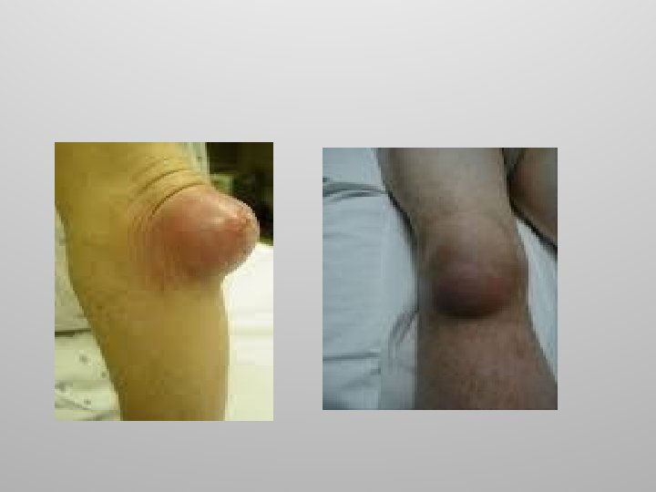
The synovial cavity of the knee has a complicated shape and over one dozen associated bursae. Some are easily injured such as the subcutaneous prepatellar bursa which lies just over the patella (house maid’s knee).
House Maid’s Knee
Knee Injuries Common knee injuries involve the 3 C’s: – Collateral ligaments,
Knee Injuries Common knee injuries involve the 3 C’s: – Collateral ligaments, – Cruciate ligaments and
Knee Injuries Common knee injuries involve the 3 C’s: – Collateral ligaments, – Cruciate ligaments and – Cartilage (menisci).
Knee Injuries Lateral blows are the most dangerous, tearing the tibial collateral ligament and the medial meniscus and the anterior cruciate ligament.
Figure 8. 9 A common knee injury. Lateral Hockey puck Medial Patella (outline) Tibial collateral ligament (torn) Medial meniscus (torn) Anterior cruciate ligament (torn) Copyright © 2010 Pearson Education, Inc.
Arthroscopic Knee Surgery Arthroscopy is a common surgical procedure in which a joint (arthro-) is viewed (-scopy) using a small camera.
Arthroscopic Knee Surgery Arthroscopy gives doctors a clear view of the inside of the knee. This helps them diagnose and treat knee problems.
Arthroscopic Knee Surgery The orthopaedic surgeon will make a few small incisions in your knee. A sterile solution will be used to fill the knee joint and rinse away any cloudy fluid. This helps your orthopaedic surgeon see your knee clearly and in great detail.
Arthroscopic Knee Surgery
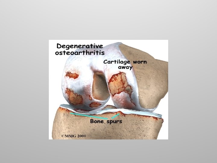
Knee Replacement People with degenerative arthritis, chronic injuries often lose that smooth articular cartilage. The result is bone on bone. The knee joints must be replaced.
Knee Replacement
Shoulder (Glenohumeral) Joint The large head of the humerus fits into the glenoid cavity of the scapula. The cavity is extended by a fibrocartilage ring called the glenoid labrum. Connective tissue support comes from three groups of ligaments.
Figure 8. 10 c The shoulder joint. Acromion Coracoacromial ligament Subacromial bursa Coracohumeral ligament Greater tubercle of humerus Transverse humeral ligament Tendon sheath Tendon of long head of biceps brachii muscle Coracoid process Articular capsule reinforced by glenohumeral ligaments Subscapular bursa Tendon of the subscapularis muscle Scapula (c) Anterior view of right shoulder joint capsule Copyright © 2010 Pearson Education, Inc.
Shoulder (Glenohumeral) Joint B) Three Glenohumeral ligaments strengthen the front of the capsule. These ligaments are weak.
Figure 8. 10 d The shoulder joint. Acromion Coracoid process Articular capsule Glenoid cavity Glenoid labrum Tendon of long head of biceps brachii muscle Glenohumeral ligaments Tendon of the subscapularis muscle Scapula Posterior Anterior (d) Lateral view of socket of right shoulder joint, humerus removed Copyright © 2010 Pearson Education, Inc.
Shoulder (Glenohumeral) Joint C) The Rotator Cuff is formed from four tendons and muscles that encircle the joint. The muscles include the Subscapularis, Supraspinatus Infraspinatus and Teres minor.
Shoulder (Glenohumeral) Joint
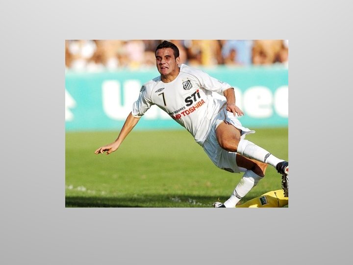
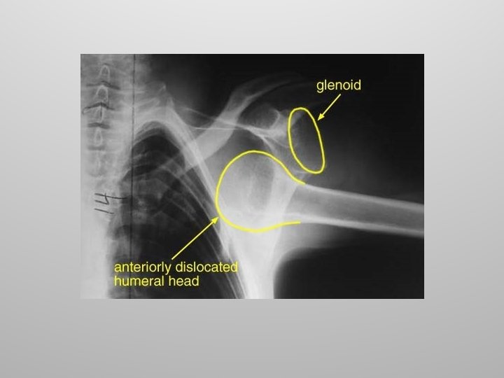
Shoulder Injuries Rotator Cuff Because of its mobility, the stability of the shoulder joint has been sacrificed. Anterior dislocations are the most common along with damage to the rotator cuff muscles due to severe circumduction.
Shoulder Dislocation and Reduction
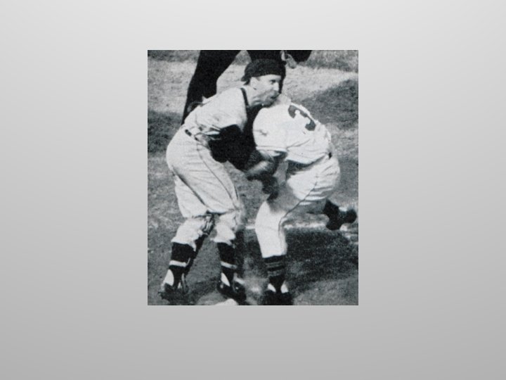
Shoulder Injuries Rotator Cuff Supraspinatus and Infraspinatus are the most commonly injured rotator cuff muscles. Due to the function of these muscles, sports which involve a lot of shoulder rotation – for example, pitching in baseball, swimming, – often put the rotator cuff muscles under a lot of stress.
Shoulder Injuries Rotator Cuff Problems with the rotator cuff muscles can be classed into two categories – Tears of the tendons/muscles, and inflammation of the tendons (often called tendinopathy or tendonitis).
Shoulder Injuries Rotator Cuff Surgery to repair a torn rotator cuff tendon usually involves: – Removing loose fragments of tendon, bursa, and other debris from the space in the shoulder where the rotator cuff moves (debridment).
Shoulder Injuries Rotator Cuff Surgery to repair a torn rotator cuff tendon usually involves: – Removing loose fragments of tendon, bursa, and other debris from the space in the shoulder where the rotator cuff moves (debridement). – Making more room for the rotator cuff tendon so it is not pinched or irritated.
Shoulder Injuries Rotator Cuff Surgery to repair a torn rotator cuff tendon usually involves: – Removing loose fragments of tendon, bursa, and other debris from the space in the shoulder where the rotator cuff moves (debridement). – Making more room for the rotator cuff tendon so it is not pinched or irritated. – Sewing the torn edges of the supraspinatus tendon together and to the top of the upper arm bone (humerus).
Shoulder Injuries Rotator Cuff
Shoulder Injuries Rotator Cuff
Elbow Joint This is a hinge joint where the radius and ulna articulate with the condyles of the humerus. The ulna’s trochlear notch forms a tight hinge with the trochlear of the humerus. This articulation allows for flexion and extension only.
Elbow Joint Side to side movement is prevented by the ulnar collateral ligament (triangular) and radial collateral ligament.
Figure 8. 11 d The elbow joint. Articular capsule Anular ligament Humerus Coronoid process Medial epicondyle Radius Ulnar collateral ligament Ulna (d) Medial view of right elbow Copyright © 2010 Pearson Education, Inc.
Figure 8. 11 c The elbow joint. Humerus Anular ligament Medial epicondyle Radius Articular capsule Ulnar collateral ligament Coronoid process Ulna (c) Cadaver photo of medial view of right elbow Copyright © 2010 Pearson Education, Inc.
Figure 8. 11 b The elbow joint. Humerus Anular ligament Lateral epicondyle Radius Articular capsule Radial collateral ligament Olecranon process (b) Lateral view of right elbow joint Copyright © 2010 Pearson Education, Inc. Ulna
Elbow Joint Injuries
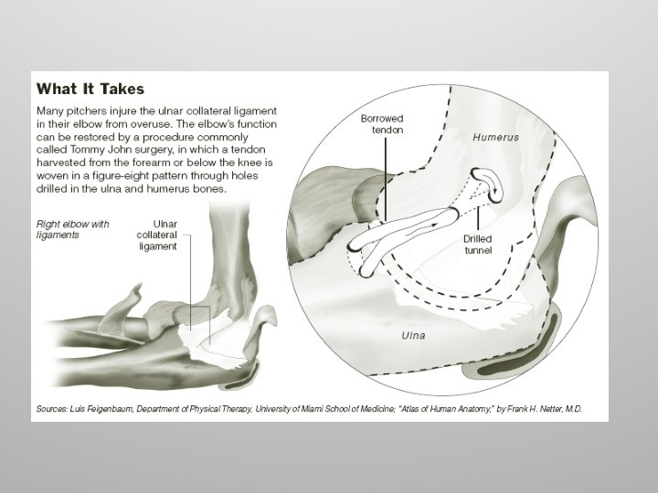
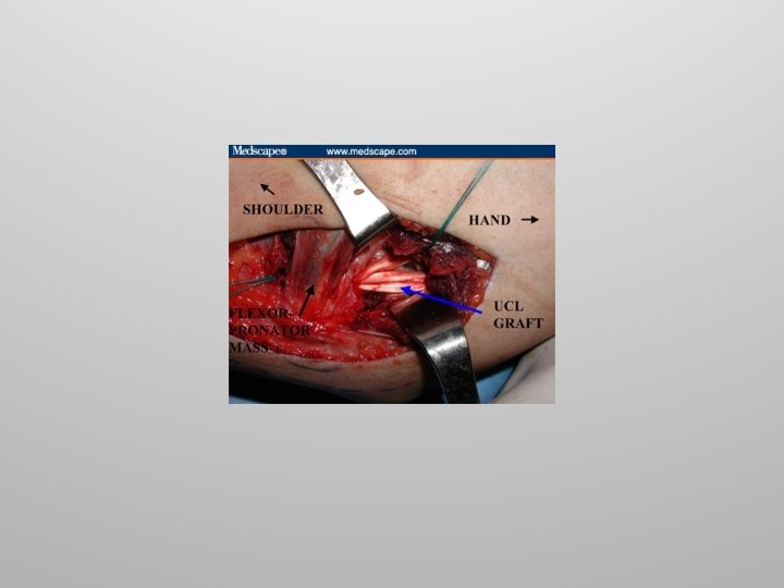
Tommy John Surgery This procedure, more formally known as UCL (Ulnar Collateral Ligament) reconstruction, is designed to repair a torn elbow ligament—an injury typically caused by strong, repetitive overhead throwing motions of the arm.
Tommy John Surgery It was first performed in 1974 on baseball pitcher Tommy John. The procedure typically lasts about an hour and a half, and patients usually leave the hospital the same day.
Elbow Dislocations
Elbow Dislocations
Hip (Coxal Joint) This is a ball and socket joint whose movement is limited by strong ligaments. It is formed from the spherical head of the femur and the deeply cupped acetabulum in the pelvis.
Figure 8. 12 b The hip joint. Acetabular labrum Synovial membrane Ligament of the head of the femur (ligamentum teres) Head of femur Articular capsule (cut) (b) Photo of the interior of the hip joint, lateral view Copyright © 2010 Pearson Education, Inc.
Figure 8. 12 c The hip joint. Ischium Iliofemoral ligament Ischiofemoral ligament Greater trochanter of femur (c) Posterior view of right hip joint, capsule in place Copyright © 2010 Pearson Education, Inc.
Hip (Coxal Joint) The ligamentum teres attaches the femur to the acetabulum.
Figure 8. 12 a The hip joint. Articular cartilage Acetabular labrum Femur Coxal (hip) bone Ligament of the head of the femur (ligamentum teres) Synovial cavity Articular capsule (a) Frontal section through the right hip joint Copyright © 2010 Pearson Education, Inc.
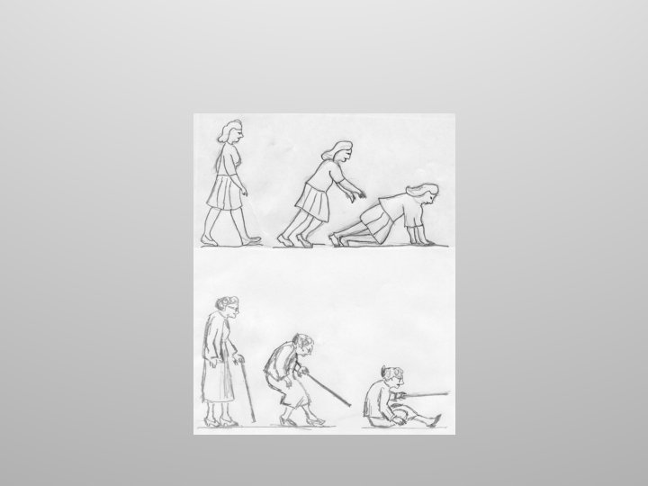
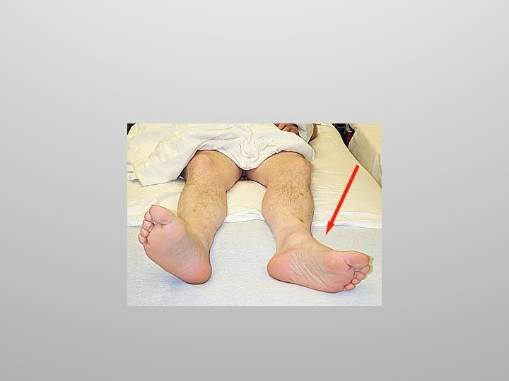
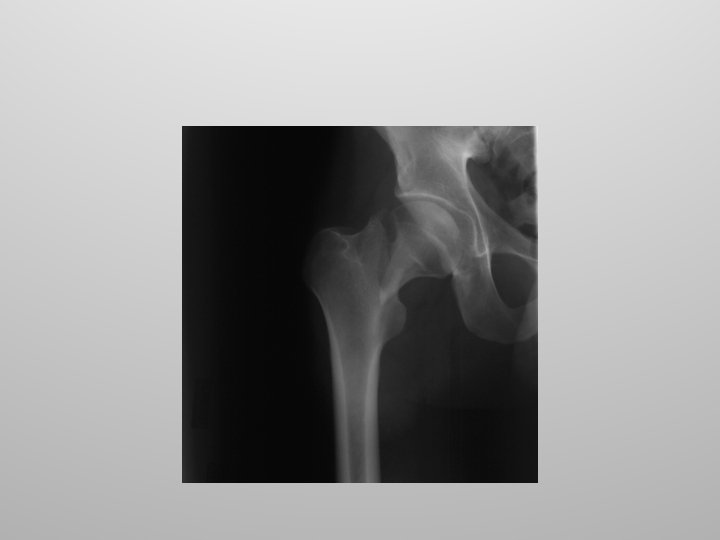
Hip Injuries Common injuries to the hip joint include fractures and dislocations. Hip fractures typically involve the neck of the femur and are the result of underlying disease such as osteoporosis.
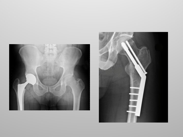
Hip Replacement Hip replacement surgery, also called total hip arthroplasty, involves removing a diseased hip joint and replacing it with an artificial joint, called a prosthesis.
Hip Replacement Hip replacement is typically used for people with hip joint damage from arthritis or an injury. Followed by rehabilitation, hip replacement can relieve pain and restore range of motion and function of your hip joint Lets do a hip replacement!
Temporomandibular Joint Two distinct movements can occur with the jaw, a hinge like movement and the second is a lateral movement.
Temporomandibular Joint
Figure 8. 13 c The temporomandibular (jaw) joint. Superior view Outline of the mandibular fossa (c)Lateral excursion: lateral (side-to-side) movements of the mandible Copyright © 2010 Pearson Education, Inc.
Temporomandibular Joint A lateral ligament attaches the ramus of the mandible to the zygomatic arch of the temporal bone.
Figure 8. 13 a The temporomandibular (jaw) joint. Mandibular fossa Articular tubercle Zygomatic process Infratemporal fossa External acoustic meatus Articular capsule Ramus of mandible Lateral ligament (a) Location of the joint in the skull Copyright © 2010 Pearson Education, Inc.
Injuries to the Jaw Injuries to the jaw include fractures and dislocations.
Injuries to the Jaw Injuries to the jaw include fractures and dislocations. A broken jaw is a break in the jaw bone. A dislocated jaw means the lower part of the jaw has moved out of its normal position at one or both joints.
Dislocation of the Jaw Symptoms of a dislocated jaw include pain in or around the jaw, the misalignment of teeth, and forward movement of the jaw beyond its regular position. Other symptoms include difficulty opening and closing the mouth.
Dislocation of the Jaw A dislocated jaw can be caused by forceful yawning or by some sort of trauma or impact with the face. Common causes of jaw dislocation include a punch to the face, sports injuries, and car accidents.
Dislocation of the Jaw
Fractures of the Jaw A broken jaw) is a common facial injury. Fractures (these are breaks in the bone) are generally the result of a direct force to the jaw.
Diseases of the joints Besides trauma, inflammation from over use or repetitive motions are the most common diseases seen in the joints.
Bursitis Inflammation of a bursal sac usually as the result of chronic irritation
Arthritis is a common term for over 100 conditions which describe degenerative processes found in the joints. Major examples include osteoarthritis, rheumatoid arthritis and gout.
Figure 8. 15 X ray of a hand deformed by rheumatoid arthritis. Copyright © 2010 Pearson Education, Inc.
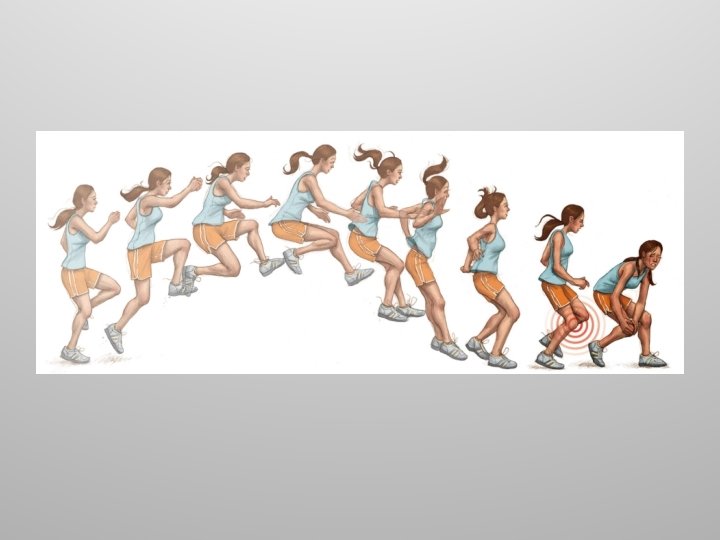
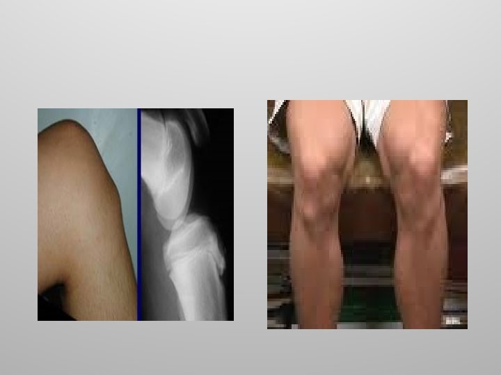
Osgood–Schlatter disease Is also known as tibial tubercle apophyseal traction injury is a rupture of the growth plate at the tibial tuberosity.
Osgood–Schlatter disease The condition occurs in active boys and girls aged 9– 16 coinciding with periods of growth spurts. It occurs more frequently in boys than in girls.
Osgood–Schlatter disease The condition is usually self-limiting and is caused by stress on the patellar tendon that attaches the quadriceps muscle at the tibial tuberosity. Treatment is conservative with rest, RICE (Rest, Ice, Compression, and Elevation),
Shin Splints Shin splints is a general medical term denoting medial tibial stress syndrome (MTSS), a slow healing and painful condition in the shins, usually caused by exercise such as running, jumping, swimming, cycling, dancing or other sports.
The onset of shin splints is most common after exercise, caused by high impact training, excessive training, poor technique
Knuckle Cracking When one cracks a knuckle, the stretching of the capsule lowers the pressure inside the joint and creates a vacuum which is filled by the gas previously dissolved in the synovial fluid. This creates a “bubble” which then bursts producing the characteristic “popping” or “cracking” sound.
Knuckle Cracking There is no evidence that cracking knuckles causes any damage such as arthritis in the joints.
Questions?