Joints Introduction to Joints A joint is a


 Joint • Biaxial (flexion extension, abduction adduction) • The joint surfaces are")
 • The bones set together as")
 • A rounded")
 Joint • Uniaxial (permits gliding movements) • The bone surfaces involved are")


 of")


")

")


- Slides: 39
Joints
Introduction to Joints • A joint is a point of connection between two bones • Strands of connective tissue, called ligaments, hold the bones together and ensure the stability of joints
Joint Classification • Joints are classified according to their motion capabilities: – Syntharoses • immovable – Amphiarthroses • Slightly movable – Diarthroses • Allow the greatest amount of motion
Joint Classification • Joints are further classified by the material that joints them: – Fibrous joint • Allow no movement • E. g. sutures of the skull – Cartilaginous joint • Allow limited movement • E. g. intervertebral disks – Synovial joint • Allow large range of movement • Eg: Hip joint
Characteristics of Synovial Joints • Hyaline cartilage – A protective layer of dense white connective tissue that covers the ends of the articulating bones • • Joint cavity Synovial membrane – Covers joint cavity, except over the surfaces of articular cartilages – Secretes the lubrication fluid • Synovial fluid – Lubricates the joints • Capsule – May or may not have thickenings called intrinsic ligaments • Extrinsic ligaments – Support the joint and connect the articulating bones of the joint
Types of Synovial Joints • There are three basic types of synovial joints: – Unilateral (rotation about only one axis) – Biaxial joints (movement about two perpendicular axes) – Multiaxial joints (movement about all three perpendicular axes)
Types of Synovial Joints • Synovial joints are further classified into: – Hinge joint – Pivot joint – Condyloid joint – Saddle shaped joint – Ball and socket joint – Plane joint
Types of Synovial Joints
Hinge Joint • Uniaxial • Has one articulating surface that is convex, and another that is concave • E. g. humero ulnar elbow joint, interphalangeal joint
Pivot Joint • Uniaxial • E. g. head of radius rotating against ulna
Condyloid (Knuckle) Joint • Biaxial (flexion extension, abduction adduction) • The joint surfaces are usually oval • One joint surface is an ovular convex shape, and the other is a reciprocally shaped concave surface
Saddle Joint • Biaxial (flexion extension, abduction adduction) • The bones set together as in sitting on a horse • E. g. carpometacarpal joint of the thumb
Ball and Socket Joint • Multiaxial rotation (rotation in all planes) • A rounded bone is fitted into a cup – like a receptacle • E. g. shoulder & hip joints
Plane (Gliding) Joint • Uniaxial (permits gliding movements) • The bone surfaces involved are nearly flat • E. g. intercarpal joints and acromioclavicular joint of the vertebrae
Joints of the Pectoral Girdle
Sternoclavicular Joint • Connects the sternum to the clavicle • The only joint connecting the pectoral girdle to the axial skeleton • True synovial joint strengthened by an intracapsular disc and extrinsic ligaments
Acromioclavicular Joint • Unites the lateral end of the clavicle with the acromion process of the scapula • Where the shoulder separations often occur in sports such as hockey, baseball, and football
Glenohumeral Joint • • Connects the upper limb and the scapula Typical multiaxial joint Wide range of movement at this joint Compromised = relative lack of stability
Upper Limb Joints
Elbow Joint There are three joints at the elbow: • Humero ulnar joint – Medial (with respect to anatomical position) – Between the trochlea of the humerus and the olecranon process of the ulna • Humero radial joint – Lateral – Between the capitulum of the humerus and the head of the radius • Radio ulnar joint – Between the radius and the ulna
Elbow Joint
Joints of the Wrist • Radio carpal joint – Between the distal end of the radius and the carpals – Movements: flexion extensionand abduction adduction
Joints of the Hand • Intercarpal joints – Between the bones of the Carpus – Gliding joints • Carpometacarpal joint – Between carpals and metacarpals – The characteristics of the carpometacarpal joint of the thumb allows the range of movement necessary for grasping
Joints of the Hand • Metacarpophalangeal joints – Joints between metacarpals and phalanges – The knuckles – Movement flexion extension, abduction adduction • Interphalangeal joints – Joints between the phalanges – Permit flexion extension
Joints of the Pelvic Girdle
Hip Joint • Between the head of the femur and the cup (acetabulum) of the hip bone (os coxae) • Like the shoulder, hip joint is: – Ball and socket joint – Mutliaxial joint that allows flexion extension, abduction adduction and circumduction
Hip Joint • Unlike shoulder joint, hip joint is very stable • Dislocation in sports is not common, but can occur via car collisions • Dislocate the head posteriorly or drive it through the posterior lip of the acetabulum • In fact, it is the body’s most stable synovial joint due to: – Deepened socket – An intrinsic and very strong extrinsic ligaments
Hip Joint
Lower Limb Joints
Knee Joint • Tibiofemoral or knee joint • Incredible range of movement (flexion extension)
Knee • However, knee joint is relatively stable due to additional structural supports from: – Menisci • Shock absorbing fibrocartilaginous discs – Anterior and posterior cruciate ligaments • In the center of the joint – Lateral and medial collateral ligaments • Extending from the sides of the femur to the tibia and fibula – The musculature that surrounds it
Knee Joint Movements: • Primary action is flexion extension (i. e. squat or jump) • When flexed, medial and lateral rotation can also occur
Ankle Joint • Talocrural or ankle joint • Involves several bones – Medial and lateral malleoli of the tibia and fibula – Head of the talus – Calcaneus (heel bone)
Foot and Toe Joints • Intertarsal joints – Between tarsals – Transverse tarsal joint • Between the proximal and distal row of the tarsal bones • Movement: inversion eversionof the sole of the foot • This action enables you to adjust to uneven ground when walking or running
Foot and Toe Joints • As in the hand, there are joints between the tarsal bones, metatarsals and phalanges: – Tarsometatarsal – Metatarsalophalangeal – Interphalangeal • They are strengthened by plantar ligaments which aid in maintaining the arc of the foot
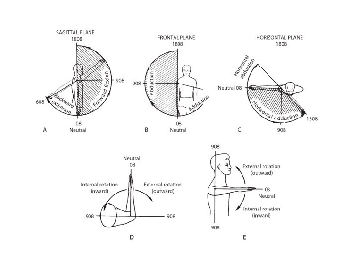
Think About it… 1. Why is it most appropriate to measure movement with tri planar motion (frontal, sagittal, transverse)? Explain. 2. Do 10 squats where you are. When you go down in a squat, there is movement at the hip joint, knee joint and ankle joint. Remember that when an angle between two bones is decreased, it is called flexion. Name the movements for each three joints. 3. Shoulder Weight lifting Exercises: The shoulder joint has a large Range of Motion. It can move in the frontal, sagittal and transverse plane. Name a weight lifting exercise that involves: – Shoulder extension (elbow straight, only sagittal plane) – Shoulder elevation (arms hanging to the side, only frontal plane) – Shoulder adduction (elbow straight, only frontal plane)
Think About it… 4. Define the following terms and find a picture to match them: a) Lateral Truck Flexion b) Hip hyperextension c) Shoulder Horizontal Flexion 5. Why is training in a single plane motion training not very beneficial for sports performance? Explain.
Think About it… 4. a. Lateral trunk flexion side bending away from the midline of the body (abduction) then straightening back to center (adduction) b. Hip hyperextension femur moves past straight, extended position to move behind the body c. shoulder horizontal flexion arms are raised out to shoulder level and then drawn toward midline