Introduction to Magnetic Resonance Imaging 07 31211017007 Magnetic

技術的歷史 MRI- basic principles Relaxation time Repetition")


人體置於強大磁場中,人體內氫原子一 律向Z軸排列 Net spinning Precession (2)給予等於Larmor")
 (外加磁場) (外加電磁波) RF = Larmor frequency")


 Normal tissues Malignant tissues T 1 and T")
 Excitation TR = repetition time")


: used as contrast agent (0. 2")


 Brownian motion of water molecules Water Ice")
 Apparent diffusion coefficient (ADC) values in ranula")


")

 2001, November 1. 1 st")
 5. Wilkinson E. MRI researchers")
- Slides: 37
主治醫師教學 Introduction to Magnetic Resonance Imaging 核磁共振影像介紹 陳玉昆醫師: 高雄醫學大學 口腔病理科 07 -3121101~7007
Magnetic Resonance Imaging 學 習 目 標 核磁共振(MRI)技術的歷史 MRI- basic principles Relaxation time Repetition time and echo time Determining image contrast MRI- cautions, advantages, disadvantages
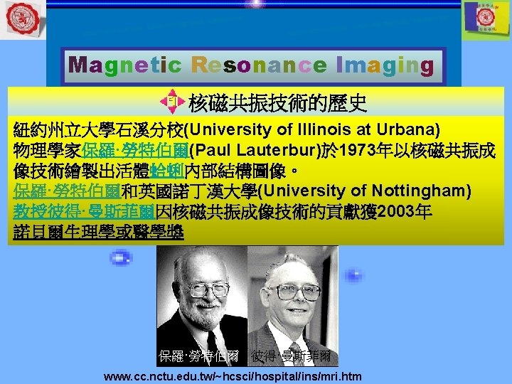
Magnetic Resonance Imaging 核磁共振技術的歷史 Dr. Raymond Damadian built the 1 st whole body MRI machine - 1977 Dr. Raymond Damadian & his associates, Drs. Larry Minkoff & Michael Goldssmith, completed the world's 1 st whole body MRI. Indomitable to capture Named Indomitable spirit of its 7 -year construction (now located at Smithsonian Ins, Washington, DC) Indomitable: 百折不撓 The first whole body transaxial image took 4 h 45 min to produce. Made on July 3, 1977, it shows the thoracic spine of Larry Minkoff Ref. 6
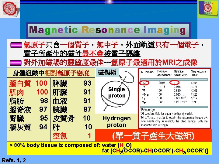
Magnetic Resonance Imaging MRI- basic principle 自由磁核 Bo 順向平行: 低能量 Bo 隨機排列無外加磁場 Ref. 7 Mo (even no) (odd no) Alignment 逆向平行: 高能量 Mo: 靜磁場 (net magnetic field) Bo: 外加磁場(external magnetic field) 平行排列外加磁場 多出的 順向平 行
Magnetic Resonance Imaging MRI 成 像 順 序 (1)人體置於強大磁場中,人體內氫原子一 律向Z軸排列 Net spinning Precession (2)給予等於Larmor frequency之射頻脈波 (RF: radiofrequency)使氫原子的自旋軸自 Z軸偏向,稱共振 Resonance (Excitation) (3)停止RF,氫原子核逐漸回到其原平行之Z 軸方向,而達到平衡狀態 Relaxation (4) 過程中釋放訊號由接受器接收,電腦解 (1) Free induction (2) decay 析組成,構成影像 MRI machine: *The coil is enveloped by liquid helium (-2730 C) to attain a super-conducting state *Strength of magnetic field is 1. 5 T or (3, 4) 3. 0 T (T: Telsa) Refs. 2, 8
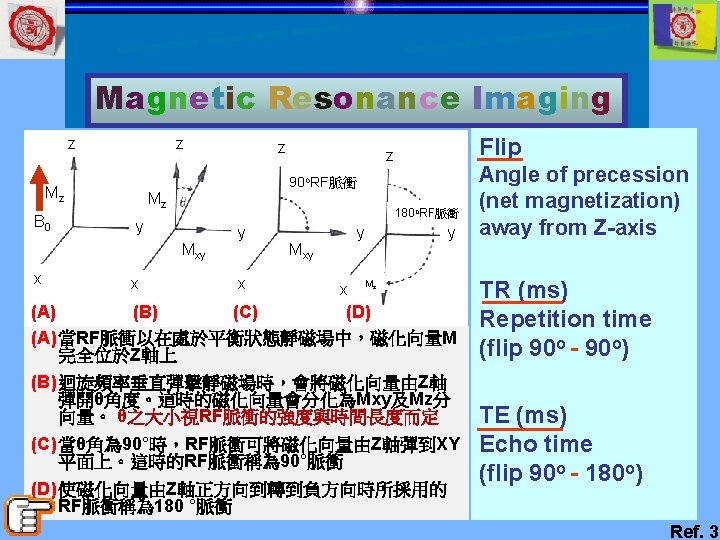
Magnetic Resonance Imaging Relaxation time Larmor frequency (precession) (外加磁場) (外加電磁波) RF = Larmor frequency excitation (resonance) (Flipping angle) n tio a x a Rel 平衡時,核磁化矩與靜磁場方 向(Z)一致 90°RF波使核磁化矩由Z方向轉 到Y方向 此時Z方向之分量為零。此後 XY平面分量逐漸消失,Z方向 分量由零漸增長而達平衡 描述核磁化矩在XY平面分量之 消失的過程稱spin-spin(T 2) relaxation或transverse relaxation (外加線圈 接受訊號Free induction decay) 描述Z方向分量之增長的過程 稱spin-lattice (T 1) relaxation 或longitudinal relaxation T 2 -time T 1 -time; T 1 - & T 2 - relaxation occur concurrently Refs. 2, 3
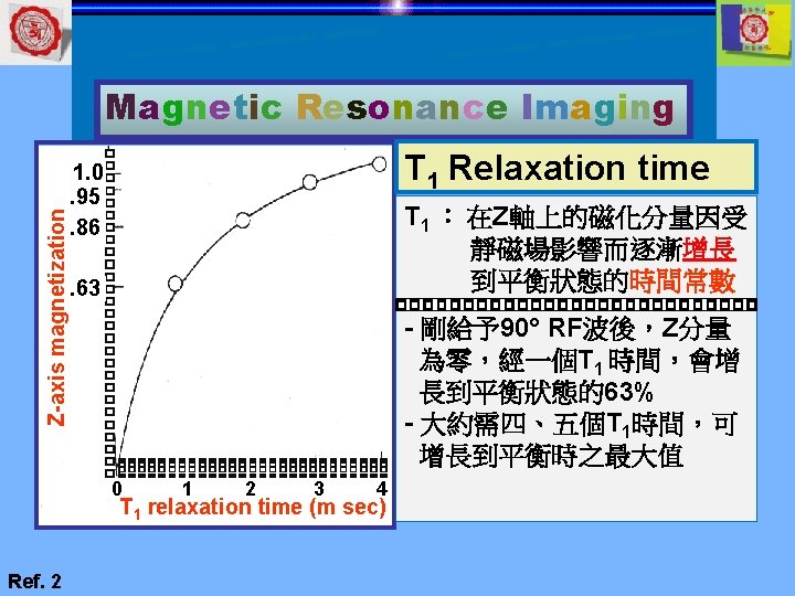
Magnetic Resonance Imaging Y-axis magnetization 1. 0 . 37. 14. 05 1 2 3 4 T 2 relaxation time (m sec) Ref. 2 T 2 Relaxation time T 2:平面上磁化矩下降, 成指數的下降 - 從 90° 波後的最大值下 降到零大約需四、五 個T 2的時間 - 經一過T 2 的時間大約 剩下開始時的37%
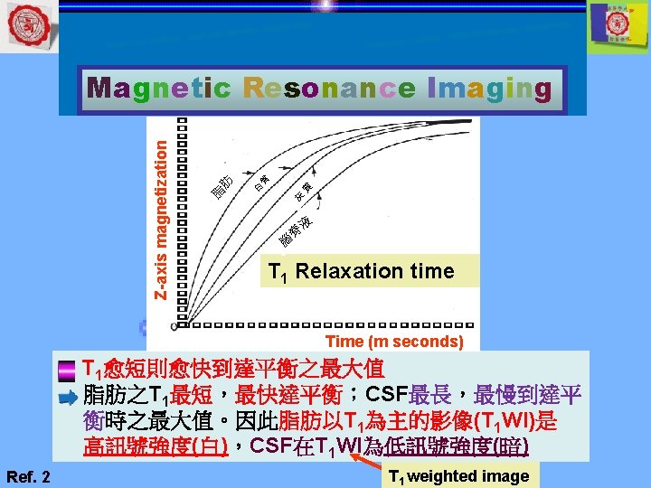
Y-axis magnetization Magnetic Resonance Imaging 腦脊液 CSF T 2 Relaxation time 白質 灰質 Time (m seconds) CSF之T 2值最長,最慢消失;白質在三者中最短 最快消失。CSF以T 2為主的影像(T 2 WI) T 2 weighted image 中為高訊號強度(白),而白質為低訊號強度(灰暗 Ref. 2 )
Magnetic Resonance Imaging T 1 (s) Normal tissues Malignant tissues T 1 and T 2 relaxation times of malignant tissues > normal tissues (exception is melanoma < normal tissues) Liver Ref. 1
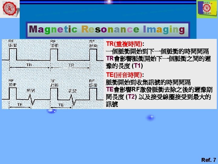
Magnetic Resonance Imaging Repetition time and echo time (Resonance) Excitation TR = repetition time TE = echo time Detection MR Image The spin-echo pulse sequence in MRI A series of 90 o & 180 o RF pulses is given at a repetition time operator (TR) selected by the operator The signal from the tissues is detected at the time of echo operator (TE), again selected by the operator Image contrast TR & TE will affect image Ref. 4
Magnetic Resonance Imaging Determining image contrast Image Contrast T 1 -weighted T 2 -weighted Time of Repetition *Short Long Time of Echo Short *Long Water T 1 -weighted & proton density-weighted: (long T 1 - &T 2(visualize the tissue anatomy) time)T 1 -relaxation time: major determinant of tissue Dark: T 1 WI image contrast of normal anatomy Bright: T 2 WI image Tissue with short T 1 -time: bright; long T 1 -time: dark T 2 -weighted: (壓抑 fat signal - additional tissue information) T 2 -relaxation time: major determinant of tissue contrast of pathological lesion Tissue with long T 2 -time: bright (high signal) short T 2 -time: dark (low signal) Ref. 4
Magnetic Resonance Imaging Time of Repetition Image Contrast Time of Echo *Short Long T 1 -weighted T 2 -weighted 脂肪和水在 沒有對比即 訊號相同、 無法區分 訊號強度 短T 1(脂肪) Short *Long 脂肪和水只有些微的 對比差異 訊 號 強 度 長T 1(水) 脂肪和水 的對比 長T 2(水) 脂肪和水 有大的對 比差 短T 2(脂肪) 短TR TR(ms) 長TR 短TE TE(ms) 長TE Refs. 4, 7
Magnetic Resonance Imaging Time of Repetition Image Contrast Time of Echo *Short T 1 -weighted Short-TI inversion recovery (STIR) Bo Bo 脂肪 第一次RF脈衝 遲豫 水 橫向平面 脂肪 Bo 脂肪和水遲 豫至Bo方向 Bo Bo 水 橫向平面 Short TR 第二次RF 脈衝 第一次RF脈衝 水 橫向平面 Bo Bo Short TE 脂肪 第二次RF脈衝 橫向平 面 脂肪和水 在橫向平面 Bo 脂肪和水的向 量表現出質子 密度 Ref. 7
Magnetic Resonance Imaging Magnetic resonance imaging signals of normal tissue T 1 WI T 2 WI High intensity (light gray)--(亮) Fat(亮) Bone marrow (adult) Fat(亮) Water(亮) Bone marrow (adult) Bone marrow (child) Low intensity (dark gray)--(暗) Water(暗) Muscle Bone marrow (child) Muscle No signal intensity (black)--(黑) Cortical bone-calcium deposition Enamel-dentin Air-blood vessel Ref. 8
Magnetic Resonance Imaging T 1 WI T 2 WI MRI of lipoma: Lesion in right cheek ---- high signal in both T 1 -weighted & T 2 -weighted images; diagnosed as fatty tissue Ref. 8
Magnetic Resonance Imaging T 1 WI T 2 WI MRI of ranula: Lesion in left submandibular region ---- low signal in T 1 -weighted images & high signal in T 2 -weighted images; defined as water Ref. 8
Magnetic Resonance Imaging T 2 WI STIR MRI of inflammation in right cheek: T 2 -weighted image: fatty tissue & water based tissue ---- high signals; range of inflammation is difficult to determine Short-TI inversion recovery: cancels signal from fatty tissue; range of inflammation is easy to ascertain Ref. 7
Magnetic Resonance Imaging Synovial chondromatosis, right TMJ T 1 WI PD KMU case
Magnetic Resonance Imaging Contrast-enhanced MRI 1. Gadolinium (钆): used as contrast agent (0. 2 ml/kg , IV) 2. Gadolinium: high T 1 -shortening effect & to increase diagnostic ability. 3. Adverse effect rate: ~1 -2%; safer than iodine contrast agent 4. Contraindicated for asthma patients 5. Caution for severe renal impairment patients due to risk of nephrogenic systemic fibrosis Ref. 8
Magnetic Resonance Imaging Contrast-enhanced MRI Time-signal intensity curves Type A Signal Intensity Type C Type B Type D Time Type A: Rapid initial enhancement followed by a gradual decrease in enhancement (e. g. SCC). Type B: Rapid initial enhancement followed by a rapid decrease in enhancement (e. g. Warthin tumor, lymphoma). Type C: Gradual increase in enhancement (e. g. pleomorphic adenoma). Type D: No enhancement (e. g. cyst) Observing signal within a lesion at regular time intervals: Blood flow state within lesion: useful for identifying diseases Ref. 8
Magnetic Resonance Imaging Contrast-enhanced MRI Signal Intensity Time-signal intensity curves of ranula Ranula 1. Ranula has no internal blood flow; so, contrast agent does not flow into lesion 2. The time-signal intensity curve consequently exhibits a plateau shape Time Ref. 8
Magnetic Resonance Imaging Contrast-enhanced MRI Time-signal intensity curves of SCC slow Squamous cell carcinoma (SCC) rapidly Signal Intensity ly 1. SCC tissue with significant blood flow inside; contrast agent flows rapidly into lesion. 2. Thereafter, contrast agent slowly flows out from tumor 3. Time-signal intensity curve exhibits a sudden increase & then a gradual decrease Time Ref. 8
Magnetic Resonance Imaging Diffusion-weighted imaging (DWI, DWMRI) Brownian motion of water molecules Water Ice Movement of water molecules in liquids & solids: Apparent diffusion coefficient (ADC) values 1. Water molecules are more easily diffused in liquids due to vigorous Brownian motion. So, ADC values are high 2. Due to lack of Brownian motion in solids, diffusion of water molecules does not readily occur & ADC values are correspondingly low Ref. 8
Magnetic Resonance Imaging Diffusion-weighted imaging (DWI, DWMRI) Apparent diffusion coefficient (ADC) values in ranula & squamous cell carcinoma Due to high cellular density in squamous cell carcinoma, ADC value is low (1. 31 × 10− 3 mm 2/s) Ranula Squamous cell carcinoma of the tongue Because ranula contains liquid, ADC value is high ( 2. 87 × 10− 3 mm 2/s) Ref. 8
Magnetic Resonance Imaging MRI- cautions, advantages, disadvantages Cautions before MRI imaging 1. Medical equipment: stretcher, wheelchair, scissor, & gas cylinders used in the MRI room must be made with nonmagnetic materials 2. Contraindicated for patient with cardiac pacemaker, implantable cardioverter defibrillator & artery clip Ref. 8
Magnetic Resonance Imaging MRI- cautions, advantages, disadvantages Cautions before MRI imaging 3. Patient with tattoo, wearing colored contact lens, mascara, or eye shadow as all these materials contained minute iron particles that cause image artifacts & can become heated due to magnetic field, resulting patient burn Thunderbolt-2 nd burn AJR 2000; 174: 1795
Magnetic Resonance Imaging Advantages of MRI 1. 2. 3. 4. Noninvasiveness & lack of radiation exposure Produce any given tomographic images Display blood vessels without contrast agent Higher tissue resolution & a lower spatial resolution than CT muscle lymph node submandibular gland blood vessel MRI has a lower spatial resolution than CT; tissue boundaries are difficult to ascertain MRI has high ability to resolve tissues. While CT scans do not indicate density differences among tissues, the differences in density among tissues are clear in MRI Ref. 8
Magnetic Resonance Imaging Disadvantages of MRI 1. Long scan time (approximately 30 -60 min) 2. Inability to obtain signal from cortical bone &calcification 3. Inability to perform the test when metal is inside body 4. Difficulty in scanning claustrophobic patients Axial Sagittal Coronal Artifacts caused by metals in CT only within slices, but they appear in 3 D in MRI Ref. 8
Magnetic Resonance Imaging Summaries Finishing the teaching, you can understand: - 核磁共振技術的歷史 - MRI-- basic principles - T 1 and T 2 relaxation time - Repetition and echo time - Determining image contrast - MRI-- cautions, advantages, disadvantages
Magnetic Resonance Imaging References for the present lecture (1) 2001, November 1. 1 st edition, p. 1 -17, 157. 3. 鄭慶明 February 4. Basic Principles 2. 沈茂忠et al, 3 rd of MR Imaging. edition, Chapter 13, 1994; 37(2): 33 -38 Sharon LB, p. 569 p. 463 -471. 84
Magnetic Resonance Imaging References for the present lecture (2) 5. Wilkinson E. MRI researchers win Nobel Prize. Lancet Oncol 2003; 4: 649 6. Peggy Woodward MRI for technologists Mc. Graw-Hill, Chapter 1 pp. 1 -12 7. 陳煥武譯 圖解核 磁共振造影學, 合記 出版社 2006 1 st pp. 12 -37 8. Oral Radiol 2017; 33: 92– 100