Section A Applied Anatomy and Physiology 2 Joint


 * the amount of movement is based on the length of")
")
 – temporary joints present in children – E. g.")


 capsule: unites the")


- Slides: 27
Section A: Applied Anatomy and Physiology 2. Joint type
Syllabus • Definitions and examples of fibrous, cartilaginous and synovial joints • The typical structure and features of a synovial joint • The type of join and the bones which articulate at the following joints: – Shoulder, elbow, radio-ulnar, wrist, hip, knee, ankle, spine
Joint Classification • Structural classification is based on anatomical characteristics. • Fibrous: no synovial cavity and bones are held together by fibrous connective tissue. • Cartilaginous: no synovial cavity and the bones are held together by cartilage. • Synovial: bones forming the joint have a synovial cavity. Held together by the articular capsule and often by accessory ligaments.
Fibrous Joint (Suture) * the amount of movement is based on the length of the tissue fibres *
Fibrous Joint (Syndesmosis)
Cartilaginous Joint Types • (Synchondroses) – temporary joints present in children – E. g. epiphyseal plates in long bones • (Sympheses) – permanent cartilaginous joints – E. g. vertebral column
Temporary Joint
Permanent Joint
Synovial Joints • There are 6 types • These are categorized according to range of movement possible • Movement determined by: – Shape of articulating surfaces – Position of ligaments – Number of ligaments
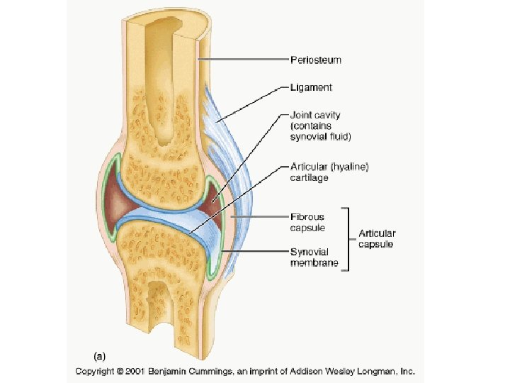
Synovial Joint Features • Common features include – A joint (articular) capsule: unites the articulating bones. – A joint cavity. – A synovial membrane: secretes synovial fluid to reduce friction/absorb shock, supply nutrients. – Articular (hyaline) cartilage.
Some Synovial Joints Also Have… • Bursae – Small sacs of synovial fluid located at points of friction • Menisci – Discs of cartilage between bone surfaces • Pads of fat – Added protection
Synovial Joints
Synovial Joints
Ball and Socket
Ball and Socket Joint • Multiaxial • Movement in all 3 planes • • Flexion/Extension Abduction/Adduction Circumduction Rotation
Hinge
Hinge Joint • Uniaxial • Only permits flexion and extension • Examples are the knee, elbow and ankle
Pivot
Pivot Joints • Uniaxial • It allows rotation only around its own longitudinal axis • Example is the atlanto-axial joint
Plane
Planar Joints • Nonaxial • The motion they allow does not occur around an axis or along a plane • Examples are intercarpal joints, intertarsal joints etc
Saddle
Saddle Joints • Biaxial • Flexion/Extension • Abduction/Adduction • Circumduction • Example is the carpometacarpal joint
Condyloid
Condyloid Joints • Biaxial • Flexion/Extension • Abduction/Adduction • Circumduction • Examples are the wrist and metacarpophalangeal joints