SYNOVIAL FLUID COMPOSITION INTRODUCTION Synovial fluid is a


 can be elevated in synovial fluid,")


 Which of the following cells in the synovium increases the viscosity of")
 Type A is derived from blood monocytes, and it removes the")
 Which test can be used to evaluate the level of synovial fluid")
 String sign is used for the measure of viscosity – 1")
 The presence of uric acid in synovial fluid is helpful in diagnosis")
 Gout arthritis is caused by the deposition of crystals of uric")
 In which joint condition does the synovial fluid examination shows WBC count")
 Osteoarthritis is the degenerative disease b) Gout arthritis is caused by")
. The highest diagnostic potential for predicting septic arthritis in synovial fluid is:")
 Normally, synovial fluid glucose levels are less than 10 mg/d. L")
- Slides: 33
SYNOVIAL FLUID COMPOSITION
INTRODUCTION • Synovial fluid is a thick, stringy fluid found in the cavities of synovial joints. • With its egg-like consistency ("synovial" partially derives from ovum, Latin for egg), synovial fluid reduces friction between the articular cartilage and other tissues in joints, to lubricate and cushion them during movement. • The inner membrane of synovial joints is called the synovial membrane and secretes synovial fluid into the joint cavity. • This fluid forms a thin layer (roughly 50 μm) at the surface of cartilage.
SYNOVIAL JOINT • Plasma constituents that enter joint fluid must cross a doublebarrier membrane. • First, the endothelial lining of the capillaries is traversed followed by movement through a matrix that surrounds synovial cells. • This ultrafiltrate is combined with a mucopolysaccharide (hyaluronate) synthesized by the synovium.
Synovial fluid electrolytes • The distribution ratio of most electrolytes between synovial fluid and serum is 1. • The average ratio of total anions of serum to total anions of synovial fluid is 1. 99. • Ionic calcium ratios are approximately 1. 18. • This higher ratio is thought to be due to the base combining power of mucin for calcium.
Secretion of Synovial Fluid Synovial tissue is composed of connective tissue that lacks a basement membrane. There are two main types of synovial lining cells: o Type A cells are macrophage-like and have primarily a phagocytic function which is important to remove microbes and the debris that results from normal wear and tear in the joint. o Type B cells are fibroblast-like and produce hyaluronate, which accounts for the increased viscosity of synovial fluid. • Other cells found in the synovium include antigen-presenting cells called dendritic cells and mast cells. • Synovial tissue also contains fat and lymphatic vessels, fenestrated microvessels, and nerve fibers derived from the capsule and periarticular tissues.
Secretion of Synovial Fluid • Synovial fluid is made of hyaluronic acid and lubricin, proteinases, and collagenases. • Normal synovial fluid contains 3 -4 mg/ml hyaluronate (hyaluronic acid), a polymer of disaccharides composed of D-glucuronic acid and D-N-acetylglucosamine joined by alternating beta-1, 4 and beta-1, 3 glycosidic bonds. • Synovial fluid also contains lubricin secreted by synovial cells. It is chiefly responsible for so-called boundary-layer lubrication, which reduces friction between opposing surfaces of cartilage. It may also have a role in synovial cell growth.
FUNCTIONS Synovial fluid is believed to have two main functions: Ø To aid in the nutrition of articular cartilage by acting as a transport medium for nutritional substances, such as glucose. • Articular cartilage has no blood, nerve, or lymphatic supply. v Supplying oxygen and nutrients to and removing carbon dioxide and metabolic wastes from the chondrocytes within articular cartilage. • Glucose for articular cartilage chondrocyte energy is transported from the periarticular vasculature to the cartilage by the synovial fluid.
FUNCTIONS Ø To aid in the mechanical function of joints by lubrication of the articulating surfaces. • Lubrication reduces frictional resistance between bearing surfaces by keeping them apart. • During movement, the synovial fluid held in the cartilage is squeezed out mechanically to maintain a layer of fluid on the cartilage surface. • The lubricating properties of synovial fluid on articular cartilage are due to the presence of hyaluronate or mucin and a glycoprotein in it.
Physical Characteristics of Normal Synovial Fluid • Normal synovial fluid is clear, pale yellow, viscid, and does not clot. • Volume: The amount of fluid contained in joints is usually small. The knee joint normally contains up to 4 m. L of fluid. v An increase in synovial fluid enough to aspirates is due to some disease.
Clotting • Normal synovial fluid: Do not clot • Clotting of synovial fluid means fibrinogen is present: 1. Damaged synovial membrane 2. Traumatic tap
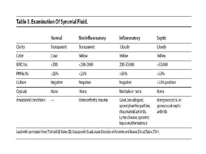
Color and clarity: • Normal synovial fluid is colorless and clear. • Other appearances may indicate various disease states. • Loss of clarity : crystals, increased cellularity, infective, cartilage debris • E. g. Yellow/clear (non inflammatory effusions), yellow/cloudy (inflammatory processes), white/ cloudy (crystals); and red brown (hemorrhage).
Physical Characteristics of Normal Synovial Fluid • Viscosity: Synovial fluid is very viscous due to its high concentration of polymerized hyaluronate. • A string test can be used to evaluate the level of synovial fluid viscosity. After removing the needle or cap from the syringe, synovial fluid is expressed into a test tube one drop at a time. • Normal synovial fluid will form a “string” approximately 5 cm long before breaking. • In addition, the fluid may cling to the side of the test tube rather than running down to the bottom. • Low viscosity of synovial indicates the presence of an inflammatory process.
Physical Characteristics of Normal Synovial Fluid • Temperature – 32 degree Celsius (peripheral joints are cooler than core body temperature). • p. H – 7. 4
Indications • Suspected septic arthritis – Any febrile person with unexplained inflammatory fluid -> presumed septic joint – Even with acute flare of arthritis (e. g. , RA), should rule out septic joint • Suspected crystal-induced arthritis – Gout or pseudogout diagnosis largely made with microscopic examination – Diagnosis requires negative Gram stain/culture • Unexplained joint or bursa swelling – Largely to permit classification into inflammatory vs noninflammatory categories – Diagnosis made along with history & physical.
BIOCHEMICAL EXAMINATION • Protein: Synovial fluid contains all proteins found in plasma, except various high–molecular weight proteins. • These high–molecular-weight proteins include fibrinogen, beta 2 macroglobulin, and alpha 2 macroglobulin, and can be absent or present in very low amounts. • Most commonly used serum protein procedures can be used to measure synovial fluid protein. • The normal range for synovial fluid protein is 1– 3 g/d. L. • Increased synovial fluid protein levels are seen in ankylosing spondylitis, arthropathies that accompany Crohn disease, gout, psoriasis, Reiter syndrome, and ulcerative colitis.
BIOCHEMICAL EXAMINATION • Glucose: Synovial fluid glucose levels should be interpreted using serum glucose levels. • A fasting specimen should be used or at least one 6– 8 hours postprandially. • Normally, synovial fluid glucose levels are less than 10 mg/d. L lower than serum levels. • Joint disorders that are classified as infectious demonstrate large decreases in synovial fluid glucose and can be as much as 20– 100 mg/d. L less than serum levels. • Other groups of joint disorders demonstrate a less of a decrease in synovial fluid glucose, 0– 20 mg/d. L
BIOCHEMICAL EXAMINATION • Uric acid: Synovial fluid uric acid normally ranges from 6 to 8 mg/d. L. • The presence of uric acid in synovial fluid is helpful in diagnosis gout. • Usually, crystal identification is used for this determination, but synovial fluid uric acid levels may be performed in laboratories that do not have a light polarizing microscope. • Lactic acid: Lactic acid is rarely measured in synovial fluid but can be helpful in diagnosing septic arthritis. v Lactate levels in the synovial fluid have the highest diagnostic potential for predicting septic arthritis. • Normally, synovial fluid lactate is less than 25 mg/d. L but can be as high as 1000 mg/d. L in septic arthritis.
BIOCHEMICAL EXAMINATION • Enzymes: Alkaline phosphatase, acid phosphatase, lactic dehydrogenase, and other enzymes are present in detectable quantities. • Synovial fluid to serum ratios of these and other enzymes vary with the presence of articular disease. • Enzymes enter the synovial fluid directly from the plasma or may be produced locally by the synovial membrane or released by synovial fluid macrophages.
BIOCHEMICAL EXAMINATION • Lactate dehydrogenase: Lactate dehydrogenase (LD) can be elevated in synovial fluid, while serum levels remain normal. • Synovial fluid LD levels are usually increased in RA, infectious arthritis, and gout. • The neutrophils that are increased during the acute phase of these disorders contribute to this increased LD level.
BIOCHEMICAL EXAMINATION • Complement levels: Normal synovial fluid complement levels in humans are approximately 10% of the serum values. • In the inflamed joint synovial fluid complement levels will vary. The long-term patterns of variation have some prognostic value in human rheumatoid arthritis patients. • Rheumatoid factor (RF) is an antibody to immunoglobulins. • RF is present in the serum of most patients with RA, whereas just more than half of these patients will demonstrate RF in synovial fluid.
CLINICAL IMPORTANCE • Synovial fluid analysis is a well-established procedure in the evaluation of joint disease. • The purpose of synovial fluid analysis is to determine the presence of arthritis and to place a fluid into one of several categories. • Appropriate treatment of joint disease depends on proper identification of disease.
Practice Questions
Q 1) Which of the following cells in the synovium increases the viscosity of synovial fluid? a) b) c) d) Type A cells (macrophage-like) Type B cells (fibroblast-like) mast cells dendritic cells
key: b a) Type A is derived from blood monocytes, and it removes the wear-andtear debris from the synovial fluid. b) Type B produces hyaluronan. Synovial fluid is made of hyaluronic acid and lubricin, proteinases, and collagenases. Synovial fluid contains lubricin (also known as PRG 4) as a second lubricating component, secreted by synovial fibroblasts c) & d). – Normally Synovial Fluid has. . NO leukocytes (white blood cells) NO red blood cells NO plasma clotting factors NO culture (no bacteria) NO chondrocytes – In pathologic fluids, chondrocytes, osteoblasts, and osteoclasts may be seen.
Q 2) Which test can be used to evaluate the level of synovial fluid viscosity? a) b) c) d) String test Gram staining Coagulation test Illumination test
key: a a) String sign is used for the measure of viscosity – 1 -2 inches ; When a drop is taken between the thumb and finger as in pinching and then they are separated, the drop forms a string till 1 -2 inches b) Gram stain is used to distinguish and classify bacterial species into two large groups c) Cogulation test is used to assess blood clotting function. d) The Illumination & Visibility Testing Laboratory is used for conducting photometric measurements and human visual performance
Q 3) The presence of uric acid in synovial fluid is helpful in diagnosis of: a) Gout b) Rheumatoid arthritis c) Septic arthritis d) Osteoarthritis
key: a a) Gout arthritis is caused by the deposition of crystals of uric acid in a joint. b) Rheumatoid arthritis (RA) is an autoimmune disease c) Septic arthritis is a penetrating injury delivers germs directly into the joint. d) Osteoarthritis is a degenerative disorder of joints.
Q 4) In which joint condition does the synovial fluid examination shows WBC count more than 50, 000 per ml? a) Osteoarthritis b) gout c) trauma d) gonococcal septic arthritis
key: d a) Osteoarthritis is the degenerative disease b) Gout arthritis is caused by the deposition of crystals of uric acid in a joint. c) Trauma does not elevate the WBC count d) Synovial fluid white blood cell count in patients with septic arthritis is usually greater than 50, 000 per mm.
Q 5). The highest diagnostic potential for predicting septic arthritis in synovial fluid is: a) Acetate b) Lactate c) Pyruvate d) Uric acid
key: b a) Normally, synovial fluid glucose levels are less than 10 mg/d. L lower than serum levels. So, Acetate will be not increased. b) The highest concentrations of lactate occurred in nongonococcal septic synovial fluids. Lactate levels in the synovial fluid have the highest diagnostic potential for predicting septic arthritis. c) Normally, synovial fluid glucose levels are less than 10 mg/d. L lower than serum levels. So, pyruvate will be not increased. d) The presence of uric acid in synovial fluid is helpful in diagnosis gout.