Internal Derangement of the Knee and Anterior Knee


 and record the")


 using stretching, compressing, and contracting to duplicate")
 for early detection of")


?")
 also known as degenerative arthritis or")


 n Some physicians refer to IDK as “I don’t")


 is a frequently encountered overuse injury")


")


 implicated increased q angle,")


- Slides: 104
Internal Derangement of the Knee and Anterior Knee Pain James J. Lehman, DC, MBA, FIANM Associate Professor of Clinical Sciences School of Chiropractic Director Community Health Clinical Education Department University of Bridgeport
Learning Objectives n Correlate anatomy and the patients’ signs and symptoms in order to locate the neuromusculoskeletal lesions of the knee and properly record the findings.
Learning Objectives n Elicit a patient history and record the subjective findings in order to perform differential diagnosis procedures and determine use of objective testing.
Learning Objectives n Perform neuromusculoskeletal evaluation procedures (Posture, orthopedic and neurological) and record the objective findings in order to make an assessment/diagnosis.
Learning Objectives n n Organize a clinical thought process while performing a neuromusculoskeletal evaluation. Identify injured and painful tissues through careful assessment and intelligent use of neuromusculoskeletal testing and document the findings.
Opening Statement … n Diagnosis is the key to successful treatment!
Knee pain accounts for approximately one third of musculoskeletal problems seen in primary care settings.
Knee pain is most prevalent in physically active patients, with as many as 54 percent of athletes having some degree of knee pain each year. Rosenblatt RA, Cherkin DC, Schneeweiss R, Hart LG. The content of ambulatory medical care in the United States. An interspecialtycomparison. N Engl J Med. 1983; 309: 892– 7.
The abbreviation, “IDK” often means “I don’t know” rather than internal derangement of the knee.
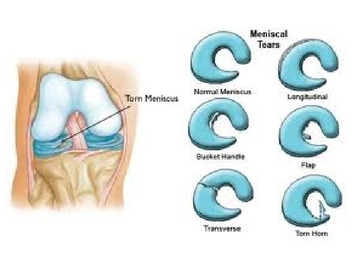
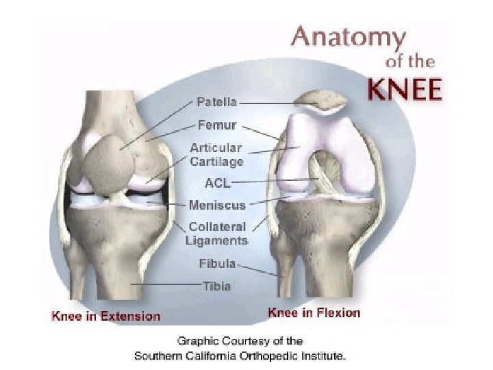
Internal Derangement of the Knee ICDA 10 M 23. 91 Internal derangement of the knee (IDK) is the term used to cover a group of disorders involving disruption of the normal functioning of the ligaments or cartilages (menisci) of the knee joint.
Unspecified Internal Derangement of Knee ICD-10 -CM Code M 23. 91
American Family Physician Evaluating Acutely Injured Patients for Internal Derangement of the Knee. Am Fam Physician. 2012 Feb 1; 85(3): 247 252. MICHAEL GROVER, DO, Mayo Clinic College of Medicine, Scottsdale, Arizona http: //www. aafp. org/afp/2012/0201/ p 247. html
History: Painful Tissue Identification Check List ü ü ü ü ü Ask the patient to point to the pain When did the pain commence? Trauma? What makes it better or worse? Quality of the pain Radiating or localized? Pain severity rating When is it painful Treatments and reaction? Previous diagnoses?
ACL Sprain Mechanism of Injury
Generalized Pain Description n Joint pain may be constant dull, deep ache but sharp upon motion Muscle pain may be a dull ache, crampy or spasm sensation Trigger points may be localized with pin point pain or diffuse with poor localization and paresthesias (formication)
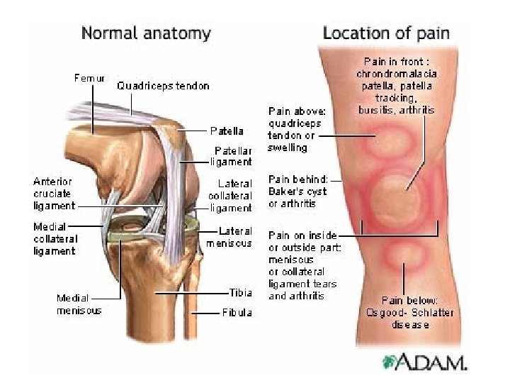
Knee Pain Can you identify the condition with the point of pain? n n n Osgood Schlatter’s Disease? Jumper’s knee? PFA? Collateral ligament sprain? Meniscal tear?
Examination Protocols 1. 2. 3. 4. 5. 6. 7. History taking or interview Observation and inspection Palpation Range of motion Special tests 3 part PNS exam Imaging
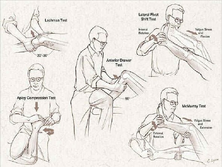
Orthopedic test A provocative maneuver (most often) using stretching, compressing, and contracting to duplicate the pain and identify the involved tissues.
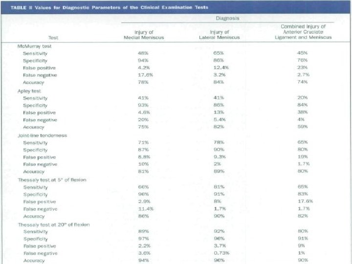
Diagnostic accuracy of a new clinical test (the Thessaly test) for early detection of meniscal tears. J Bone Joint Surg Am. 2005 May; 87(5): 955 62. Available from: http: //www. ncbi. nlm. nih. gov/pubmed/15866956 n Thessaly test at 20 degrees of knee flexion had a high diagnostic accuracy rate of 94% in the detection of tears of the medial meniscus and 96% in the detection of tears of the lateral meniscus, and it had a low rate of false positive and false negative recordings. Other traditional clinical examination tests, with the exception of joint line tenderness, which presented a diagnostic accuracy rate of 89% in the detection of lateral meniscal tears, showed inferior rates.
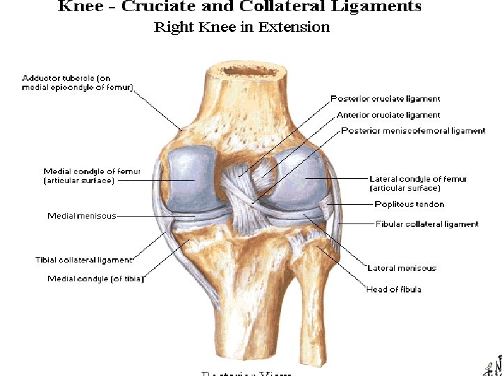
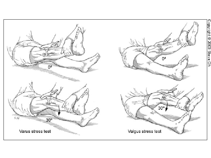
Ligament Injuries Medial collateral and anterior cruciate ligaments are the most commonly injured.
Anterior Drawer Sign n n Anterior cruciate Medial collateral ligament ITB Capsules & ligaments Arcuate politeus complex
Ligament Instability Anterior and Posterior Drawer Signs
Ligament Instability Lachman’s Test n n Anterior and posterior cruciate ligament sprains Most reliable test for anterior cruciate ligament rupture
Anterior Drawer Sign and Lachman’s Anterior Cruciate & Posterior Oblique n Anterior translation of more than 5 mm indicates injury
Meniscus and Ligament Instability n n Apley’s compression tests meniscus Apley’s distraction tests nonspecific ligaments
Meniscal Injury Mc. Murray’s Test n n n Flex and extend with internal and external rotation. Stresses distorted meniscus Palpable or audible click is positive
Thessaly’s Test New test for the early detection of meniscal injury. http: //www. ejbjs. org/cgi/content/abs tract/87/5/955
Thessaly Test for Meniscus Tear Five degree of knee flexion Unaffected knee first
Thessaly Test for Meniscal Tear External rotation assisted Internal rotation
Thessaly Test for Meniscal Tear Positive findings n n n Pain medial or lateral Clicking or locking Most accurate at 20 degrees of knee flexion Repeat process at 20 degrees
Did I mention… n Diagnosis is the key to successful treatment!
Persistent Anterior Knee Pain n A 20 year old military recruit presented for follow up of anterior knee pain. Previously, he presented with three weeks of right knee pain without trauma, swelling, locking, or instability. At that visit, he was referred for physical therapy, but the knee did not improve during treatment. On recent examination, the patient had a positive “theater sign” (i. e. , knee pain upon arising after prolonged sitting), positive patellar compression and inhibition tests, stable ligaments, pain with Mc. Murray's test, and tenderness of the medial tibial plateau.
Question n n n Based on the patient's history, physical examination, and radiography results, which one of the following is the most likely diagnosis? A. Medial plica syndrome. B. Patellar tendinopathy. C. Patellofemoral pain syndrome. D. Pes anserine bursitis. E. Proximal tibial stress fracture.
Atraumatic, Persistent, Anterior Knee Pain
Discussion n The answer is E: proximal tibial stress fracture. Most tibial stress fractures occur in the tibial diaphysis. However, proximal tibial stress fractures, typically involving the medial condyle, can occur in athletes. Stress fractures may result from weaker bone that fails under normal loading (i. e. , an insufficiency fracture) or normal bone that fails under new or increased repetitive loading (i. e. , a fatigue fracture). Athletes are more likely to experience a fatigue fracture; military recruits are also at risk. Drabicki RR, Greer WJ, De. Meo PJ. Stress fractures around the knee. Clin Sports Med. 2006; 25: 105– 15. Raasch WG, Hergan DJ. Treatment of stress fractures: the fundamentals. Clin Sports Med. 2006; 25: 29– 36.
Discussion n Medical history that suggests a stress fracture will include persistent pain following an acute increase in physical activity (i. e. , time, distance, or pace). Physical examination may reveal tenderness at or near the medial joint line, tenderness with a leverage motion, and tenderness with hopping on the affected leg. A joint effusion may also be present.
Discussion n Plain radiographs can appear normal for weeks or even months. Because the metaphyseal portions of long bones are mostly cancellous bone, the typical periosteal/endosteal reaction will not be visible. In this type of bone, the features of a stress fracture manifest as a band-like area of sclerosis. If a stress fracture is suspected but radiographic results are normal, then magnetic resonance imaging (MRI) or a bone scan is the next diagnostic study. Of the two, MRI is more sensitive for diagnosing stress fracture.
Patellofemoral Dysfunction Patella Grinding Test n n n Chrondomalacia patellae Patellofemoral arthralgia Chondral fracture
Patellofemoral Dysfunction Patella Apprehension Test n n Pain and apprehension are present Positive test indicates lateral patellar dislocation
Patellofemoral Dysfunction Clarke’s Patellar Scrape Test n Pain and crepitation may indicate patellofemoral arthralgia or chondromalcia patellae
Noble Compression Test for ITBS 1. 2. 3. 4. 5. Patient supine Apply thumb pressure to the lateral femoral condyle with knee flexed to 75 degrees. Maintain thumb pressure while extending knee At 30 degrees of flexion, patient will complain of severe pain over ITB This is a positive test for ITBS
Ober Test Iliotibial Band Syndrome The patient lies down with the unaffected side down and the unaffected hip and knee at a 90 degree angle. If the iliotibial band is tight, the patient will have difficulty adducting the leg beyond the midline and may experience pain at the lateral knee.
Ober Test Iliotibial Band Syndrome
Wilson Sign for Osteochondritis Patient is supine Knee is passively flexed to 90 degrees and then extended with medial rotation of knee. Near 30 degrees of flexion, the pain increases and patient may resist further extension. Rotating the knee laterally eliminates the pain. This is a positive test for osteochondritis of the femur.
Radiographic Decision Rules n The Pittsburgh decision rules were 99 percent sensitive and 60 percent specific for the diagnosis of knee fractures and could have reduced the use of radiography by 52 percent, with one missed fracture. If the rules indicated a fracture, 24. 1 percent of patients actually had a knee fracture (positive predictive value); if the rules indicated no fracture, 99. 8 percent of patients did not have a knee fracture (negative predictive value). The Ottawa knee rules were 97 percent sensitive and 27 percent specific for knee fractures, with three fractures missed. The authors of the comparative study concluded that the Pittsburgh decision rules were more specific, with no loss of sensitivity. Seaberg DC, Yealy DM, Lukens T, Auble T, Mathias S. Multicenter comparison of two clinical decision rules for the use of radiography in acute, high risk knee injuries. Ann Emerg Med 1998; 32: 8 13
Pittsburgh Rules Patient should receive knee films if: Blunt trauma or a fall as mechanism of injury AND either of the following: Age younger than 12 years or older than 50 years Inability to walk four weight bearing steps in the emergency department
Ottawa Rules Age ≥ 55? Isolated tenderness of the patella (no other bony tenderness)? Tenderness at the fibular head? Inability to flex knee to 90° ? If any of the above criteria are met, this patient may need knee imaging.
Degenerative Joint Disease of the Knee Osteoarthritis (OA) also known as degenerative arthritis or degenerative joint disease, is a group of mechanical abnormalities involving degradation of joints, including articular cartilage and subchondral bone.
Knee Aspiration and Injection Knee joint aspiration and injection are performed to establish a diagnosis, relieve discomfort, drain off infected fluid, or instill medication. Because prompt treatment of a joint infection can preserve the joint integrity, any unexplained monarthritis should be considered for arthrocentesis http: //www. aafp. org/afp/2002/1 015/p 1497. html
Lateral knee joint injection. Entry should be in the soft tissue between the patella and femur. For the medial approach, the needle enters the medial side of the knee under the middle of the patella (midpole) and is directed toward the opposite patellar midpole. In the anterior approach, the knee is flexed 60 to 90 degrees, and the needle is inserted just medial or lateral to the patellar tendon and parallel to the tibial plateau. This technique is preferred by some physicians for its ease of joint entry in advanced osteoarthritis. http: //www. aafp. org/afp/2003/0 515/p 2147. html
Symptoms of DJD of the Knee The main symptom is pain, causing loss of mobility and often stiffness. "Pain" is generally described as a sharp ache, or a burning sensation in the associate muscles and tendons. OA /DJD can cause a crackling noise or crepitus when the affected joint is moved or touched, and patients may experience muscle spasm and contractions in the tendons.
Orthopedic Evaluation n n History Observation Palpation Range of motion Special tests
Treatment of DJD of the Knee Modification of lifestyle Moderate exercise Self management (weight loss) is more beneficial than medications Smoking cessation Medications: Acetaminophen preferred over NSAIDS Manipulation/mobilization
Internal Derangement of Knee (IDK) n Some physicians refer to IDK as “I don’t know. ”
Internal Derangement of the Knee ICDA 10 Code M 23. 91 Internal derangement of the knee (IDK) is the term used to cover a group of disorders involving disruption of the normal functioning of the ligaments or cartilages (menisci) of the knee joint.
Most Common IDK Conditions The commonest derangement met with is injury to the medial collateral ligament. The medial meniscus and anterior cruciate ligament are next in frequency. The lateral ligament, lateral meniscus and posterior cruciate ligament are less liable to damage.
Atraumatic Degenerative IDK Age related degeneration of a semilunar cartilage may be met with in an older patient, say over age 50. It may present as spontaneous occurrence of knee pain without any history of injury.
n n Internal derangement of the knee is the term used to describe the various types of disruption of the ligaments and cartilages of the knee. By far the most frequent cause is sports injury, with footballers especially at risk. Some disorders of the cartilages may occur without evidence of prior injury. Osteoarthritis of the knee is commonly a late sequel of cartilage or cruciate ligament damage.
Question? How do you manage internal derangement of the knee?
Iliotibial Band Syndrome The iliotibial band is a thick band of fascia that extends along the lateral thigh from the iliac crest to the knee.
Iliotibial Band Syndrome Iliotibial band friction syndrome (ITBFS) is a frequently encountered overuse injury caused by repetitive friction between the iliotibial band the lateral femoral epicondyle during active flexion and extension at the knee.
History PAIN CHARACTERISTICS The patient's description of knee pain is helpful in focusing the differential diagnosis. It is important to clarify the characteristics of the pain, including its onset (rapid or insidious), location (anterior, medial, lateral, or posterior knee), duration, severity, and quality (e. g. , dull, sharp, achy). Bergfeld J, Ireland ML, Wojtys EM, Glaser V. Pinpointing the cause of acute knee pain. Patient Care. 1997; 31(18): 100– 7.
Iliotibial Band Syndrome The primary initial complaint in patients with iliotibial band syndrome is diffuse pain over the lateral aspect of the knee.
Iliotibial Band Friction Syndrome Patients with iliotibial band syndrome often demonstrate tenderness on palpation of the lateral knee approximately 2 cm above the joint line.
Ober’s Test n Failure to descend smoothly indicates a positive test for contracture of the TFL or ITB.
Iliotibial Band Syndrome Swelling may be noted at the distal iliotibial band thorough palpation of the affected limb may reveal multiple trigger points in the vastus lateralis, gluteus medius, and biceps femoris. Palpation of these trigger points may cause referred pain to the lateral aspect of the affected knee.
Iliotibial Band Syndrome The T 1 -weighted coronal image demonstrates intermediate signal intensity (arrows) replacing normal fat signal intensity deep to the iliotibial band (arrowhead).
Patellofemoral Arthralgia Fortunately, true chondromalacia patella is relatively rare, but crepitus and subpatellar pain are probably the most common complaints seen in a sports medicine clinic, where most knee problems will have some associated patellofemoral dysfunction. Welsh RP. Can Fam Physician. 1985 March; 31: 573– 576.
Patellofemoral Arthralgia Anterior knee pain, pain with stair climbing, prolonged sitting, absence of radiographic pathology
Mechanism The cause of pain and dysfunction often results from either abnormal forces (e. g. increased pull of the lateral quadricep retinaculum with acute or chronic lateral PF subluxation/dislocation) or prolonged repetitive compressive or shearing forces (running or jumping) on the PF joint.
Patellofemoral Arthralgia n n Magnetic resonance imaging determination of tibial tubercle lateralization and patellar tilt correlates positively with the clinical diagnosis of anterior knee pain, suggesting that patellofemoral pain is caused by subtle malalignment. LEVEL OF EVIDENCE: Level III, development of diagnostic criteria on basis of nonconsecutive patients. n Arthroscopy. 2007 Mar; 23(3): 333 -4; author reply 334.
Patellofemoral Arthralgia v 28 of 30 athletic patients demonstrated positive thermography findings. v. Exercise and faradic stimulation to the vastus medialis v 3 months post treatment produced normal thermograhic findings in all 28 patients Thermography was performed on 30 patients if all the following symptoms and physical signs were present: (a) retropatellar pain related to exercise and aggravated by using stairs or by prolonged sitting with flexed knees; (b) pain on patellar compression against the femoral condyles with the knee extended; (c) medial patellar tenderness; and (d) pain on resisted patellar movement when the quadriceps was contracted whilst the patella was forcibly held downwards. Patients were excluded if they had any history of direct trauma to the patella or had an episode of patellar dislocation. M. D. DEVEREAUX, G. R. PARR, S. M. LACHMANN. D. P. PAGE THOMAS. B. L. HAZLEMAN THERMOGRAPHIC DIAGNOSIS IN ATHLETES WITH PATELLOFEMORAL ARTHRALGIA. Journal of Bone and Joint Surgery http: //web. jbjs. org. uk/cgi/reprint/68 B/1/42
Patellofemoral Arthralgia Abnormal findings Normal Symmetry
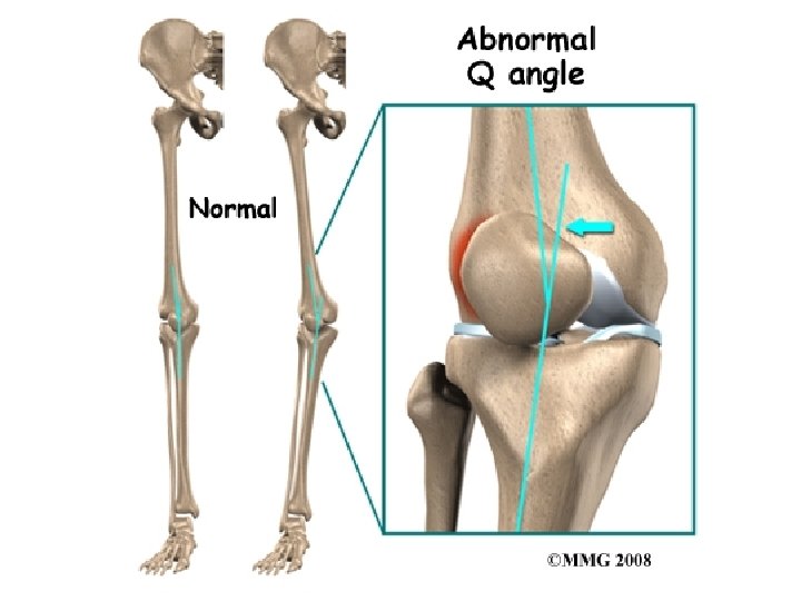
Quadriceps Angle “Q Angle” n n Adults typically 15 degrees Increases or decreases in the q angles are associated in cadaver models with increased peak patellofemoral contact pressures (Huberti & Hayes, 1984).
Increased Q Angle n n Femoral anteversion External tibial torsion Laterally displaced tibial tubercle Genu valgus
Quadriceps Angle “Q Angle” n Insall, Falvo, & Wise (1976) implicated increased q angle, along with patella alta, in a prospective study of patellofemoral pain.
Patellofemoral Dysfunction Patella Grinding Test n n n Chrondomalacia patellae Patellofemoral arthralgia Chondral fracture
Patellofemoral Dysfunction Clarke’s Patellar Scrape Test n Pain and crepitation may indicate patellofemoral arthralgia or chondromalcia patellae
M-Brace® Jumper’s Knee Brace For apophysitis, patellar chondromalacia, patellofemoral arthralgia, or jumper’s knee. • Alleviates pain by diminishing tension on muscles and ligaments. •
Management 1. 2. 3. 4. 5. 6. 7. PRICEMM NSAIDS HVG with ice PRE: Vastus medialis last 20 30 degrees of extension Spinal manipulation Myofascial trigger point pressure release Orthotics
Did I mention… n Diagnosis is the key to successful treatment!
Persistent Anterior Knee Pain n A 20 year old military recruit presented for follow up of anterior knee pain. Previously, he presented with three weeks of right knee pain without trauma, swelling, locking, or instability. At that visit, he was referred for physical therapy, but the knee did not improve during treatment. On recent examination, the patient had a positive “theater sign” (i. e. , knee pain upon arising after prolonged sitting), positive patellar compression and inhibition tests, stable ligaments, pain with Mc. Murray's test, and tenderness of the medial tibial plateau.
Question n n n Based on the patient's history, physical examination, and radiography results, which one of the following is the most likely diagnosis? A. Medial plica syndrome. B. Patellar tendinopathy. C. Patellofemoral pain syndrome. D. Pes anserine bursitis. E. Proximal tibial stress fracture.
Atraumatic, Persistent, Anterior Knee Pain
Discussion n The answer is E: proximal tibial stress fracture. Most tibial stress fractures occur in the tibial diaphysis. However, proximal tibial stress fractures, typically involving the medial condyle, can occur in athletes. Stress fractures may result from weaker bone that fails under normal loading (i. e. , an insufficiency fracture) or normal bone that fails under new or increased repetitive loading (i. e. , a fatigue fracture). Athletes are more likely to experience a fatigue fracture; military recruits are also at risk. Drabicki RR, Greer WJ, De. Meo PJ. Stress fractures around the knee. Clin Sports Med. 2006; 25: 105– 15. Raasch WG, Hergan DJ. Treatment of stress fractures: the fundamentals. Clin Sports Med. 2006; 25: 29– 36.
Discussion n Medical history that suggests a stress fracture will include persistent pain following an acute increase in physical activity (i. e. , time, distance, or pace). Physical examination may reveal tenderness at or near the medial joint line, tenderness with a leverage motion, and tenderness with hopping on the affected leg. A joint effusion may also be present.
Discussion n Plain radiographs can appear normal for weeks or even months. Because the metaphyseal portions of long bones are mostly cancellous bone, the typical periosteal/endosteal reaction will not be visible. In this type of bone, the features of a stress fracture manifest as a band-like area of sclerosis. If a stress fracture is suspected but radiographic results are normal, then magnetic resonance imaging (MRI) or a bone scan is the next diagnostic study. Of the two, MRI is more sensitive for diagnosing stress fracture.
One Final Thought… n Diagnosis is the key to successful treatment!
American Association of Family Practice Journal n n http: //www. aafp. org/afp/2005/0315/p 1169. html http: //www. aafp. org/afp/20000415/2391. ht ml
http: //www. aafp. org/afp/20000 315/1795. html n Management of Osteoarthritis of the Knee Nonpharmacologic treatment (e. g. , patient education and support, exercise, weight loss, joint protection) plus Acetaminophen (Tylenol) in a dosage of up to 4 g per day to control pain and other symptoms, and before activity Add topical capsaicin cream (e. g. , Arthri. Care) applied four times daily, if needed. n If joint effusion is present, consider aspiration and intra articular injection of a corticosteroid, such as 40 mg of triamcinolone (Aristocort). n If more pain or symptom control is needed, add an NSAID in a low dosage, such as 400 mg of ibuprofen (e. g. , Advil) taken four times daily, or a nonacetylated salicylate such as choline magnesium trisalicylate (Trilisate) or salsalate (Disalcid). n If more pain or symptom control is needed, use the full dosage of an NSAID, plus misoprostol (Cytotec) or a proton pump inhibitor if the patient is at risk for upper gastrointestinal tract bleeding or ulcer disease, or substitute a cyclo oxygenase 2 inhibitor for the NSAID; some patients may benefit from intra articular injections of a hyaluronic acid like product. n If the response is inadequate, consider referring the patient for joint lavage, arthroscopic debridement, osteotomy or joint replacement.
MUA tkr n http: //xnet. kp. org/permanentejournal/NIR/ Publications/Articles/Early_Late_Manipulat ions_TKA. pdf
Evaluation and Management of PFA by Hammer n n http: //www. dynamicchiropractic. com/mpacms/dc/article. p hp? id=44165 http: //www. dynamicchiropractic. com/mpacms/dc/article. p hp? id=44278 http: //www. dynamicchiropractic. com/mpacms/dc/article. p hp? id=44232 http: //www. dynamicchiropractic. com/mpacms/dc/article. p hp? id=17905