Brain Vascularization Arterial blood supply of the brain

Brain Vascularization

Arterial blood supply of the brain • The cerebral blood supply is derived from : Ø The internal carotid artery (Anterior circulation): • delivers blood to the to anterior 2/3 of the cerebral hemispheres • major branches : the middle and anterior cerebral arteries and the anterior choroidal artery. Ø The vertebral arteries (posterior circulation) • unite in the midline at the caudal border of the pons to form the basilar artery. • delivers blood to the brainstem and cerebellum, part of the cerebral hemispheres. • Branches : the posterior cerebral arteries. • The anterior and posterior circulations communicate with each other through the arterial circle of Willis. • Arteries of the brain lie in the subarachnoid space
 Anterior circulation (carotid)")
Posterior circulation (vertebrobasiler) Anterior circulation (carotid)
 Posterior circulation (vertebrobasiler)")
Anterior circulation (carotid) Posterior circulation (vertebrobasiler)

Carotid-vertebrobasilar anastomosis
")
Carotid-vertebrobasilar anastomosis (circle of willis)

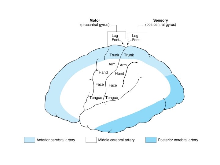
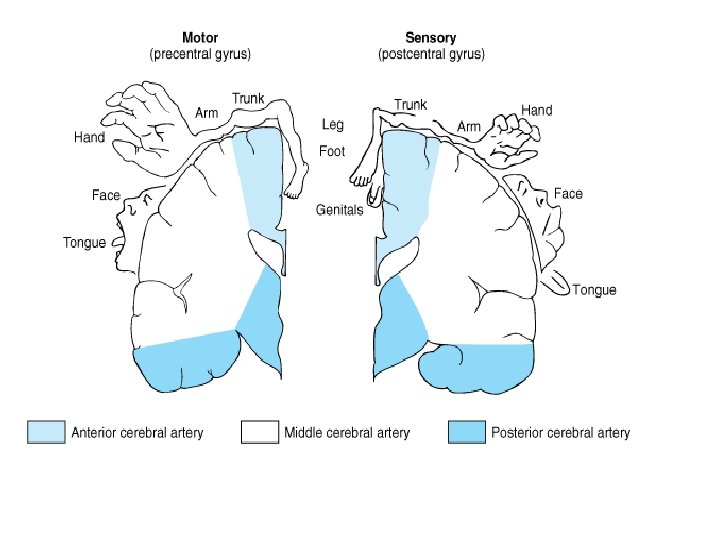
Anterior Cerebral Artery Areas supplied by anterior cerebral artery: • Septal area • Primary motor cortex for legs, foots, urinary bladder • Additional motor planning areas in the medial frontal lobe anterior to precentral gyrus • Most of the corpus callosum except its posterior part Branches : Ø Proximal branches : • small perforating branches that supply the paraseptal region, rostral portion of the basal ganglia and diencephalon, and the anterior limb of the internal capsule. • The recurrent artery of Heubner supplies the basal ganglia. Ø Cortical branches

Major cortical branches of ACA No Branch TERITORY I Orbitofrontal Orbital surface of frontal lobe II frontopolar Frontal pole III Frontal pericallosal Corpus callosum No Branch TERITORY IV Callosomarginal Cingulate and superior frontal gyri, paracentral lobule V Internal parietal

Middle Cerebral Artery Central branches: Lenticulostriate artery To: lentiform nucleus, caudate nucleus and internal capsule

Middle Cerebral Artery Cortical branches: Superior division to: • Primary motor cortex for face and arm • Broca’s area • Frontal eye fields (for “looking at” eye movements to the opposite) • Primary somatosensory cortex for face & arm • Parts of lateral frontal & parietal lobe for 3 -D visual perceptions and for ability to interpret & express emotion Inferior division to • Wernick’s area • Parts of posterior parietal lobe for 3 -D visual perceptions and for ability to interpret & express emotion • Optic radiation particularly fibers that represent information from the contralateral superior quadrants of the visual field
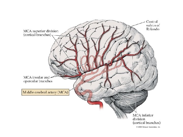

Major cortical branches of MCA No CABANG TERITORIAL I II IV Orbito frontal Prerolandic Rolandic Anterior parietal Posterior arietal Prefrontal cortex Premotor areas Pre-and postcentral gyri Post central and anterior parietal cortex Posterior parietal cortex VI Temporal and occipital cortex Angular and neighboring gyri Temporal cortex anterior V Temporoocci pital VII Posterior temporal VIII Anterior temporal

CEREBRI MEDIA ARTERIES

Branches of Vertebral & Basilar arteries Vertebral: - posterior & anterior spinal - posterior inferior cerebellar Basilar: - pontine - labyrinthine - anterior inferior cerebellar - superior cerebellar - posterior cerebral - posterior communicating
 The PICA supplies : • The basal portion of")
Posterior inferior cerebellar artery (PICA) The PICA supplies : • The basal portion of the cerebellar hemispheres, • The lower portion of the vermis, • Part of the cerebellar nuclei, and • The choroid plexus of the fourth ventricle • Dorsolateral portion of the medulla.

Basilar artery • Arises from the union of the right and left vertebral arteries in front of the brainstem at a lower pontine level. • Its major branches are : Ø Anterior inferior cerebellar artery (AICA). Ø Superior cerebellar artery (SCA). Ø Posterior cerebral arteries (PCA).

Anterior inferior cerebellar artery • Supplies the flocculus and the anterior portion of the cerebellar hemisphere. • The AICA also gives off the labyrinthine artery to the inner ear. Superior cerebellar artery • supplies the rostral portion of the cerebellar hemisphere and the upper portion of the vermis. • As it curves around the midbrain, it gives off branches to the tegmentum.
 • - PCA supplies : Diencephalon Midbrain Optic radiation &")
Posterior Cerebral Artery (PCA) • - PCA supplies : Diencephalon Midbrain Optic radiation & striate cortex (primary visual cortex) Splenium of the corpus callosum Hippocampal formation & the posterior of fornix. Branches : Ø a. thalamoperforating Ø a. Thalamogeniculatum Ø medial and lateral posterior choroidal arteries Ø Cortical branches

Thalamoperforating arteries and thalamogeniculate artery • The anterior thalamoperforating artery mainly supplies the rostral portion of the thalamus. • The posterior thalamoperforating artery supplies the basal and medial portions of the thalamus, as well as the pulvinar. • Thalamogeniculate artery supplies the lateral portion of the thalamus.

Medial and lateral posterior choroidal artery • They supply the geniculate bodies, medial and posteromedial thalamic nuclei, and pulvinar. • The medial posterior choroidal artery gives off branches to the midbrain and supplies the choroid plexus of the third ventricle. • The lateral posterior choroidal artery supplies the choroid plexus of the lateral ventricle.

Cortical branches of PCA No Arteri Teritorial I` Anterior temporal cortex Posterior temporal and occipital cortex II` III` Anterior temporal Posterior occipital No Arteri Teritorial IV` V` Calcarina cortex Cuneus and precuneus Splenium of corpus callosum Calcarina Parietooccipit al Callosal

Arterial supply of brain : summary

Arterial supply of brain : summary

• Superficial veins • Deep veins

Superficial veins of the brain Venous blood from the brain parenchyma crosses the subarachnoid and subdural spaces in short cortical veins which include : • Superior anastomotic vein (of Trolard) • Dorsal superior cerebral vein, • Superficial middle cerebral vein, • Inferior anastomotic vein (of Labbé)

Deep veins of the brain Venous blood from deep regions of the brain, drains into : Internal cerebral veins Basal veins of Rosenthal Great vein of Galen Confluens sinuum.

Dural venous sinuses • Blood-filled spaces situated between layers of dura mater: – Superior & inferior, straight, transverse, sigmoid, and occipital sinuses - Confluens of sinuses - Cavernous sinuses - Superior and inferior petrosal sinuses

Dural venous sinuses

Thank You

Frontal Lobe • Blood supply - ACA and MCA • Major functions: – personality, behaviour – motor function – judgement/problem solving – micturation – expressive speech - Broca’s word formation, articulation and speech production – concentration, reasoning 30 Neuroanatomy and Cerebral Circulation Review, West GTA Stroke Network, 2003

Parietal Lobe • Blood supply – ACA, MCA and PCA • Major functions: – sensory function – body part awareness – visual spatial information 31 Neuroanatomy and Cerebral Circulation Review, West GTA Stroke Network, 2003

Temporal Lobe • Blood supply - MCA and PCA • Major Functions: – understanding speech -Wernickes – visual, olfactory and auditory perception – learning, memory, emotional affect 32 Neuroanatomy and Cerebral Circulation Review, West GTA Stroke Network, 2003

Occipital Lobe • Blood supply - MCA, PCA • Major Functions: – primary visual area – some visual reflexes – involuntary smooth eye movements – recognition & identification of objects 33 Neuroanatomy and Cerebral Circulation Review, West GTA Stroke Network, 2003
- Slides: 36