ANTERIOR TRIANGLE 2 Boundaries Front of sternocleidomastoid Anterior

ANTERIOR TRIANGLE -2

Boundaries Front of sternocleidomastoid. Anterior. Posterior. Base. Apex. Roof.

Submental. Digastric. Muscular. Carotid.

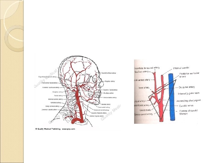
CAROTID TRIANGLE Anteriosuperiorly- post belly of digastric. Anterioinferiorly sup belly of omohyoid. Posteriorly ant border of sternocleidomastoid.

Roof Skin. Superficial fascia Investing layer of deep cervical fascia Floor Thyrohyoid Hyoglossus Middle and inferior constrictors

Contents • Arteries o o • o o Carotid sheath with its contents Carotid arterial system Veins Internal jugular vein Nerves Glossopharyngeal nerve Vagus nerve Accessory nerve Hypoglossal nerve Ansa cervicalis Sympathetic chain Other contents Apex of parotid gland Deep cervical lymph nodes

ARTERIES: 1. 2. 3. 4. • • • Termination of common carotid artery. Proximal part of external carotid artery. Proximal part of internal carotid artery. Five branches of external carotid artery: Ascending pharyngeal, Superior thyroid, Lingual, Facial & Occipital.

VEINS: o o o o Internal jugular veins. Five tributaries to internal jugular veins pharyngeal veins common facial, lingual, superior thyroid, middle thyroid,
 Internal")
CAROTID SHEATH: A condensation of deep fascia. Contents; Common& Internal carotid artery. (med) Internal jugular vein. (lat) Vagus nerve: between & little posterior to arteries & vein. Extent; Base of the skull. Arch of the aorta. Relations; Ant –ansa cervicalis. Post – sympathetic chain.

FIVE NERVES: TWO NERVES RELATED TO CAROTID SHEATH: 1. Ansa cervicalis : embedded in the anterior wall of sheath Sympathetic trunk: embedded in the posterior wall of sheath 2.

THREE NERVES BETWEEN INTERNAL JUGULAR VEIN & INTERNAL CAROTID ARTERY: 3. Vagus nerve: descends between internal jugular vein & common carotid artery 4. Spinal part of accessory nerve: passes deep to sternomastoid to reach posterior triangle 5. Hypoglossal nerve: passes deep to posterior belly of digastric to reach digastric triangle

DEEP CERVICAL LYMPH NODES: Along the internal jugular vein. • Jugulodigastric • Juguloomohyoid •

Ansa cervicalis It is nerve loop plastered on the anterior wall of carotid sheath Formation- superior and inferior root. Sup rootdescendens hypoglossi. C 1 through hypoglosal nerve.

Distribution - Infrahyoid muscles Sup root sup belly of omohyoid Loop/Ansa sternohyoid Sternothyroid Inf belly of omohyoid

External carotid artery Common carotid artery Left-arch of aorta. Rightbrachiocephalic artery.

CCA-runs upwards in the neck up to the upper border of thyroid cartilage at the level of intervertebral disc of C 3 -C 4. Divides into terminal branches Internal & External carotid artery. ICA-supplies- brain. no branches in the neck.

External carotid artery Terminal branch of the common carotid artery. Origin - Level of the upper border of the thyroid cartilage. Terminates within the parotid gland at the level of the neck of the mandible.

course taking a slightly curved course, passes upward and forward, and behind the neck of the mandible, where it divides into Superficial temporal artery Maxillary artery

Branches-8 Anterior Superior thyroid Lingual Facial Posterior Occipital Post auricular Medial Ascending pharyngeal Terminal Superficial temporal Maxillary.

1. Superior thyroid artery ◦ Origin- below the level of hyoid bone. ◦ Passes along with the external laryngeal nerve. ◦ Supplies thyroid gland. ◦ Gives imp branch superior laryngeal artery.

2. Lingual artery: ◦ Origin –level of hyoid bone ◦ Passes deep to the hyoglossus muscle. ◦ Supplies the tongue through its undersurface.

3. Facial artery Origin -above the level of hyoid bone. Course in the neck Ascends deep to the posterior belly of the digastric muscle. Winds around the post part of the submandibular salivary gland. Reaches –base of mandible

Course in the face Enters the face through the anteroinferior part of the masseter muscle. Reaches 1. 25 cms lateral to the angle of the mouth Ascends along the lateral side of the nose. Terminates - medial angle of the eye anatomizing with the Dorsal nasal branch of ophthalmic artery.

Branches in the neck. 1. Ascending palatine artery. 2. Tonsillar artery 3. Submental artery 4. Glandular branches to submandibular gland. Branches in the face. 1. Inferior labial artery. 2. Superior labial artery. 3. Lateral nasal artery.

4. Occipital artery: ◦ Origin – opp to facial artery. ◦ Passes posteriorly along the lower border of the posterior belly of the digastric muscle. ◦ Supplies posterior quadrant of the scalp.

5. Posterior auricular artery: ◦ Passes posteriorly along the upper border of the posterior belly of the digastric muscle. ◦ Supplies the external ear and posterior quadrant of the scalp.

6. Ascending pharyngeal artery: ◦ Origin – medially Ascends the wall of the pharynx. ◦ Supplies pharynx and palatine tonsil.

7. Superficial temporal artery: ◦ Terminal branch ◦ Origin –behind the neck of mandible ◦ Gives the transverse facial artery which appears on the face. ◦ Supplies the temporal region and anterior quadrant of the scalp.

8. Maxillary Artery: ◦ Origin - behind the neck of mandible ◦ Larger Terminal branch. ◦ Passes anteriorly in the infratemporal fossa. ◦ Devided into 3 parts but the lateral pterygoid muscle.

Applied anatomy Carotid sinus Dilatation –termination of CCA/beginning of ICA. Glossopharyngeal & symp nerves. Barareceptor/pressure receptcr. Carotid sinus syndrome: syncope and slowing of heart rate.

Carotid body Oval, reddish massbifurcation of CCA. Supplied by glossopharyngeal, vagus & symp nerves. Chemoreceptor, responds to changes O 2 &CO 2 changes in blood. Potato tumor: enlargement of the carotid body. Pressure on carotid sinus.

THANK YOU
- Slides: 33