Anterior Compartment of Leg Anterior Compartment of Leg

 compartment, is located anterior")


- Slides: 29
Anterior Compartment of Leg
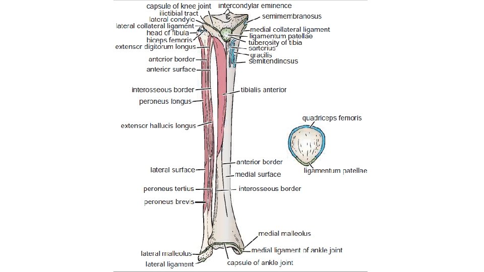
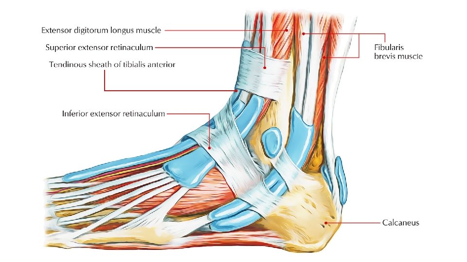
Anterior Compartment of Leg The anterior compartment, or dorsiflexor (extensor) compartment, is located anterior to the interosseous membrane, between the lateral surface of the tibial shaft and the medial surface of the fibular shaft. Inferiorly, two bandlike thickenings of the deep fascia form retinacula that bind the tendons of the anterior compartment muscles, preventing them from bow-stringing anteriorly during dorsiflexion of the ankle joint. The superior extensor retinaculum is a strong, broad band of deep fascia passing from the fibula to the tibia, proximal to the malleoli. The inferior extensor retinaculum, a Y-shaped band of deep fascia, attaches laterally to the anterosuperior surface of the calcaneus and medially to the medial malleolus and medial cuneiform.
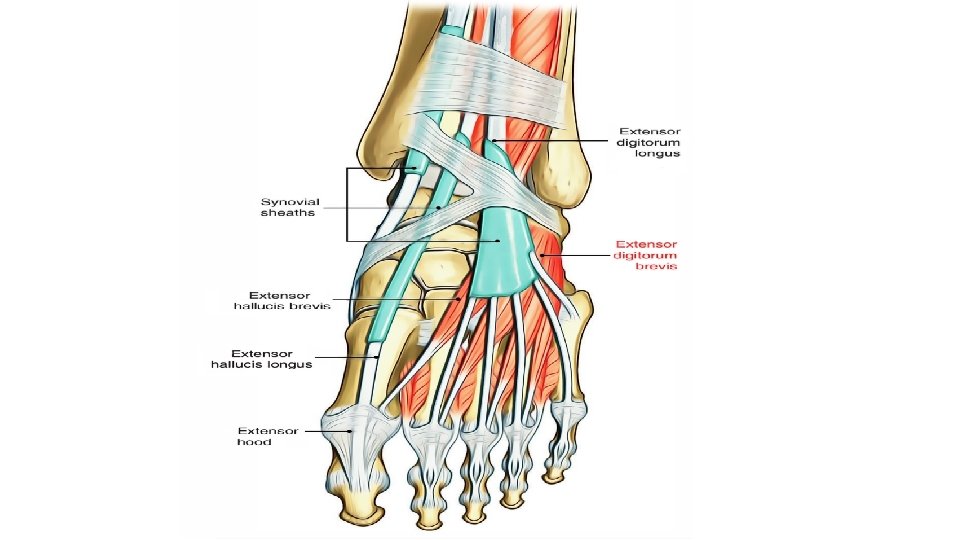
The four muscles in the anterior compartment are: • Tibialis anterior • Extensor digitorum longus • Extensor hallucis longus • Fibularis tertius These muscles are mainly dorsiflexors of the ankle joint and extensors of the toes
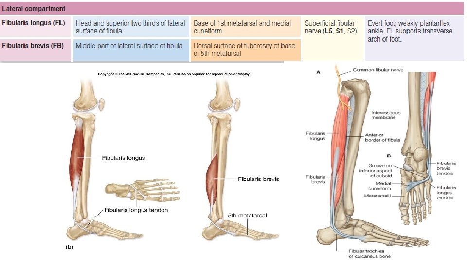
Lateral Compartment of Leg The lateral compartment, or evertor compartment, is bounded by the lateral surface of the fibula, the anterior and posterior intermuscular septa, and the deep fascia of the leg. The lateral compartment contains two muscles—the fibularis longus and brevis—that pass posterior to the lateral malleolus
Posterior Compartment of Leg
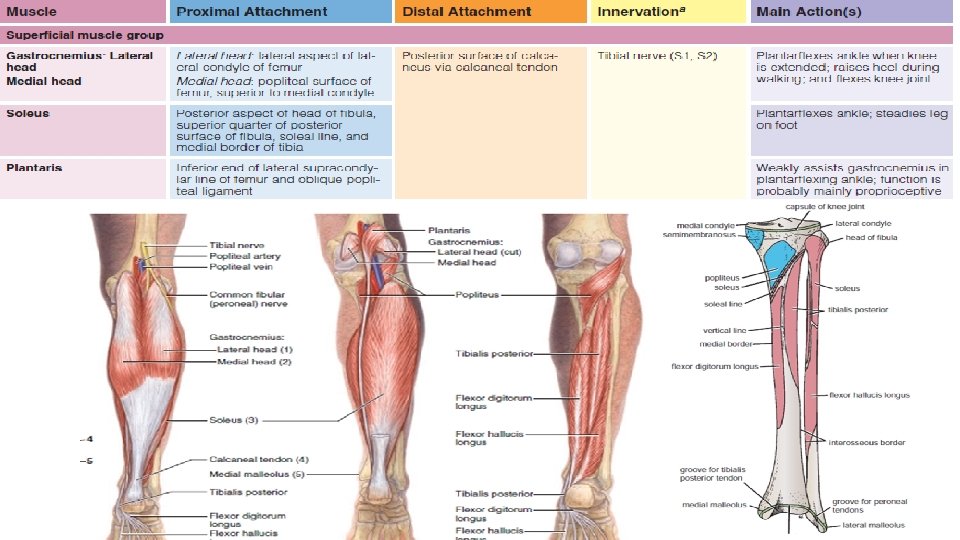
Posterior Compartment of Leg Or plantarflexor compartment, is the largest of the three leg compartments. The posterior compartment and the calf muscles within it are divided into superficial and deep muscle groups by the transverse intermuscular septum. The tibial nerve and posterior tibial and fibular vessels supply both parts of the posterior compartment but run in the deep part, just deep (anterior) to the transverse intermuscular septum. SUPERFICIAL MUSCLE GROUP The superficial group of plantarflexors, including the gastrocnemius, soleus, and plantaris, forms a powerful muscular mass in the calf. The two-headed gastrocnemius and the soleus share a common tendon, the calcaneal tendon ( tendo calcaneus ), which attaches to the calcaneus. Collectively, these two muscles form the three-headed triceps surae ( calf). The triceps surae elevates the heel and thus depresses the forefoot, generating as much as 93% of the plantar flexion force
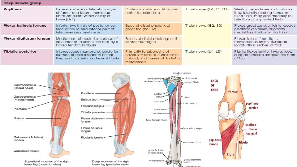
DEEP MUSCLE GROUP Four muscles make up the deep group in the posterior compartment: • Popliteus • Flexor digitorum longus • Flexor hallucis longus • Tibialis posterior The popliteus is a thin, triangular muscle in the floor of the popliteal fossa. The popliteus acts to unlock the fully extended knee joint, whereas the other muscles act on the ankle and foot joints. The flexor hallucis longus is the powerful flexor of all the joints of the great toe.
Proximal Tibiofibular Joint Ø Articulation Between the lateral condyle of the tibia and the head of the fibula. The articular surfaces are flattened and covered by hyaline cartilage. Ø Type This is a synovial, plane, gliding joint. Ø Capsule The capsule surrounds the joint and is attached to the margins of the articular surfaces. Ø Ligaments Anterior and posterior ligaments strengthen the capsule. The interosseous membrane, also greatly strengthens the joint.
Synovial Membrane The synovial membrane lines the capsule and is attached to the margins of the articular surfaces. Nerve Supply The common peroneal nerve supplies the joint. Movements A small amount of gliding movement takes place during movements at the ankle joint.
Distal Tibiofibular Joint Articulation is between the fibular notch at the lower end of the tibia and the lower end of the fibula. The opposed bony surfaces are roughened. Type The distal tibiofibular joint is a fibrous joint. Capsule There is no capsule.
Ø Ligaments The interosseous ligament is a strong, thick band of fibrous tissue that binds the two bones together. The interosseous membrane, which connects the shafts of the tibia and fibula together, also greatly strengthens the joint. The anterior and posterior ligaments are flat bands of fibrous tissue connecting the two bones together in front and behind the interosseous ligament. The inferior transverse ligament runs from the medial surface of the upper part of the lateral malleolus to the posterior border of the lower end of the tibia. Ø Nerve Supply Deep peroneal and tibial nerves supply the joint. Ø Movements A small amount of movement takes place during movements at the ankle joint.
Bones of the Foot The bones of the foot are the tarsal bones, the metatarsals, and the phalanges. Tarsal Bones The tarsal bones are the calcaneum, the talus, the navicular, the cuboid, and the three cuneiform bones. Only the talus articulates with the tibia and the fibula at the ankle joint.
Calcaneum The calcaneum is the largest bone of the foot and forms the prominence of the heel. It articulates above with the talus and in front with the cuboid. It has six surfaces: ■■ The anterior surface is small and forms the articular facet that articulates with the cuboid bone. ■■ The posterior surface forms the prominence of the heel and gives attachment to the tendo calcaneus.
■■ The superior surface is dominated by two articular facets for the talus, separated by a roughened groove, the sulcus calcanei. ■■ The inferior surface has an anterior tubercle in the midline and a large medial and a smaller lateral tubercle at the junction of the inferior and posterior surfaces. Inferior view
■■ The lateral surface is almost flat. On its anterior part is a small elevation called the peroneal tubercle, which separates the tendons of the peroneus longus and brevis muscles. ■■ The medial surface possesses a large, shelflike process, termed the sustentaculum tali, which assists in the support of the talus and allow passage of flexor hallucis longus
Talus The talus articulates above at the ankle joint with the tibia and fibula, below with the calcaneum, and in front with the navicular bone. It possesses a head, a neck, and a body. Navicular Bone The tuberosity of the navicular bone can be seen and felt on the medial border of the foot 1 in. (2. 5 cm) in front of and below the medial malleolus; it gives attachment to the main part of the tibialis posterior tendon
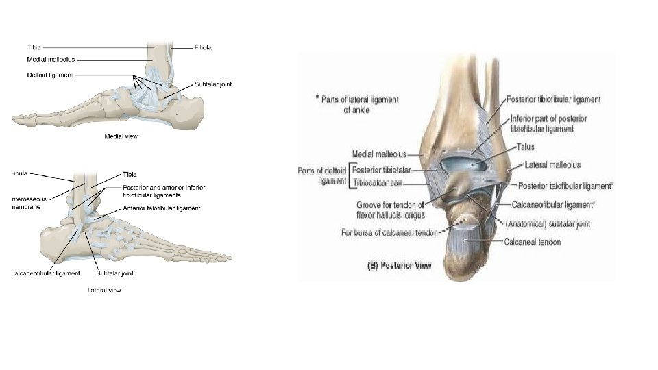
Ankle Joint The ankle joint consists of a deep socket formed by the lower ends of the tibia and fibula, into which is fitted the upper part of the body of the talus. The talus is able to move on a transverse axis in a hingelike manner. The shape of the bones and the strength of the ligaments and the surrounding tendons make this joint strong and stable. Articulation is between the lower end of the tibia, the two malleoli, and the body of the talus. The inferior transverse tibiofibular ligament, which runs between the lateral malleolus and the posterior border of the lower end of the tibia, deepens the socket into which the body of the talus fits snugly. The articular surfaces are covered with hyaline cartilage. Type The ankle is a synovial hinge joint.
Capsule The capsule encloses the joint and is attached to the bones near their articular margins. Ligaments The medial, or deltoid, ligament is strong and is attached by its apex to the tip of the medial malleolus. Below, the deep fibers are attached to the nonarticular area on the medial surface of the body of the talus; the superficial fibers are attached to the medial side of the talus, the sustentaculum tali, the plantar calcaneo-navicular ligament, and the tuberosity of the navicular bone. The lateral ligament is weaker than the medial ligament and consists of three bands. 1. The anterior talofibular ligament runs from the lateral malleolus to the lateral surface of the talus. 2. The calcaneofibular ligament runs from the tip of the lateral malleolus downward and backward to the lateral surface of the calcaneum. 3. The posterior talofibular ligament runs from the lateral malleolus to the posterior tubercle of the talus.
v Synovial Membrane : The synovial membrane lines the capsule. v Nerve Supply : Deep peroneal and tibial nerves supply the ankle joint. v Movements : Dorsiflexion (toes pointing upward) and plantar flexion (toes pointing downward) are possible. The movements of inversion and eversion take place at the tarsal joints and not at the ankle joint. Ø Dorsiflexion is performed by the tibialis anterior, extensor hallucis longus, extensor digitorum longus, and Fibularis (peroneus) tertius. It is limited by the tension of the tendo calcaneus, the posterior fibers of the medial ligament, and the calcaneofibular ligament. Ø Plantar flexion is performed by the gastrocnemius, soleus, plantaris, peroneus longus, peroneus brevis, tibialis posterior, flexor digitorum longus, and flexor hallucis longus. It is limited by the tension of the opposing muscles, the anterior fibers of the medial ligament, and the anterior talofibular ligament
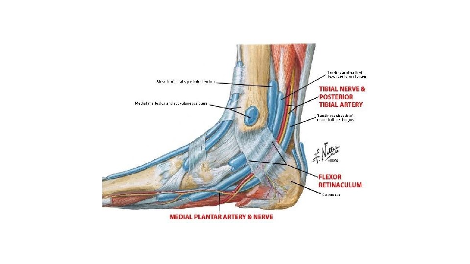
Important Relations ■■ Anteriorly: The tibialis anterior, the extensor hallucis longus, the anterior tibial vessels, the deep peroneal nerve, the extensor digitorum longus, and the peroneus tertius ■■ Posteriorly: The tendo calcaneus and plantaris ■■ Posterolaterally (behind the lateral malleolus): The peroneus longus and brevis. ■■ Posteromedially (behind the medial malleolus): The tibialis posterior, the flexor digitorum longus, the posterior tibial vessels, the tibial nerve, and the flexor halluces Longus.
Thank You