Anterior and posterior compartment of arm Dr Qudusia


- Slides: 36
Anterior and posterior compartment of arm Dr. Qudusia Sultana
Objectives Anterior compartment muscles Posterior Compartment: Muscles Brachial artery Profunda Brachii artery
THE ARM/BRACHIUM
Arm /Brachium Deep fascia of arm extends to the humerus as medial and lateral intermuscular septa and divide arm into Anterior compartment Posterior compartment
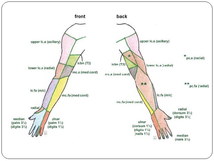
Cutaneous Innervation of Arm • The upper medial surface of the arm -the lateral branch of the second intercostal nerve (the intercostobrachial nerve). • The lower medial surface of the arm-the medial cutaneous nerve of the arm. • The lateral aspect of the arm – §upper lateral cutaneous nerve (a branch of the axillary nerve) § lower lateral cutaneous nerve (a branch of the radial nerve). • The posterior aspect is supplied by the posterior cutaneous nerve of the arm, a branch of the radial nerve
ANTERIOR COMPARTMENT
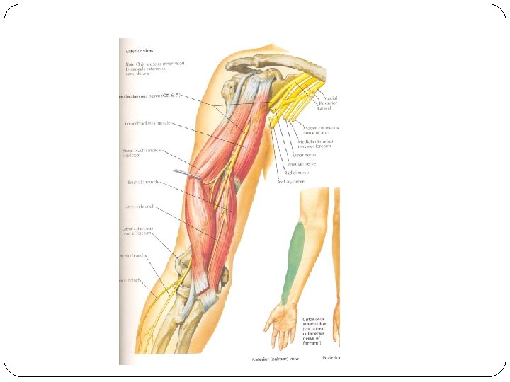
Anterior compartment of arm Muscles: Coracobrachialis Biceps brachii Brachialis Nerves: Musculocutaneous Nerve Artery: Brachial Artery. Structures passing through arm Median, Radial, Ulnar, Medial cutaneous nerve of arm and forearm and basilic vein
Posterior compartment of arm Muscles: Triceps Brachii Nerve: Radial Nerve. Artery: Profunda brachii Artery.
Coracobrachialis Origin: Tip of the coracoid process of scapula. In common with short head of biceps Insertion: Middle 5 cm of the medial border of the shaft of humerus. Nerve Supply: Musculocutaneous Nerve. Action: Flexion of arm. Weak adduction of shoulder
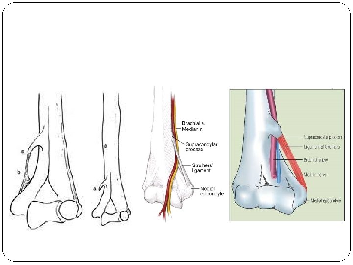
Ligament of Struthers? ? ?
Biceps brachii Origin: Short Head: Tip of the coracoid process of Scapula. Long Head: Supraglenoid tubercle of the scapula and labrum
Biceps brachii Insertion: Posterior rough part of the radial tuberosity. Bicipital Aponeurosis inserted into the subcutaneous posterior border of Ulna through deep fascia from the medial side of the forearm.
Nerve Supply: Musculocutaneous nerve. Actions: Strong supinator when forearm is flexed. Screwing movement. Short head is a flexor of the arm. Flexor of the elbow. Long head prevents upwards displacement of the head of the humerus.
Brachialis Origin: Lower half of the front of the humerus, including anteriomedial and anteriolateral surfaces. Medial and lateral intermuscular septa. Insertion: Ulnar tuberosity. Rough anterior surface of the coronoid process of the ulna.
Nerve Supply: Musculocutaneou s Nerve, Radial Nerve is proprioceptive Actions: Flexes forearm at the elbow joint.
Triceps Origin – Long head –infra glenoid tubercle of the scapula. Lateral head –lateral lip of the radial groove Medial head – posterior surface of humerus below radial groove. Medial and lateral intermuscular septa.
Insertion – superior surface of the olecrenon process of ulna. Actions – Powerful extensor of the elbow. Long head supports the head of the humerus in the abducted position. long medial lateral
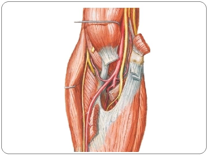
Brachial artery Continuation of the Axillary artery. Origin - at the lower border of Teres major. It runs downwards and laterally in front of arm and crosses the elbow joint Terminates – in the cubital fossa at the neck of the radius as radial and ulnar arteries.
Relations: Anteriorly: Superficial throughout its extent accompanied by venae commitants. coracobr achialis Middle Part: Crossed by median nerve from laterally to medially. Median n Triceps Elbow –bicipital aponeurosis Bicipital aponeurosis
Relations: Posteriorly: Insertion of the Coracobrachialis Triceps Brachialis The radial nerve Profunda Brachii artery. coracobr achialis Median n Triceps Bicipital aponeurosis
Medially Upper Part: Ulnar nerve, Basilic vein. Lower Part: Median Nerve. Laterally Upper Part: Coracobrachialis, biceps and median Nerve. Lower Part: Tendon of Biceps.
At Elbow –med to lat Median nerve Brachial artery Biceps brachii tendon. Radial nerve
Branches: Muscular Branches. Profunda brachii artery. Superior Ulnar Collateral branch Nutrient Artery. Inferior Ulnar Collateral. Terminal - Radial and Ulnar artery.
Applied anatomy Recording blood pressure. Brachial pulseinsertion of Coracobrachialis Volkmann’s Ischaemic contracturefractures-rupture of arteries-necrosis
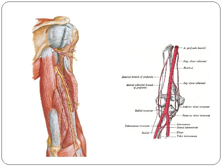
Profunda brachii artery Large branch of brachial artery. Accompanies the radial nerve in the radial groove. Terminates by dividing into radial collateral and middle collateral branches.
Bbranches Radial collateral/ Anterior descending artery. Continuation of profunda brachii artery. Ends by anastomosing with the radial recurrent artery in front of the lateral epicondyle of the humerus.
Middle collateral/ posterior descending artery. Largest terminal branch. Ends by anastomosing with the interosseous recurrent artery behind the lateral epicondyle of the humerus.
Deltoid/Ascending branch ascends up and anastomose with posterior circumflex humeral artery Nurtrient artery to the humerus.
Applied Anatomy Anastomosis around the elbow joint and thus helpful in collateral circulation