The peritoneum peritoneal cavity Dr Kumar Satish Ravi


 Lesser Sac q. It is a part of the peritonial")


")
 -lies lateral to the descending colon. It is")


- Slides: 56
The peritoneum & peritoneal cavity Dr. Kumar Satish Ravi M. B. B. S. , M. D. (JIPMER), MAMS
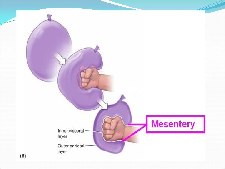
Peritoneum Tough layer of elastic areolar tissue Lined with simple squamous epithelium Largest of the serous sacs of the body Has 2 layers- the parietal and visceral Layers separated from each other by a thin film of fluid
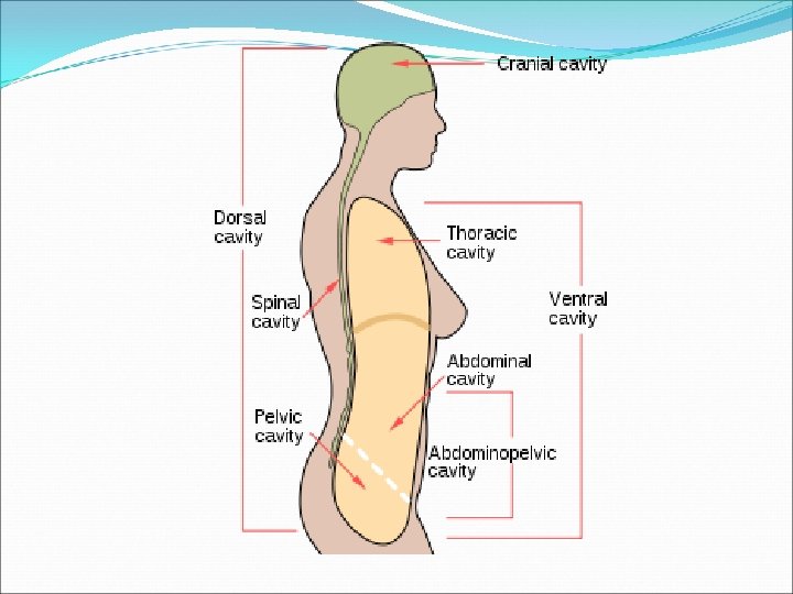
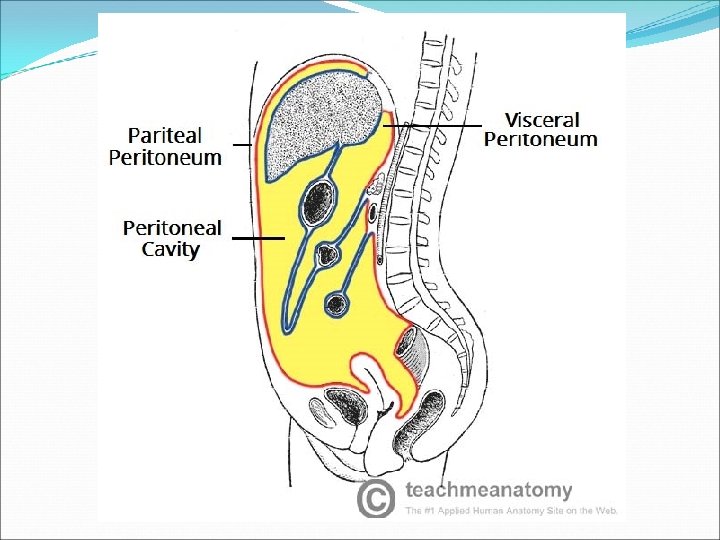
General features The peritoneum is a thin serous membrane that line the walls of the abdominal & pelvic cavities & cover the organs within these cavities Parietal peritoneum -lines the walls of the abdominal & pelvic cavities
General features Visceral peritoneum -covers the organs Peritoneal cavity -the potential space between the parietal and visceral layer of peritoneum, ♂, is a closed sac, but in ♀, there is a communication with the exterior through the uterine tubes, the uterus, and the vagina
Function Secretes a lubricating serous fluid that continuously moistens the associated organs Absorb Support viscera Hepatorenal Pouch & Rectouterine Pouch
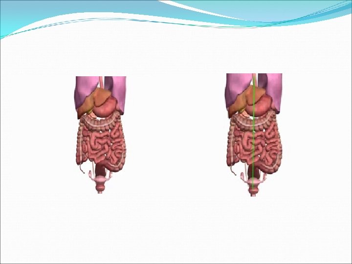
• • • Superficial view of the abdominal organs
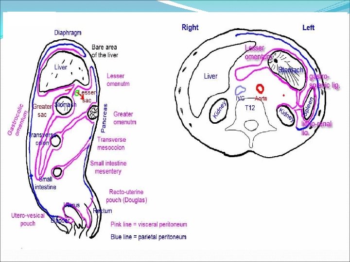
The peritoneum v. Is a thin serous membrane, §Lining the wall of the abdominal and pelvic cavities, (the parietal peritoneum). §Covering the existing organs, (the visceral peritoneum). §The potential space between the two layers is the peritoneal cavity. Parietal Visceral
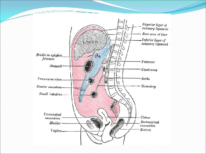
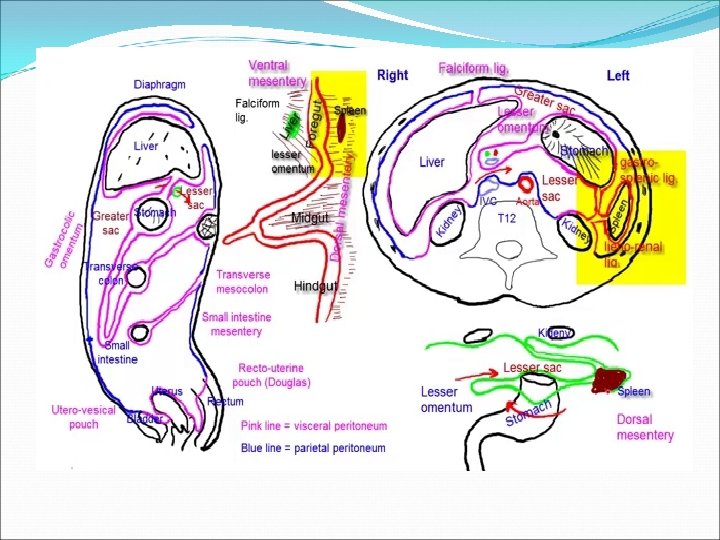
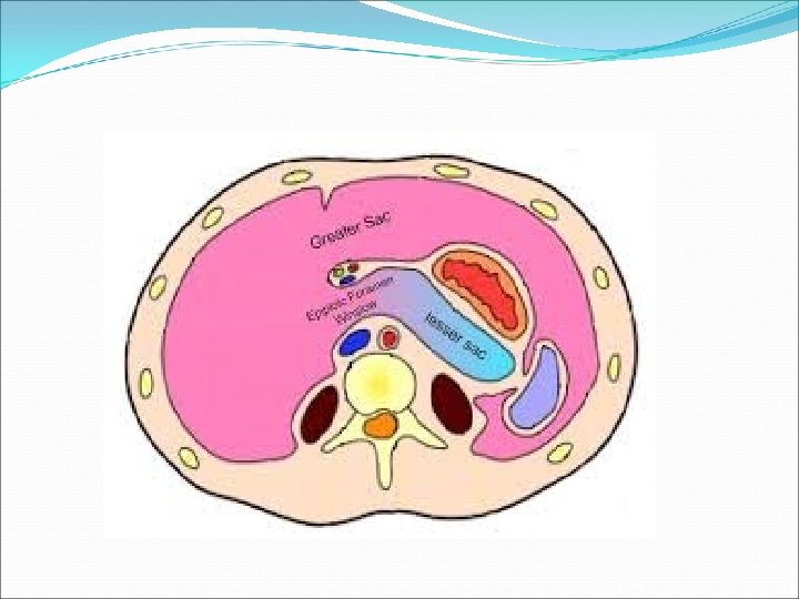
The peritoneum Lesser Sac Greater Sac v. The peritoneal cavity is the largest one in the body. v. Divisions of the peritoneal cavity : §Greater sac; extends from diaphragm down to the pelvis. §Lesser sac; lies behind the stomach. §Both cavities are interconnected through the epiploic foramen. §In male : the peritoneum is a closed sac. §In female : the sac is not completely closed because it communicates with the exterior through the uterine tubes, uterus and vagina.
The peritoneum q. Intraperitonial and retroperitonial; describe the relationship between various organs and their peritoneal covering; §Intraperitonial structure; which is nearly totally covered by visceral peritoneum. §Retroperitonial structure; lies behind the peritoneum, and partially covered by visceral peritoneum. Intraperitoneal viscera Retroperitoneal viscera
Intraperitoneal organ : Is surrounded by the peritoneum and has a supporting mesentery : stomach & 1 st part of duodenum, liver, gall bladder, spleen, jejunum, ileum, transverse colon, sigmoid colon, uterus, and ovaries. Extraperitoneal or retroperitoneal organ : Structure that lies behind the peritoneum or An organ, which is only partially covered by the peritoneum and has no supporting mesentery. Primarily retroperitoneal organs develop and remain outside the peritoneal cavity: kidneys, suprarenal glands, aorta, inferior vena cava, urinary bladder, prostate, vagina, and rectum. Secondarily retroperitoneal organs develop in mesenteries, but get pushed against the body wall (parietal peritoneum) during growth so that only half of their surface or less is covered by peritoneum : pancreas, duodenum, ascending and descending colon.
Folds of the peritoneum q. Types of peritoneal folds : Omenta. Mesenteries. Ligaments.
Omenta Lesser omentum v Two layered fold of peritoneum connecting the stomach to another viscus. § The lesser omentum attaches the lesser curvature of the stomach to the liver. § The greater omentum connects the greater curvature of the stomach to the transverse colon. Greater omentum
Lesser omentum q Extends between the liver and the lesser curvature of the stomach. It is continuous with the two layers of peritoneum which cover the anterior & posterior surfaces of stomach and 1 st part of the duodenum. Ascend as a double fold to the porta hepatis of liver, and fissure for ligamentum venosum. To the left of porta hepatis it is carried to the diaphragm. Its right border is a free margin; constitutes the anterior boundary of the epiploic foramen. q Contents between the two layers of the lesser omentum : Close to the right free margin, are the hepatic artery, the common bile duct, the portal vein, lymphatics, and the hepatic plexus of nerves. At the attachement to the stomach, run the right and left gastric vessels.
Greater omentum
Greater omentum The largest peritoneal fold, with cribriform appearance, contains some adipose tissue. It consists of a double sheet of peritoneum, folded on itself so that it is made up of four layers (anterior 2 layers + posterior 2 layers). The two layers which descend from the greater curve of the stomach and commencement of the duodenum, pass downward in front of the small intestines, then turn upon themselves, and ascend to the transverse colon, where they separate and enclose it. The left border of the greater omentum is continuous with the gastrosplenic ligament. Its right border extends as far as the commencement of the duodenum. Contents : the anastomosis between the right and left gastroepiploic vessels.
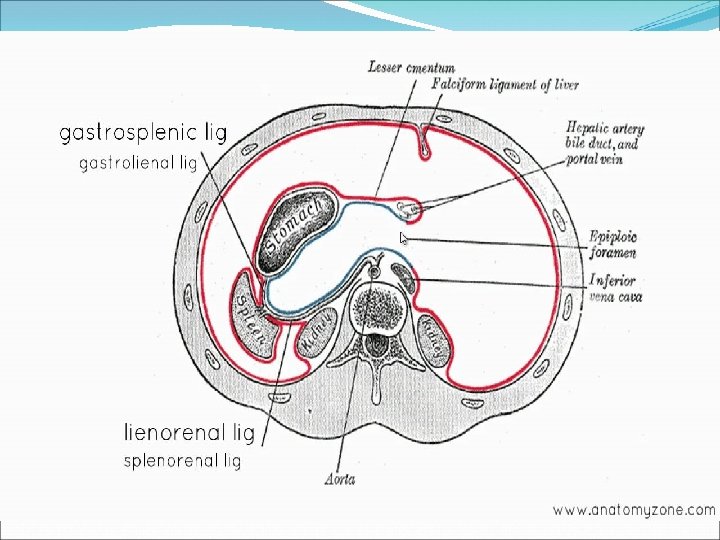
Omental bursa, (Lesser Sac) Lesser Sac q. It is a part of the peritonial cavity behind the stomach. q. Boundaries of the omental bursa ; §Anterior wall, from above downward, by the caudate lobe of the liver, the lesser omentum, back of the stomach, and the anterior two layers of the greater omentum. §Posterior wall, from below upward, by the posterior two layers of the greater omentum, the transverse colon, and the ascending layer of the transverse mesocolon, the upper surface of the pancreas, the left suprarenal gland, and the upper end of the left kidney.
Epiploic foramen It is the communication between the greater and lesser sacs. It is bounded by; In front by the free border of the lesser omentum, with its contents : hepatic artery, common bile duct, and portal vein between its two layers. Behind by the peritoneum covering the inferior vena cava. Above (roof) by the peritoneum on the caudate process of the liver. Below (floor) by the peritoneum covering the commencement of the duodenum and the hepatic artery, before ascending between the two layers of the lesser omentum.
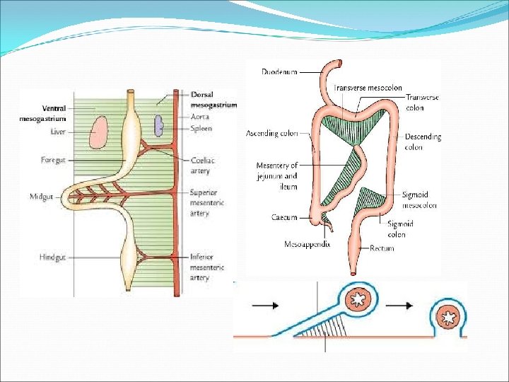
Mesenteries or mesocolons -two-layered fold of peritoneum that attach part of the intestines to the posterior abdominal wall
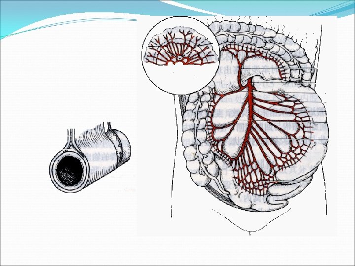
Mesentery -suspends the small intestine from the posterior abdominal wall Broad and a fan-shaped Consists of two peritoneal layers Intestinal border-folded, 7 m long Radix of mesentery 15 cm long Directed obliquely from left side of L 2 to in front of right sacroiliac joint
Structure crossed by the root of mesentry
Mesoappendix Triangular mesentery- extends from terminal part of ileum to appendix Appendicular artery runs in free margin of the mesoappendix
Transverse mesocolon -a double fold of peritoneum which connects the transverse colon to the posterior abdominal wall
Sigmoid mesocolon - a trianguar fold of peritoneum. inverted V-shaped, with apex located in front of left ureter and division of common iliac artery
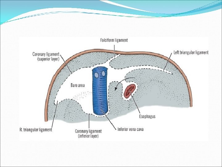
Ligaments -two-layered folds of peritoneum that attached the lesser mobile solid visera to the abdominal wall Ligaments of liver Falciform ligament of liver Consists of double peritoneal layer Extends from anterior abdominal wall (umbilicus) to liver Free border of ligament site of ligamentum teres
Coronary ligament -the area between upper & lower parts of the coronary ligament is the bare area of liver, this area is devoid of peritoneum and lies in contact with the diaphragm Left and right triangular ligament -formed by right extremity of coronary ligament and left leaf of falciform ligament, respectively
Hepatogastric ligament Hepatoduodenal ligament Ligamentum teres hepatis
Ligaments of spleen Gastrosplenic ligament -a double layer of peritoneum that connects the fundus of stomach to hilum of spleen. In this double layer of peritoneum are the short gastric and left gastroepiploic vessels Splenorenal ligament -extends between the hilum of spleen and anterior aspect of left kidney. The splenic vessels lies within this ligament, as well as the tail of pancreas Phrenicosplenic ligament Splenocolic ligament
Ligaments of stomach Hepatogastric ligament Gastrosplenic ligament Gastrophrenic ligament Gastrocolic ligament
Folds and recesses of posterior abdominal wall Superior duodenal fold and recess Inferior duodenal fold and recess Intersigmoid recess -formed by the inverted V attachment of sigmoid mesocolon
Retrocecal recess -in which the appendix frequenty lies Hepatorenal recess -lies between the right lobe of liver, right kidney, and right colic flexure, and is the lowest parts of the peritoneal cavity when the subject is supine
Folds and fossas of anterior abdominal wall Median umbilical fold -contain the remnant of urachus (median umbilical ligaments) Medial umbilical fold -contains remnants of the umbilical arteries (medial umbilical ligaments) Lateral umbilical fold -contains the inferior epigastric vessels
Pouches In male-rectovesical pouch In female Rectouterine pouch -between rectum and uterus Vesicouterine pouch -between bladder and uterus
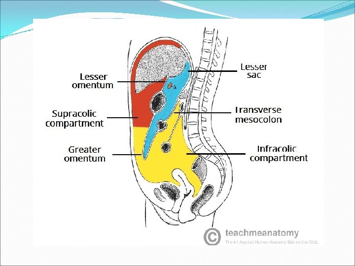
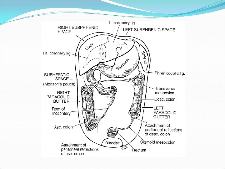
Peritoneal subdivisions The transverse colon and transverse mesocolon divides the greater sac into supracolic and infracolic compartments. Supracolic compartments (subphrenic space)-lies between diaphragm and transverse colon and transverse mesocolon Suprahepatic recess lies between the diaphragm and liver -the falciform ligament divides it into right and left suprahepatic recesses
Left suprahepatic recesses left anterior suprahepatic spaces left posterior suprahepatic spaces Right suprahepatic recesses right anterior suprahepatic spaces right posterior suprahepatic spaces bare area of live (extraperitoneal space)
Infrahepatic recess lies between the liver and transverse colon & transverse mesocolon-the ligamentum teres hepatic divides it into right and left infrahepatic recesses Right infrahepatic recesses (hepatorenal recess) Left infrahepatic recesses left anterior infrahepatic space left posterior infrahepatic space
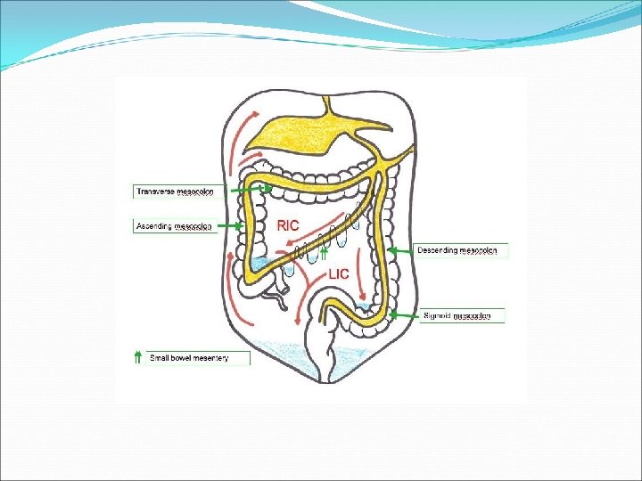
Infracolic compartments -lies below the transverse colon and transverse mesocolon Right paracolic sulcus (gutter) -lies lateral to the ascending colon. It communicates with the hepatorenal recess and the pelvic cavity. It provides a route for the spread of infection between the pelvic & the upper abdominal region.
Infracolic compartments Left paracolic sulcus (gutter) -lies lateral to the descending colon. It is separated from the area around the spleen by the phrenicocolic ligament, a fold of peritoneum that passes from the colic flexure to the diaphragm.
Right mesenteric sinus -triangular space, lies between root of mesentery, ascending colon, right 2/3 of transverse colon and transverse mesocolon Left mesenteric sinus -lies between root of mesentery, descending colon, right 1/3 of transverse colon and transverse mesocolon, its widens below where it is continuous with the cavity of the pelvis
Applied Anatomy Peritoneum & surgical procedures Peritonitis & Ascites Abdominal paracentesis Intraperitoneal injection peritoneal dialysis
Paracenteis abdominis is done to relieve abdominal ascites
Please revise!