Posterior COMPARTMENTS OF THE LEG Posterior Compartment The


 • Nerve supply: Tibial nerve A-The")


 It receives insertion of Gastrocnemius and soleus")


 phalanx of the big toe")


- Slides: 29
Posterior COMPARTMENTS OF THE LEG
Posterior Compartment. The posterior compartment (superficial & deep) • Nerve supply: Tibial nerve A-The Superficial posterior compartment • Gastrocnemius (popliteal fossa) • Plantaris (popliteal fossa) • Soleus (popliteal fossa) B-The deep posterior compartment • Popliteus (popliteal fossa) • Tibialis posterior (back of leg) • Flexor digitorum longus (back of leg) • Flexor hallicus longus (back of leg)
Tibial nerve Posterior tibial artery & vein Flexor digitorum longus soleus Tendon of plantaris Medial head of gasrocnemius Tibialis posterior T F Flexor hallucis longus Lateral head of gasrocnemius
Superficial group
Plantaris Soleus TRICEPS SURAE Gastrocnemius
Popliteus Tibialis posterior Flexor hallucis longus Flexor digitorum longus
Plantaris lateral head of gastrocnemius medial head of gastrocnemius Tendocalcaneus Soleus
Origin of the superficial group Femur Lateral condyle Fibula Popliteus Tibia
Gastrocnemius 1 - Medial head: From the popliteal surface of the femur just above the medial condyle. 2 - Lateral head: from the lateral surface of the lateral condyle of the femur above the lateral epicondyle. It contains sesamoid bone called Fabella. Plantaris ** Origin: from the popliteal surface of the femur just above the lateral condyle. The muscle may be absent. Soleus ** Origin: from 1 - Back of the head of the fibula. 2 - The upper 1/3 of the posterior surface of the fibula. 3 - Tendinous arch (between head of the fibula and soleal line). 4 - Soleal line of the tibia 5 - Middle 1/3 of the medial border of the tibia.
Insertion Tendocalcaneus Gastrocnemius Plantaris Tendo-Achilis Soleus
** Insertion of Gastrocnemius and soleus: • Tendocalcaneus into the middle of the posterior surface of the calcaneus. ** Insertion of Plantaris: either Into the tendocalcaneus. OR separately in the posterior surface of the calcaneus. v Actions of three muscles • 1 - Powerful Plantar flexion of the foot (at ankle joint). • Raising the heel and foot from the ground in walking, running and jumping) • 2 - Flexion of the knee joint during sitting (gastrocnemius).
• • • v Tendocalcaneus (Tendo-Achilis) It receives insertion of Gastrocnemius and soleus muscles. It ends in the middle of the posterior surface of the calcaneus. Tendocalcaneus is the strongest and thickest tendon of the body. Soleus muscle has a very strong but slow action (like the 1 st gear of car). When movement is under way, the quicker acting gastrocnemius increases the speed (like the top gear of the car) e. g. in running. The 2 heads of gastrocnemius and soleus are called triceps surae. The soleus muscle contains a rich venous plexus which drains the superficial veins and pumps it to the deep veins against gravity (peripheral heart). So, it liable to deep venous thrombosis especially with old age, bed rest for a long time, sitting for long time, or fracture neck of femur Rupture of the tendocalcaneus leading to walking disability and running is impossible. Rupture of tendon of plantaris leading to sudden and severe pain.
Deep group
v Popliteus ** Origin: from groove on lateral surface of lateral condyle of femur below lateral epicondyle. It is intracapsular extrasynovial It separates lateral meniscus from capsule and lateral (fibular) collateral ligament ** Insertion: triangular area on the posterior surface of tibia above soleal line. ** Nerve supply: Tibial nerve. It descends superficial to muscle and then hooks on lower border to supply it through deep surface. Unlocking of knee joint At the beginning of flexion of knee joint Lateral rotation of femur on tibia when the foot is fixed on the ground Or medial rotation of tibia on femur when the foot is raised from the ground Protection of the lateral meniscus.
Capsule of knee joint Lateral collateral ligament Popliteus muscle
Medial meniscus Knee joint Cruciate ligaments Popliteus Lateral meniscus Lateral or Fibular collateral ligament
Longitudinal Deep group
S o le al li ne Pop So Flexor Digitorum Longus ** Origin: posterior surface of the tibia below the soleal line and medial to the vertical line. Flexor Hallucis Longus ** Origin: lower 2/3 of posterior surface of fibula lateral to the median crest. Tibialis Posterior 1 - Posterior surface of tibia below soleal line and lateral to the vertical line 2 - Posterior surface of fibula medial to median crest 3 - Interosseus membrane. Vertical line Origin Median crest
Insertion Flexor Hallucis Longus: plantar surface of terminal (distal) phalanx of the big toe Flexor Digitorum Longus - They divide into 4 tendons which are inserted into plantar surface of the distal (terminal) phalanges of the lateral 4 toes. - Each tendon passing through an opening in corresponding tendon of Flexor digitorum brevis opposite the proximal phalanx. Flexor hallucis longus Flexor digitorum longus
x x x Tibialis Posterior ** The tendon descends in the groove behind the medial malleolus to reach the sole of the foot. - It divided into 2 slips: A- Medial slip to the tuberosity of navicular bone (main insertion). B- Lateral slip divided into several slips to: 1 - All tarsal bones except talus. 2 - Bases of all metatarsal bones except the first and the fifth.
** Actions of Flexor Hallucis Longus 1 - Flexion of all Joints of the big toe. 2 - Plantar flexion of the foot. 3 - Supporting the longitudinal arch of the foot 4 - Inversion of the foot. ** Actions of Flexor Diestrum Longus 1 - Flexion of all joints of the lateral 4 toes. 2 - Plantar flexion of the foot. 3 - Supporting the longitudinal arch of the foot. 4 - Inversion of the foot. ** Actions of Tibialis Posterior 2 - Plantar flexion of the foot. 3 - Supporting the longitudinal arch of the foot. 4 - Inversion of the foot.
Effect of injury of tibial nerve : • Paralysis of muscles of posterior compartment of leg leading to Loss of planter flexion, loss of flexion of toes & weak inversion of foot • Paralysis of muscles of sole of the foot (clawing of the toes). • Deformity calcaneovalgus -The foot is dorsi flexed & everted.
Tendo-Achilis Flexor hallucis longus Flexor digitorum longus
Medial malleolus Flexor retinaculum Calcaneus
Flexor hallucis longus Tibialis posterior Flexor digitorum longus Posterior tibial artery Posterior Tibial nerve Tom Designs Very Nice House
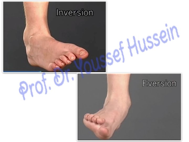
Inversion and eversion of the foot 1 - Inversion; medial rotation of the foot and the sole looks inwards (medially). It is done by a) Tibialis anterior supplied by anterior tibial nerve. ibialis posterior supplied by posterior tibial nerve. c) Flexor hallicus longus supplied by posterior tibial nerve. tensor hallicus longus supplied by anterior tibial nerve. 2 - Everson: Lateral rotation of the foot and the sole looks outwards (laterally); It is done by a) Peroneus longus supplied by superficial peroneal nerve. b) Peroneus brevis supplied by superficial peroneal nerve. c) Peroneus tertius supplied by anterior tibial nerve. ** Joints: Talo-calcaneo-navicular joint ** Significance: They occur mainly during walking on the rough ground. N. B: - The Lateral malleolus is Lower than the medial malleolus; So this bony projection resists the eversion.
Th ank Qu you est ion s