Intracellular Compartments and Protein Sorting Intracellular Compartments and


- Slides: 68

Intracellular Compartments and Protein Sorting

Intracellular Compartments and Protein Sorting ►Functionally distinct membrane bound organelles ► 10 billion proteins of 10, 000 -20, 00 diff kinds ►Complex delivery system

Compartmentalization of Cells Membranes ► ► ► Partition cell Important cellular functions Impermeable to most hydrophilic molecules contain transport proteins to import and export specific molecules Mechanism for importing and incorporating organelle specific proteins that define major organelles

Compartmentalization of Cells All Eucaryotic Cells Have Same Basic Set of Membrane Bound Organelles

Compartmentalization of Cells

Compartmentalization of Cells

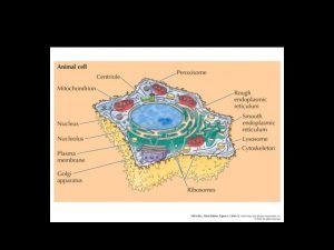
Compartmentalization of Cells Major Organelles ►Nucleus ►Cytosol ►ER ►Golgi Apparatus ►Mitochondria and Chloroplast ►Lysosomes ►Endosomes ►Peroxisomes

Compartmentalization of Cells ►Occupy 50% cell volume ►Perform same basic function ►Vary in size and abundance ►May take on additional functions ►Position dictated by cytoskeleton

Compartmentalization of Cells Topology governed by evolutionary origins Invagination of pm creates organelles such as nucleus that are topologically equivalent to cytosol and communicate via pores

Compartmentalization of Cells Topology governed by evolutionary origins Endosymbiosis of mito and plastids creates double membrane organelle (have own genome)

Compartmentalization of Cells Topology governed by evolutionary origins Organelles arising from pinching off of pm have interior equivalent to exterior of cell

Compartmentalization of Cells 3 Types of Transport Mechanisms 1. Gated Transport: gated channels topologically equivalent spaces 2. Transmembrane Transport: protein translocators topologically distinct space 3. Vesicular transport: membrane enclosed intermediates topologically equivalent spaces


Compartmentaliztion of Cells Families of Intracellular Compartments: 1. nucleus and cytosol 2. organelles in the secretory pathway 3. mitochondria 4. plastid Transport guided by: 1. sorting signals in transported proteins 2. complementary receptor proteins

Compartmentalization of Cells 2 Types of Sorting Signals in Proteins 1. Signal Sequence continuous sequence of 15 -60 aa sometimes removed from finished protein sometimes a part of finished protein 2. Signal Patch specific 3 d arrangement of atoms on protein surface; aa’s distant persist in finished protein

Compartmentalization of Cells

Compartmentalization of Cells Signal Sequences/Patches Direct Proteins to Final Destination Signal patches direct proteins to: 1. nucleus 2. lysosomes Signal Sequences direct proteins to: 1. ER proteins possess N-terminal signal of 5 -10 hydrophobic aa 2. mito proteins have alternating + chg aa w/ hydrophobic aa 3. proxisomal proteins have 3 aa at C-terminus

Compartmentalization of Cells Sorting signals recognize complementary sorting receptors Receptors unload cargo ► Function catalytically and are reusable ►

Compartmentalization of Cells Organelles Cannot be Constructed Denovo ► Organelles reproduced via binary fission ► Organelle cannot be reconstructed from DNA alone ► Info in form of one protein that pre-exists in organelle mem is required and passed on from parent to progeny ► Epigenetic information essential for propogation of cell’s compartmental organization

Transport of Molecules Btwn Nucleus and Cytosol Nuclear Envelope ►Two concentric membranes -Outer membrane contiguous w/ER -Inner membrane contains proteins that act as binding sites for chromatin and nuclear lamina ►Perforated by nuclear pores for selective import and export

Transport of Molecules Btwn Nucleus and Cytosol Nuclear Pore Complex ►mass of 125 million; ~50 different proteins arranged in octagon ►Typical mammalian cell 3, 000 -4, 000 ►Contains >1 aqueous channels thru which sm molec can readily pass <5, 000; molec > 60, 000 cannot pass ►Functions ~diaphram ►Receptor proteins actively transport molec thru nuclear pore

Transport of Molecules Btwn Nucleus and Cytosol

Transport of Molecules Between Nucleus and Cytosol Nuclear Localization Signal ► ► ► ► Generally comprised of two short sequences rich in + chged aa lys & arg Can be located anywhere Thought to form loops or patches on protein surface Resident, not cleaved Transport thru lg aqueous pores as opposed to translocator proteins Transports proteins in folded state Energy requiring process

Transport of Molecules Btwn Nucleus and Cytosol Nuclear Import- the players ► Importins = cytosolic receptor protein binds to NLS of “cargo” proteins ► Nucelar Export Receptors = binds macromolecules to be exported from nucelus ► Adaptors = sometimes required to bind target protein to nuclear receptor ► Ran = cytosolic GTP/GDP binding protein complexes with importins in the cytosol. ► Fibril proteins and nucleoporins contain phenylalanine/glycine repeats (FG) repeats. Repeats transiently bound and released by importin/cargo/Ran-GDP, causing the complex to “hop” into the nucleus Import Receptors release cargo in nucleus and return to cytosol Export Receptors release cargo in cytoplasm and return to nucleus

Transport of Molecules Btwn Nucleus and Cytosol Ran GTPase= molecular switch ► Drives directional transport in appropriate directin ► Conversion btwn GTP and GDP bound states mediated by Ran specific regulatory proteins GAP converts RNA-GTP to Ran-GDP via GTP hydrolysis GEF promotes exchg of GDP for GTP converting Ran-GDP to Ran-GTP ► Ran GAP in cytosol thus more Ran-GDP in cytosol ► Ran GEF in nucleus thus more Ran-GTP in nucleus

Importin is a type of karyopherin that transports protein molecules into the nucleus by binding to specificr ecognition sequences called nuclear localization sequences(NLS). Importin has two subunits, importin α and importin β. Members of the importin-β family can bind and transport cargo by themselves, or can form heterodimers with importin-α. As part of a heterodimer, importin-β mediates interactions with the pore complex, while importin-α acts as an adaptor protein to bind the nuclear localisation signal (NLS) on the cargo. The NLS-Importin α-Importin β trimer dissociates after binding to Ran GTP inside the nucleus with the two importin proteins being recycled to the cytoplasm for further use.

Transport of Molecules Btwn Nucleus and Cytosol Nuclear Export ► Works like import in reverse ► Export receptors bind export signals and nucleoporins to guide cargo thru pore ► Import and export receptors member of same gene family

Transport of Molecules Btwn Nucleus and Cytosol Regulation Afforded by Access to Transport Machinery ► ► Controlling rates of import and export determines steady state location phosphorylation/dephosphorylation of adjacent aa may be required for receptor binding Cytosolic anchor or mask proteins block interaction w/ receptors Protein made and stored in inactive form as ER transmembrane protein

Transport of Molecules Btwn Nucleus and Cytosol Control of m. RNA Export ► ► ► Proteins w/ export signals loaded onto RNA during transcription and processing (RNP) Export signals guide RNA out of nucleus thru pores via exportin proteins than bind RNP Export mediated by transient binding to FG repeats Imature m. RNAs retained by anchoring to transcription and splicing machinery Proteins disassociate in cytosol and return to nucleus

Transport of Molecules Btwn Nucleus and Cytosol Nuclear Lamina ►Meshwork of intermediate filaments ►Maintenance of nuclear shape ►Spacial organization of nuclear pores ►Regulation of transcription ►Anchoring of interphase chromatin ►DNA replication ►Phosphorylation causes depolymerizes during mitosis when nucleus disassembles

Transport of Molecules Btwn Nucleus and Cytosol Nuclear envelop disassembles during mitosis and reassembles when ER wraps around chromosomes and begins to Fuse


When a nucleus disassembles during mitosis, the nuclear lamina depolymerizes. The disassembly is at least partly a consequence of direct phosphorylation of the nuclear lamins by the cyclin-dependent protein kinase Cdk that is activated at the onset of mitosis. At the same time, proteins of the inner nuclear membrane are phosphorylated, and the NPCs disassemble and disperse in the cytosol. During this process, some NPC proteins become bound to nuclear import receptors, which play an important part in the reassembly of NPCs at the end of mitosis. Nuclear envelope membrane proteins—no longer tethered to the pore complexes, lamina, or chromatin—disperse throughout the ER membrane. The dynein motor protein, which moves along microtubules , actively participates in tearing the nuclear envelope off the chromatin. Together, these processes break down the barriers that normally separate the nucleus and cytosol, and the nuclear proteins that are not bound to membranes or chromosomes intermix completely with those of the cytosol

Later in mitosis, the nuclear envelope reassembles on the surface of the chromosomes. In addition to its crucial role in nuclear transport, Ran-GTPase also acts as a positional marker for chromatin during cell division, when the nuclear and cytosolic components intermix. Because Ran-GEF remains bound to chromatin when the nuclear envelope breaks down, Ran molecules close to chromatin are mainly in their GTP-bound conformation. By contrast, Ran molecules further away have a high likelihood of encountering Ran-GAP, which is distributed throughout the cytosol; these Ran molecules are therefore mainly in their GDP-bound conformation. Chromatin in mitotic cells is therefore surrounded by a cloud of Ran-GTP. This cloud locally displaces nuclear import receptors from NPC proteins, which starts the assembly process of NPCs attached to the chromosome surface. At the same time, inner nuclear membrane proteins and dephosphorylated lamins bind again to chromatin. ER membranes wrap around groups of chromosomes and continue fusing until they form a sealed nuclear envelope. During this process, the NPCs start actively reimporting proteins that contain nuclear localization signals. Because the nuclear envelope is initially closely applied to the surface of the chromosomes, the newly formed nucleus excludes all proteins except those initially bound to the mitotic chromosomes and those that are selectively imported through NPCs. In this way, all other large proteins are kept out of the newly assembled nucleus.

Protein Transport into the Mitochondria and Chloroplast Subcompartments of the Mitochondria and Chloroplast

Protein Transport into the Mitochondria and Chloroplast Translocation into Mitochondrial Matrix Governed by: 1. Signal Sequence (amphipathic alpha helix cleaved after import) 2. Protein Translocators

Protein Transport into the Mitochondria and Chloroplast Players in Protein Translocation of Proteins in Mitochondria ► ► ► TOM- functions across outer membrane TIM- functions across inner membrane OXA- mediates insertion of IM proteins syn w/in mito and helps to insert proteins initially transported into matrix Complexes contain components that act as receptors and others that form translocation channels


Protein Transport into the Mitochondria and Chloroplast Import of Mitochondrial Proteins ►Post-translational ►Unfolded polypeptide chain 1. precursor proteins bind to receptor proteins of TOM 2. interacting proteins removed and unfolded polypetide is fed into translocation channel ►Occurs contact sites joining IM and OM TOM transports mito targeting signal across OM and once it reaches IM targeting signal binds to TIM, opening channel complex thru which protein enters matrix or inserts into IM


Protein Transport into the Mitochondria and Chloroplast Import of Mitochondrial Proteins ►Requires energy in form of ATP and H+ gradient and assitance of hsp 70 -release of unfolded proteins from hsp 70 requires ATP hydrolysis -once thru TOM and bound to TIM, translocation thru TIM requires electrochemical gradient

Protein Transport into the Mitochondria and Chloroplast Protein Transport into IM or IM Space Requires 2 Signal Sequences 1. Second signal =hydrophobic sequence; immediately after 1 st signal sequence 2. Cleavage of N-terminal sequence unmasks 2 nd signal used to translocate protein from matrix into or across IM using OXA 3. OXA also used to transport proteins encoded in mito into IM 4. Alternative route bypasses matrix; hydrophobic signal sequence = “stop transfer”


Protein Transport into the Mitochondria and Chloroplast Protein Transport into Chloro Similar to Transport into Mito 1. 2. 3. 4. 5. 6. occur posttranslationally Use separate translocation complexes in ea membrane Translocation occurs at contact sites Requires energy and electrochemical gradient Use amphilpathic N-terminal signal seq that is removed Like the mito a second signal sequence required for translocation into thylakoid mem or space

Protein Transport into the Mitochondria and Chloroplast

Peroxisomes and Protein Import Peroxisomes ►Use O 2 and H 2 O 2 to carry out oxidative rxns ►Remove H from specific organic compounds RH 2 + O 2 R + H 2 O 2 ►Catalases use H 2 O 2 to oxidize other substances, particularly in liver and kidney detoxification H 2 O 2 + R’H 2 R’ + H 2 O ►Beta Oxidation ►Formation of plasmalogens (abundant class of phospholipids in myelin) ►Photorespiration and glyoxylate cycle in plants

Peroxisomes and Protein Import Peroximsomes in Plants ►Site of Photorespiration= glycolate pathway in leaves ►Called glyoxysomes in seeds where fats converted into sugar

Proxisomes and Protein Import ►Peroxisomes arise from pre-existing peroxisomes ►Signal sequence of 3 aa at COOH end of peroxisomal proteins= import signal ►Some have signal sequence at N-terminus ►Involves >23 distinct proteins ►Driven by ATP hydrolysis ►Import mechanism distinct, not fully characterized ►Oligomeric proteins do not unfold when imported ►Zellweger Disease= peroxisomal deficiency

ER and Protein Trafficking Endoplasmic Reticulum Occupies >= 50% of cell volume ► Continuous with nuclear membrane ► Central to biosyn macromolecules used to construct other organelles ► Trafficking of proteins to ER lumen, Gogli, lysosome or those to be secreted from cell ►

ER and Protein Trafficking ER Central to Protein Synthesis and Trafficking Removes 2 Types of Proteins from Cytosol: 1. transmembrane proteins partly translocated across ER embedded in it 2. water soluble proteins translocated into lumen

ER and Protein Trafficking Quantity of SER and ER Dependent Upon Cell Type RER assoc. w/ protein synthesis SER assoc. lipid biosynthesis, detoxification, steroid synthesis, Ca 2+ storage

ER and Protein Trafficking Import of Proteins into ER ►Occurs co-translationally ►Signal recognition sequence recognized by SRP ►SRP recognized by SRP receptor ►Protein Translocator

ER and Protein Trafficking Hydrophobic signal sequence of diff sequence and shape ► SRP lg hydrophobic pocket lined by Met having unbranched flexible side chains ► Binding of SRP causes pause in protein synthesis allowing time for SRP-ribosome complex to bind to SRP receptor ►

ER and Protein Trafficking Protein to be imported passes through an aqueous pore in the translocator that is a dynamic structure ►Sec 61 protein translocator ►Signal sequence triggers opening of pore ►Translocator pore closes when ribo not present

ER and Protein Trafficking Some proteins are imported in to ER by a posttranslational mechanism ►Proteins released into cytoplasm ►Binding of chaperone proteins prevents them from folding ►Translocation occurs w/out ribo sealing pore ►Mechanism whereby protein moves through pore unkwn

ER and Protein Trafficking Signal Sequence is Removed from Soluble Proteins Two signaling functions: 1) directs protein to ER membrane 2) serves as “start transfer signal” to open pore ► Signal peptidase removes terminal ER signal sequence upon release of protein into the lumen ►

ER and Protein Trafficking Single Pass Transmembrane Proteins 1. N-terminal signal sequence initiates translocation and additional hydrophobic “stop sequence anchors protein in membrane 2. Signal sequence is internal and remains in lipid bilayer after release from translocator 3. Internal signal sequence in opposite orientation 4. Orientation of start-transfer sequence governed by distribution of nearby chg aa

ER and Protein Trafficking Multipass Transmembrane Proteins Combinations of start- and stop-transfer signals determine topology ► Whether hydrophobic signal sequence is a start- or stop-transfer sequence depends upon its location in polypeptide chain ► All copies of same polypeptide have same orientation ►

Positively-charged segment determines the orientation of integral membrane proteins

ER and Protein Trafficking Folding of ER Resident Proteins ER resident proteins contain an ER retention signal of 4 specific aa at C-terminus ► PDI protein disulfide isomerase oxidizes free SH grps on cysteines to from disulfide bonds S-S allowing proteins to refold ► Bi. P chaperone proteins, pulls proteins posttranslationally into ER thru translocator and assists w/ protein folding ►

ER and Protein Trafficking Glycolsylation of ER Proteins Most soluble and transmembrane proteins made in ER are glycolsylated by addition of an oligosaccharide to Asn ► Precursor oligosaccharide linked to dolichol lipid in ER mem, in high energy state ► Transfer by oligosaccharyl transferase occurs almost as soon as polypeptide enters lumen ►

ER and Protein Trafficking Oligosaccharide assembled sugar by sugar onto carrier lipid dolichol

ER and Protein Trafficking Retrotranslocation Improperly folded ER proteins are exported and degraded in cytosol ► Misfolded proteins in ER activate an “Unfolded Protein Response” to increase transcription of ER chaperones and degradative enzymes ►

ER and Protein Trafficking The Unfolded Protein Response

ER and Protein Trafficking Assembly of Lipid Bilayers on ER ER synthesizes nearly all major classes of lipids ► Phospholipid synthesis occurs on cytoplasmic face by enzymes in mem ► Acyl transferases add two FA to glycerol phosphate producing phosphatidic acid ► Later steps determine head group ►

ER and Protein Trafficking Assembly of Lipid Bilayers on ER ►Scramblase phospholipid translocator equilibrates phospholipids distribution ►Flipasses of PM responsible for asymmetric distribution of phospholipids

ER and Protein Trafficking Phospholipid Exchange Proteins ►Transfer individual phosphlipids between membranes at random btwn all membranes ►Exchange protein specificity ►Extracts phospholipid and diffuses away w/ it buried w/in lipid binding site; discharges phospholipid when it encounters another membrane
 Intracellular compartments and protein sorting
 Internal vs external sorting
 Carrier vs channel proteins
 Protein-protein docking
 Intracellular and extracellular
 Fluid compartments in the body
 Ecf icf and interstitial fluid
 Intracellular extracellular fluid
 Titanic watertight compartments
 Posterior compartment of thigh
 Derine
 Intracellular accumulation meaning
 Interstitial vs intracellular
 Hand compartments
 V
V Abdomen regions
 Lipofuscin
 Cubital fossa veins
 For offical use only
 Intracellular hormone binding
 Definition of community bag
 Temperate phage
 Youtube.com
 Intracellular ph regulation
 Breakdown of food
 Intracellular structure
 Intracellular proteins
 Dry vs wet gangrene
 Cl extracellular concentration
 Intracellular receptors
 Intracellular accumulations
 Blood loss estimation gauze
 Body fluid compartments
 Osmolarity vs tonicity
 Potato grading standards
 Bubble sort vs selection sort
 Searching and sorting arrays in c++
 Big o java
 Orale charakterstruktur
 Unit operation in food processing
 Lesson 2 assignment a sort of sorts answers
 Searching and sorting in java
 Searching and sorting java
 Searching and sorting in java
 Physical and chemical properties sorting activity
 Lego sorting machine
 Tujuan dari sorting adalah
 What is stable sorting
 Evaporation example mixture
 How is chert formed
 Rake classifier working principle
 Grain size sorting formula
 Sediment sorting
 Inplace sorting
 Harry potter outline
 External sorting techniques
 Stable sorting algorithm
 Sorting circuit
 Draw two different quadrilaterals
 Changes in wave properties sorting activity
 Special quadrilaterals
 Searching struktur data
 Pengertian searching dalam struktur data
 Sorting struktur data
 Spike sorting tutorial
 Proses mengatur
 Pigeon sort
 Recursive sorting algorithms
 Quadratic sorting algorithms
