Fluid Management and Blood Transfusion Dr Ahmed Shahin

Fluid Management and Blood Transfusion Dr. Ahmed Shahin M. D

Outline � Body Fluid Compartements � Body Fluid Composition, Estimated Blood Volume, Allowable Blood Loss � Osmolality and Tonicity � Daily Intake and Output � Types of Fluid � Blood Transfusion

Body Fluid Compartments �TBW: 55 -60% of the BW in men and 45 -50% in young women

Body Fluid Composition in Age Groups AGE Neonate TBW AS % OF TOTAL BODY WEIGHT 80 6 months 70 1 year 60 Young adult Elderly 60 50
 �Na+ �K+ �Ca 2+ �Mg 2+ �Cl�HCO")
Composition of Body Fluid Compartments �Ion (mmol/L) �Na+ �K+ �Ca 2+ �Mg 2+ �Cl�HCO 3�HPO 42�Sulphate�Proteinate- Plasma (mmol/L) ICF 143 5 1. 3 0. 9 103 24 0. 4 1. 14 9 135 <0. 8 25 9 9 74 19 64

ESSENTIAL PRINCIPLES

Solvents, solutes, and solutions �Mixture, Solution, solvent, solute. �Osmole, mole

Osmolarity and Osmolality �These are ways of quantifying how much of a solute is dissolved in a solution. • Osmola(R)ity No. of osmoles of solute particles per unit VOLUME of solution and has units osmoles/litre. In the body we use milliosmole • Osmola(L)ity No. of osmoles of solute particles per unit WEIGHT of solvent and has units osmoles/kilogram.
 + glucose + urea =")
Plasma Osmolality Plasma osmolality = 2 (Na + K) + glucose + urea = 2 (137 + 4. 0) + 5. 0 + 4 = 291 mosmol/kg H 20 Glu: /18 Urea: /2. 8

Tonicity �A way of describing the relative solute concentrations of two solutions which are separated by a selectively-permeable membrane (often called a semi-permeable membrane).
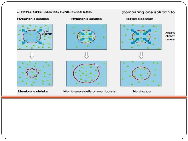

�In clinical practice the tonicity of fluid administered intravenously is described relative to the tonicity of the internal environment of the red blood cell and with reference to the red blood cell membrane.

WHAT IS THE ‘NORMAL’ DAILY INTAKE AND OUTPUT OF FLUID AND ELECTROLYTES?

OUTPUT, INTAKE �Input: Oral, Enteral, Intravenous �Output: ‘Sensible’: that it is easily seen and measured e. g. urine output and loses from the gastrointestinal tract. ‘Insensible’: not seen and not easy to quantify e. g. sweat, and water vapor in exhaled gases.
 200 ml Skin(sweat) 400 ml Respiratory 400 ml Total")
OUTPUT Urine 1500 ml Gastrointestinal(faeces) 200 ml Skin(sweat) 400 ml Respiratory 400 ml Total 2500 INTAKE Drinking 1500 ml Eating 750 ml Metabolism 250 ml Total 2500

�The above volumes do not contain just water but also electrolytes….

ELECTROLYTE DAILY LOSS / REQUIREME NT 75 Kg PERSON PER DAY Sodium 1 -1. 5 mmol/kg 75 - 112. 5 mmol Potassium 1 -1. 5 mmol/kg 75 – 112. 5 mmol Magnesium 0. 1 -0. 2 mmol/kg 7. 5 – 15 mmol Calcium 0. 1 -0. 2 mmol/kg 7. 5 – 15 mmol Chloride 0. 07 -0. 22 mmol/kg 5. 25 – 16. 5 mmol 20 -40 mmol/kg 1500 – 3000 mmol Phosphate

Maintenance and Deficit �Maintenance vs deficit �Rule of 4 /2/ 1 �Ex. 70 kg patient 1 st 10 kg: 10 kg * 4 ml = 40 ml / kg 2 nd 10 kg: 10 kg * 2 ml = 20 ml / kg 3 rd 10 kg: 50 kg * 1 ml = 50 ml / kg Total=110 ml/kg �Ex. Fasting for 10 hr without any intake: 10*110= 1100 ml
 � Men 75 ml / kg �Women 65 ml /")
Estimated Blood Volume (EBV) � Men 75 ml / kg �Women 65 ml / kg � Infants 80 ml / kg � Neonates 85 ml / kg � Premature Neonates 96 ml / kg
 �EBV = weight (kg) * Average blood volume �Allowable Blood")
Allowable Blood Loss (ABL) �EBV = weight (kg) * Average blood volume �Allowable Blood Loss = [EBV*(Hi-Hf)]/Hi �Where: �EBV=Estimated Blood Volume �Hi= initial hemoglobin (Hct) �Hf= final hemoglobin (Hct) �Normal Hct Values �Men 42 -52% �Women 37 -47%

Example � Q: Before surgery is to take place, what is the EBV of a female patient weighing 50 kg? Also, what is the ABL of this patient if her Hct is 45? � EBV = 50 kg x 65 = 3250, The final lowest acceptable Hct (Hf) = 30% � (3250 x (45 – 30))/45 = 1083 Using this rough estimate, the patient in this example could loose 1083 m. L of blood without needing a transfusion. � Replacing Blood Loss � “Ideally, blood loss should be replaced with crystalloid or colloid solutions to maintain intravascular volume (normovolemia) until the danger of anemia outweighs the risks of transfusion. For most patients, that point corresponds to a hemoglobin between 7 and 10

Intravenous Fluids

Types �Three main types �Crystalloids �Colloids �Blood products

�Solutions that contain a combination of water and electrolytes. �Divided into "balanced" salt solutions (e. g. Ringer's lactate) and hypotonic solutions (e. g. D 5 W).

�Classified into three groups based on their predominant use �Replacement Solutions �Maintenance Solutions �Special Solutions

Replacement Solutions � Used to replace ECF �All isotonic, usually replace losses that involves both water and electrolytes � Have a [Na+] similar to that of the ECF which effectively limits their fluid distribution to the ECF compartment. �Distributes between the ISF ¾ and the plasma ¼ in proportion to their volumes

� 1 cc blood with 3 -4 cc crystalloids

Ringer Lactate �Na+ = 131 mmol/L �Cl- = 111 mmol/L �Lactate = 29 mmol/L �K+ = 5 mmol/L �Ca++ = 2 mmol/L �PH = 6. 5 �Osmolality = 279 mosm/L �Potential problem = potassium may accumulate, lactate metabolism causes alkalosis

Maintenance Solutions �Isosmotic as administered but not necessarily isotonic �Usually used when the loss involves mainly pure water �Ex. D 5 W, Normal Saline
 � 9 g of Na. Cl/L water �")
Normal saline (0. 9% saline solution) � 9 g of Na. Cl/L water � 154 mmol/L sodium � 154 mmol/L chloride �Osmolality = 308 mosm/L �PH = 5. 0 �Potential problem = hyperchloraemic metabolic acidosis, more likely with renal insufficiency
 saline. . hyponatremia � 30 gm Na. Cl, 1027, 4.")
Special Solutions �Hypertonic (3%) saline. . hyponatremia � 30 gm Na. Cl, 1027, 4. 5 to 7. 0 �Half normal saline. . hypernatremia � 77 meq/L � 8. 4% Bicarbonate solution. . . acidosis �Mannitol 20%. . brain oedema, pulmonary

Albumin Dextran Gelatins Starches
 �Exerts")
Colloids �Colloid: a large molecule that does not diffuse across semipermeable membranes (capillary) �Exerts an osmotic pressure in the blood, causing fluid to remain within the vascular system. The result is an increase in intravascular volume.
 �Artificial (e.")
�Two categories of colloid may be defined: �Natural (e. g. human albumin) �Artificial (e. g. gelatins, dextran and hydroxyethyl starches [HES]).
 = 1. 6 hours in plasma �Stays within the intravascular space")
Albumin �Half-life (t½) = 1. 6 hours in plasma �Stays within the intravascular space unless the capillary permeability is abnormal � 5% solution - isooncotic; 10% and 25% solutions - hyperoncotic �Expands volume 5 x its own volume in 30 minutes �Effect lasts about 24 -48 hours �Side effects - volume overload, fever (pyrogens in albumin), defects of haemostasis

Dextran �High MW polysaccharide �Dextran 40 - MW 40, 000 (greater effects on coagulation than D 70) �Dextran 70 - MW 70, 000 � 10% solution in NS or D 5 W �Side effects: anaphylaxis, coagulopathy, renal failure �Dose: limit to 20 ml/kg/day �Used as antiaggregant in patients undergoing vascular and microvascular surgical
 �Synthetic hydroxy-substituted amylopectin, a highly branched glucose polymer �Variable MW")
Hetastarch (HES= hydroxyethyl starch) �Synthetic hydroxy-substituted amylopectin, a highly branched glucose polymer �Variable MW � 6% and 10% solution in normal saline solution �Plasma t½ is 5 days � 90% eliminated in 40 days �Side effects: coagulopathy (coated platelets, increased fibrinolysis, decreased factor VIII level) but usually not a major clinical problem �Dose: limit the amount to 20 ml/kg/d.

PRIOPERATIVE BLOOD TRANSFUSION

Purpose of Infusion of Fluids and Blood Products �Maintain organ transfusion �Normal blood pressure and heart rate, normal mental status (in non-comatose patients), normal oxygen saturation, normal urine output, well perfused extremities

Major Blood Components �Cellular �Fluid

Blood Products �Whole blood �Cryoprecipitate �Packed Red Cells �Human Albumin �Platelets �Fresh Frozen Plasma

Blood Bank Practices

Preparation of Blood Components �Blood donors: �Approximately 17 million units of blood are donated in Europe each year. �Each donor is interviewed for medical history of known infectious diseases �Each unit is screened for antibodies to: � Syphilis � Hepatitis B and C � HIV 1 and 2 � +/- CMV
 for allogeneic or 32% for autologous donors.")
Exclusion Criteria �Hct less than 37%(Hgb<12. 5) for allogeneic or 32% for autologous donors. �Weight less than 50 kilograms �Temperature above 37. 5°C �Blood pressure above 180 systolic or 100 diastolic �Pulse rate outside the limits of 50 to 100 beats per minute �Arrhythmia detected on pulse examination

Centrifugation �Collect 500 m. L whole blood �Divert the first 40 m. L to reduce risk of bacterial contamination from donor skin �The 40 m. L are used for donor unit testing �Blood is centrifuged and separated into 3 parts: �◆ Red Blood Cells �◆ Plasma �◆ Buffy coat

�The Buffy coat units from four donors are further processed to separate the platelets �The red blood cell and platelet components are leukoreduced
: � 250")
�Separated of blood components by 1 unit of Whole blood: �PRBCS(hematocrit 70%): � 250 m. L+saline preservative=350 m. L. � 1– 6°C. �May be frozen in a hypertonic glycerol solution for up to 10 years(rare phenotypes)

�A preservative–anticoagulant solution is added. The most commonly used solution is CPDA-1: �Citrate as an anticoagulant (by binding calcium) �Phosphate as a buffer �Dextrose as a RC energy source �Adenosine as a precursor for ATP synthesis. � 35 days �AS-1 (Adsol) or AS-3 (Nutrice) extends the shelf-life to 6 weeks. �ADSOL (Adenine, glucose, mannitol and sodium chloride) �NUTRICE (Adenine, glucose, citrate, phosphate and Na. Cl)

�Platelet: � 50– 70 m. L. � 20– 24°C for 5 days. �Plasma: �The remaining plasma supernatant is further processed and frozen to yield fresh frozen plasma; rapid freezing helps prevent inactivation of labile coagulation factors (V and VIII). Slow thawing of fresh frozen plasma yields a gelatinous precipitate (cryoprecipitate) that contains high concentrations of Factor VIII and fibrinogen. � 200 m. L. �Once thawed it must be transfused within 24 h.

Blood Groups �At least 20 separate blood group antigen systems are known; fortunately, only the ABO and the Rh systems are important in the majority of blood transfusions.

The ABO System �Simply speaking, the chromosomal locus for this system produces two alleles: A and B. Each represents an enzyme that modifies a cell surface protein. Naturally Occurring Incidence Type Antibodies in Serum %45 Anti-B A %8 Anti-A B %4 — AB (Universal recipient) %43 Anti-A, anti-B O (Universal donor)

The Rh System and others �The Rh system is encoded by two genes located on chromosome 1. �There about 46 Rh-related antigens, but in most clinical settings, the five principal antigens (D, C, c, E, and e). �The most common and most immunogenic allele is the D antigen (80– 85% of white). �Other systems include the Lewis, P, Ii, MNS, Kidd, Kell, Duffy, Lutheran, Xg, Sid, Cartright, YK, and Chido Rodgers antigens. �Fortunately, with some exceptions (Kell, Kidd, Duffy, and S), alloantibodies against these systems rarely cause serious hemolytic reactions.

Compatibility Testing
 �The patient's RCs are tested with serum known to")
�ABO-Rh Testing (Group and Save) �The patient's RCs are tested with serum known to have anti A and anti B antibodies to determine blood type. �Confirmation of blood type is then made by testing the patient's serum against RCs with a known antigen type. �The patient's RCs are also tested with anti-D antibodies to determine Rh. �Crossmatching �Mimics transfusion: donor cells are mixed with recipient serum. �(1) confirms ABO and Rh typing (<5 min) �(2) detects antibodies to the other blood group systems (45 min) �(3) detects antibodies in low titers or those that do not

Intra-operative Transfusion Practices

Packed Red Blood Cells �Allows optimal utilization of blood bank resources. �Ideal for patients requiring RCs but not volume replacement (eg, anemia pt in compensated CHF). � Hgb 7 -8 g/d. L (<6, most people require blood; >10 most people do not) �Each unit raise Hgb by 1 g/dl � 170 -µm filter to trap any clots or debris. �Warming to 37°C during infusion. �Hypothermia and low levels of 2, 3 -diphosphoglycerate (2, 3 - DPG) in stored blood can cause a marked leftward shift of the hemoglobin–oxygen dissociation curve

Fresh Frozen Plasma �FFP contains all plasma proteins, including all clotting factors. �Indications: �Isolated factor deficiencies. �Reversal of warfarin therapy. �Coagulopathy associated with liver disease. �CABG, bleeding+NL ACT. �Massive blood transfusions. �Antithrombin III def. �The initial therapeutic dose is usually 10– 15 m. L/kg �ABO-compatible units are mandatory. � Coagulation factors INR 1. 4 -1. 6 (INR>1. 6, most people require FFP transfusion for major surgery; INR<1. 4, most people do not

Platelets �Thrombocytopenia or dysfunctional platelets. �Surgery or invasive procedures: 70, 000 x 109/L. �Vaginal delivery and minor surgical procedures: 50, 000 x 109/L. �Each unit expected to increase the count by 10, 000– 20, 000 x 109/L. �ABO-compatible platelet transfusions are desirable but not
 contains fibrinogen 150 mg, factor VIII 100 units, von")
Cryoprecipitate �Each unit (15 ml) contains fibrinogen 150 mg, factor VIII 100 units, von Willebrand factor (v. WF) (100 units) �DIC, hemophilia A, von Willebrand disease, quick reversal of thrombolytic therapy � Fibrinogen (most people require cryoprecipitate for major surgery if fibrinogen < 1 g/d. L)

Complications of Blood Transfusion

�Hemolytic reactions �Acute Vs. Delayed �Febrile Non hemolytic reactions �Transfusion Related Acute Lung Injury (TRALI) �Infectious complications

Hemolytic Reactions �Classified as either acute or delayed �Acute Hemolytic Reactions � 1: 38, 000. fatal in 100, 000 (severity depends on volume of incompatible blood) �The most common cause is misidentification … ABO mostly �Often severe. �In awake: chills, fever, nausea, and chest and flank pain. �In anesthetized: rise in temperature, unexplained tachycardia, hypotension, hemoglobinuria, and diffuse oozing in the surgical field. DIC, shock, and renal shutdown can develop rapidly

Management of hemolytic reactions 1. Once suspected, the transfusion should be stopped immediately. 2. The unit should be rechecked against the blood slip and the patient's identity bracelet. 3. Blood should be drawn to identify hemoglobin in plasma, to repeat compatibility testing, and to obtain coagulation studies and a platelet count. 4. A urinary catheter should be inserted, and the urine should be checked for hemoglobin. 5. Osmotic diuresis should be initiated with mannitol and intravenous fluids. 6. In the presence of rapid blood loss, platelets and FFP are indicated.

�Delayed Hemolytic Reactions � 1: 12, 000 �Antibodies to non-D antigens of the Rh system or to foreign alleles in other systems such as the Kell, Duffy, or Kidd antigens. � 1– 1. 6% chance even if ABO and Rh compatabale. �By the time significant amounts of these antibodies have formed (weeks to months), the transfused RCs have been cleared from the circulation. Moreover, the titer of these antibodies subsequently decreases and may become undetectable.

�The hemolytic reaction is therefore typically delayed 2– 21 days after transfusion, and mild, consisting of malaise, jaundice, and fever, failure of the patient's Hct to rise in spite of the transfusion and the absence of bleeding, and serum unconjugated bilirubin increases. �Diagnosis by direct Coombs test and re-examination of pre-transfusion specimens from both the patient and the donor. �The treatment is primarily supportive.
 �Acute (<24 hours) �Cytokine accumulation during storage of cellular")
Febrile non-haemolytic transfusion reactions (FNHTR) �Acute (<24 hours) �Cytokine accumulation during storage of cellular components (especially in platelet units) �Recipient antibodies (raised as a result of previous transfusions or pregnancies) reacting to donor human leucocyte antigens (HLA) �Unexpected temperature rise (≥ 38ºC or ≥ 1ºC above baseline), Chills, rigors, increased respiratory rate, hypotension, anxiety and a headache �leucocyte depletion

TRALI �ARDS following blood transfusion �High morbidity … mechanical ventilation �Lung injury is generally transient with PO 2 levels returning to pretransfusion levels within 48 -96 hours and CXR returning to normal within 96 hours. �Mortality rate, often approximated at 5 to 10% �Treatment as ARDS

Approaches to Fluid Management

The “Classic” Approach �Step 1: Calculate Ongoing Maintenance Requirements 4/2/1 rule: 4 cc/kg/hr for the first 10 kg, 2 cc/kg/hr for the second 10 kg, and 1 cc/kg/hr for every kg above 20. Ex. 70 kg 10 * 4 ml = 40 ml 10 * 2 ml = 20 ml 50 * 1 ml = 50 ml 110 ml / hour given each hour as oral or enteral intake is stopped Ex. 15 kg 10 * 4 ml = 40 ml + 5 * 2 ml = 10 ml = 50 ml / hour

�Step 2: Calculate Preoperative Fluid deficit Maintenance * the time without intake what so ever Ex. 70 kg fasting for 10 hours pre-operatively M = 110 ml/hour * time 10 hour = 1100 ml �Step 3: Calculate Anticipated Surgical Fluid Losses � Minimal tissue trauma (ex. herniorrhaphy): 0 -2 cc/kg/hr � Moderate tissue trauma (ex. cholecystectomy): 4 -6 cc/kg/hr � Severe tissue trauma (ex. bowel resection): 8 -10 cc/kg/hr �Ex. 70 kg undergoing major laparotomy 10 ml * 70 kg = 700 ml/hour as long as surgery is

�Step 4: Adjust for Blood Losses �A common recommendation is to give 3 -4 cc of crystalloid for every 1 cc of blood loss �Remember to add up suction volume, lap pads (100 - 150 cc each if fully soaked) and 4 x 4 small pads (10 cc each if fully soaked) Ex. In the 1 st hour of laparotomy there was 200 ml of pure blood in the suction jar, 2 fully soaked lap pads, and 10 fully soaked small gauses 200 ml + 2 * 150 ml + 10 * 10 ml = 600 ml ( in that hour) to be replaced with either 600 * 4 of cryst. or -if indicated-

� 1 st hour = Maintenance + ½ Deficit + Blood loss + Ongoing loss � 2 nd hour = Maintenance + ¼ Deficit + Blood loss + Ongoing loss � 3 rd hour = Maintenance + ¼ Deficit + Blood loss + Ongoing loss �Maintenance continued post-operatively as long as fasting �Blood loss replaced as long as there is bleeding

THANK YOU
- Slides: 74