Anterior Lateral Compartments of the Leg Dorsum of


")


- Slides: 18
Anterior, Lateral Compartments of the Leg & Dorsum of the Foot Dr Jamila EL & Dr Saeed Vohra Medany
At the end of the lecture, student should be able to: �Identify the deep fascia of leg �Identify the fascial compartments of the leg. �Describe the anatomy of the anterior & lateral compartments. �List the contents of each compartment (muscles, vessels & nerves). �Describe the anatomy and contents of the dorsum of the foot
• The deep fascia surrounds the leg and attached to anterior & medial borders of tibia. • Intermuscular Septa • Two pass from the deep aspect of this fascia to be attached to : • Anterior border of fibula (Anterior fascial septum) • Posterior border of fibula (Posterior fascial septum) • Interosseous membrane: A thin& strong membrane, that binds the interosseous borders of tibia & fibula. It binds the two bones and provides attachment for muscles.
• Together with the interosseus membrane, the septa divide the leg into 3 compartments : 1 -Anterior • 2 -Lateral (Peroneal) • 3 -Posterior • Each compartment has its own muscles, blood supply and nerve supply.
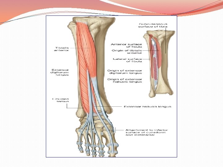
Anterior Compartment Muscles 1. Tibialis Anterior 2. Ext Dig Longus 3. Ext Hllucius Longus 4. Peroneus Tertius Artery Nerve Anterior Tibial Deep Peroneal
Origin Insertion Action Tibialis anterior Lateral surface of shaft of tibia & interosseous membrane. Medial cuneiform & base of 1 st metatarsal bone Dorsi flexion &Inversion of foot and maintains Medial Long arch Extensor dig longus Anterior surface of shaft of fibula Extensor expansion of lat (4) toes Extension of toes & Dorsi flexion of foot Extensor hallucis longus Anterior surface of shaft of fibula Base of distal phalanx of great toe Dorsi flexion& Inversion of the foot and extension of big toe Peroneus tertius Anterior surface of shaft of fibula Base of 5 th metatarsal bone Dorsiflexion and Eversion of the foot.
�Thickening of deep fascia that keep the long tendons around ankle joint in position �Superior Extensor Retinaculum : �Attached to anterior borders of tibia & fibula above ankle �Inferior Extensor Retinaculum: �Y-shaped band located inferior to ankle
From Medial to Lateral: 1. Tibialis anterior 2. Extensor Hallucis longus. 3. Anterior tibial Vessels 4. Deep peroneal Nerve. 5. Extensor Digitorum longus. 6. Peroneus tertius.
Muscles 1. Peroneus Longus 2. Peroneus Brevis Nerve Superficial Peroneal Artery Peroneal (Fibular)
Insertion Action Peroneus Lateral longus surface of shaft of fibula Medial cuneifor m & base of 1 st metatars al bone Plantar flexion & Eversion of the foot and supports lateral longitudina l arch Peroneus Lateral surface of brevis Base of 5 th Plantar metatars flexion & al bone Eversion of the foot and supports lateral longitudinal arch Origin shaft of fibula
Superior & Inferior peroneal retinacula Connect the lateral malleolus to calcaneum & hold the tendons of peroneus longus & brevis • Tendons of peronei are surrounded by a single common tubular synovial sheath, deep to inferior peroneal retinaculum they have separate sheaths
It is very thin, but just distal to ankle joint, it is thickened to form Inferior extensor retinaculum
Muscle Ext Dig Brevis Artery Dorsalis Pedis Nerve Deep Peroneal
Ext Digitorum Brevis Origin Insertion Inferior Extensor extensor R Hallucius & Brevis to base Calcaneum of proximal ph of big toe. Other (3) to EX Expansion Action Extension of IP& Metatarsoph J of 1 st, 2 nd, 3 rd& 4 th toes (during Dorsi flex).
Insertion of Long Extensor Tendons � The tendon of Ex dig longus divides into (4) to the lateral four toes. � Each tendon to the 2 nd , 3 rd & 4 th toes is joined on its lateral side by a tendon of Ex dig brevis. � The extensor tendons form � A fascial Expansion (Extensor Expansion) on the dorsum of each toe. � The expansion divides into (3) parts: � Central: inserted into the base of middle phalanx. � Two lateral : inserted into the base of distal ph. � The EX receives insertion of Interossei & Lumbrical muscles.
A separate sheath for each of: Tibialis anterior Extensor hallucis longus A common sheath for : Extensor digitorum longus & peroneus tertius, It extends to the level of base of 5 th metatarsal bone.
THANK YOU