Blood supply of brain By Dr Ch N

Blood supply of brain By Dr. Ch. N. V. Bharath

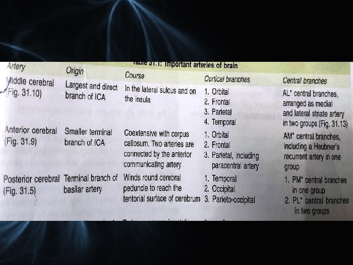
Main arteries Vertebro basillar system Internal carotid system

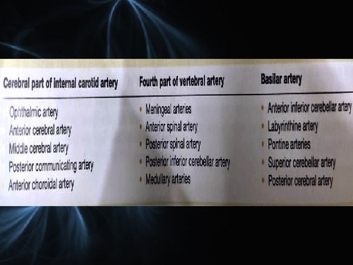
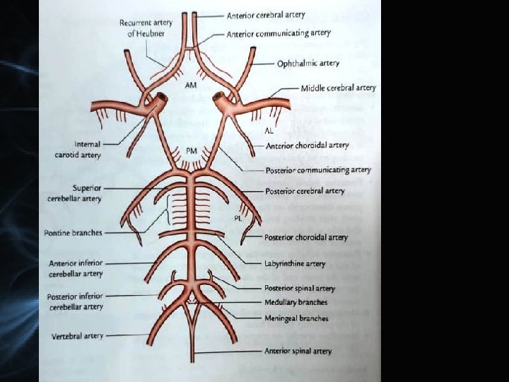
Vertebral artery • A branch of subclavian artery • Course - 4 parts Ø Ø 1 st 2 nd 3 rd 4 th

Vertebral artery Posterior spinal Meningeal Posterior inferior cerebellar Anterior spinal Medullary

• Anterior inferior cerebellar • Labyrinthine • Pontine • Superior cerebellar • Posterior Basilar artery

Posterior cerebral artery ØAnt. temporal, ØPost. temporal Øparieto-occipital Øcalcarine

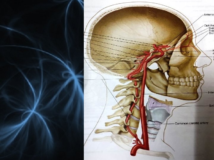
Internal carotid artery • Origin- From CCA at the level of upper border of thyroid cartilage
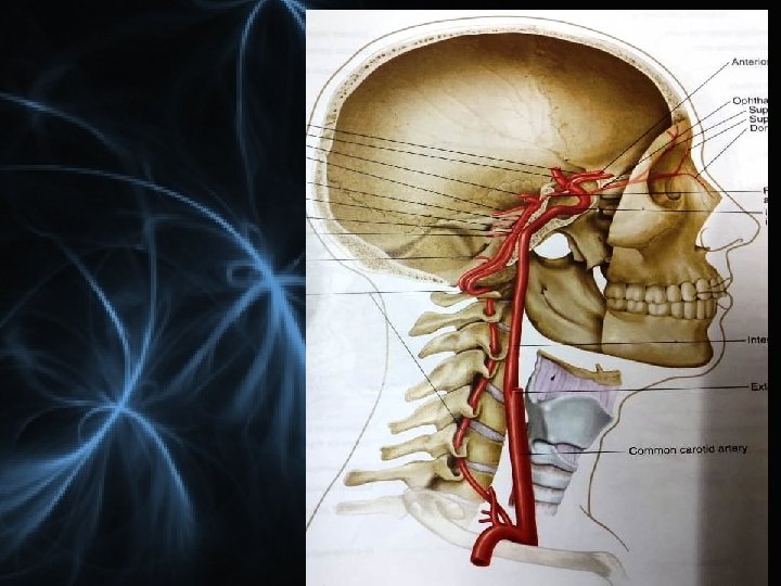

Terminal branches - Ant cerebral and middle cerebral

1. Posterior communicating artery • Runs backward to join PCA • Gives branches to hypophysis cerebri and hypothalamus

2. Anterior choroidal artery • Runs with optic tract • Enters inferior horn of LV thro choroid fissure • Supplies choroid plexus • Artery of cerebral thrombosis.

3. Anterior cerebral artery • Smaller terminal • Course – runs forward and medially. • Above the optic nerve– connected by ant. Communicating art. • Runs above the CC

Anterior cerebral arteries

• Orbital • Frontal • Parietal

4. Middle cerebral artery • Larger terminal • Runs laterally thro lateral sulcus. • Gives deep perforating brs

Temporal Frontal Parietal

5. Opthalmic artery

CIRCLE OF WILLIS • Circulus arteriosus • Location – Interpeduncular cistern • Shape – polygonal • Equalize the flow of blood • Collateral circulation

Formation

Six groups Anteromedial Right and left anterolateral Posteromedial Right and left Posterolateral Central branches

Central branches • 1. AM group from ant com, ACA and ICA Recurrent branch of ACA/Artery of HEUBNER Thrombosis of this art leads to contralateral upper monoplegia
 Charcot’s artery of cerebral hemorrhage.")
• 2. AL group from MCA (striate brs) Charcot’s artery of cerebral hemorrhage. • 3. posteromedial group from PCA and PCA • Also called thalamo perforators • 4. PL group from PCA • Also called thalamogeniculate

Branching pattern • Types of branches from ACA, MCA AND PCA ØCentral ØCortical ØChoroidal

Central thin walled , slender perforating End arteries Anteromedial Right and left anterolateral Posteromedial Right and left Posterolateral

Cortical • End arteries • 2 types 1. Short 2. Long

• Orbital Anterior cerebral artery • Frontal • Parietal

Orbital Middle cerebral artery Temporal Frontal Parietal

Posterior cerebral artery ØAnt. temporal, ØPost. temporal Øparieto-occipital Øcalcarine

Choroidal Anterior choroidal artery • Runs with optic tract • Enters inferior horn of LV thro choroid fissure • Supplies choroid plexus • Artery of cerebral thrombosis.

Posterior choroidal artery Choroid plexus of 3 rd ventricle and lateral ventricles.
 • ACA • PCA")
Supply of different surfaces • Superolateral surface MCA (2/3) • ACA • PCA
 • MCA • PCA")
Medial surface • ACA (2/3) • MCA • PCA

Inferior surface PCA MCA ACA

Cortical branches

Cortical and central branches do not anastomose. No anastomosis inside the brain tissue.

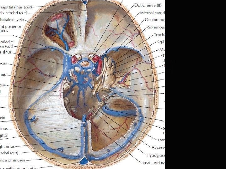
Venous drainage q Devoid of muscle q No valves q Veins of cerebrum – Superficial and deep veins

Venous drainage • Superior cerebral veins • Inferior cerebral veins • Superficial middle cerebral vein • Superior anastomotic vein • Inferior anastomotic vein

• Deep middle cerebral vein • Anterior cerebral vein

Deep veins • Internal cerebral veins •
 Basal vein")
Great cerebral vein (Galen) Basal vein

Thalamo striate vein Choroid vein Internal cerebral vein Great cerebral vein

ID AM T O R R G A C GIO AN Anterior view

D I T O CAR RAM G O I G N A Lateral view

CAROTID ANGIOGRAM

ANEURYSM

- Slides: 51