Chapter 16 The Knee Related Structures Bones Patella

Chapter 16 The Knee & Related Structures

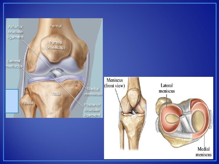
Bones • • Patella Femur Tibia Fibula

Cartilage • Fibrocartilage • Meniscus • Distribute impact and act as shock absorbers • Hyaline Cartilage • Lines articular surface of bone & provides a smooth surface for bones to glide

Bursa in the Knee • Bursae reduce friction and cushion pressure points between your bones and the tendons, muscles and skin near your joints.

Anterior Ligament • Anterior Cruciate Ligament, ACL • Protects against anterior tibial translation & posterior femoral translation • Connects the Femur & Tibia • Very important for knee stability

Posterior Ligament • Posterior Cruciate Ligament, PCL • Protects against posterior tibial translation & anterior femoral translation • Connects the Femur & Tibia • An isolated PCL tear doesn’t always require surgery to repair

Medial Ligament • Medial Collateral Ligament, MCL • Protects against Valgus force • Valgus force comes from the lateral side & transfers to the medial side • Connects the Femur & Tibia

Lateral Ligament • Lateral Collateral Ligament, LCL • Protects against Varus Force • Varus force comes from the medial side & transfers to the lateral side • Connects the Femur & Fibula • Usually damaged in combination with other structures which is called a posterior lateral corner injury

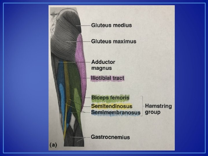
Motions & Muscles • External Tibial Rotation • Biceps Femoris • Internal Tibial Rotation • Popliteus • Semitendinosis • Semimembranosis • Sartorius • Gracilis

https: //www. youtube. com/watch? v=s. I 1 le. B 4 ke 3 U

Motions & Muscles • Flexion • Gracilis • Sartorius: Longest muscle in body, tailor’s muscle • Gastrocnemius: crosses knee • Popliteus • Plantaris • Hamstring Group • Biceps Femoris (1 -2) • Semitendinosis (3) • Semimembranosis (4)

Motions & Muscles • Extension • Quadriceps • • Vastus Medialis Vastus Intermedius Vastus Lateralis Rectus Femoris
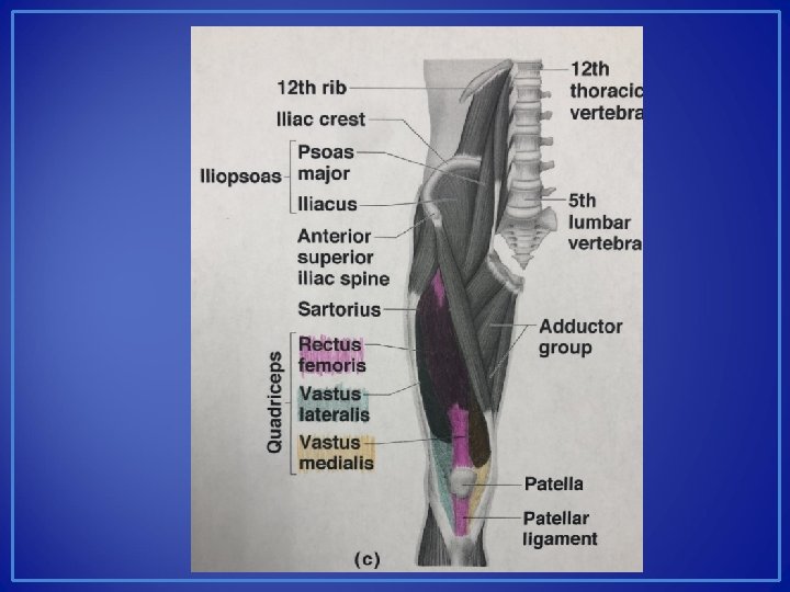

Muscles • Muscles of the Pes Anserine • Sartorius • Gracilis • Semitendinosis

Knee Posture

Knee Posture

MCL Sprain • Cause: Valgus Force or External Rotation • Signs & Symtoms: Stretching or tearing of the ligament. 3 grades of Sprain. Swelling, Bruising, Instability, loss of ROM & or function • Care: Depends on the severity of the sprain. PRICE, PT for strengthening & ROM, Brace. Rarely fixed with surgical intervention. Platelet-Rich Plasma injections could be an

LCL Sprain • Cause: Varus Force or internal rotation • Signs & Symptoms: Stretching or tearing of the ligament. 3 grades of Sprain. Swelling, Bruising, Instability, loss of ROM & or function. May see a deformity with an LCL Sprain • Care: Depends on the severity of the sprain. PRICE, PT for strengthening & ROM, Brace. Usually requires surgical intervention because it’s combined with other ligament injuries &

LCL Sprain MRI

ACL Sprain • Cause: Deceleration, rotation & valgus stress. Landing with knee in extension. Trauma • Signs & Symptoms: “POP”, swelling, pain, “feel like my knee is coming apart” • Care: Immediate care involves Ice, immobilization, Crutches. Surgery is required to repair this injury. Brace to return to play • Females are at more risk for tearing their ACL • This injury is often combined with injuries to other structures • ACL, MCL, & Medial Meniscus is AKA the Unhappy Triad

PCL Sprain • Cause: Hyperflexion with full weight on the anterior aspect of the knee. Posteriorly directed force to the front of a bent knee. Rotation. • Signs & Symptoms: ”POP” in the back of the knee, tenderness & relatively little swelling in the popliteal fossa • Care: PRICE, Non-operative Rehab. Surgical intervention may be required for Grade 3 Sprains, but is not always necessary. Brace to return to play. Likelihood of surgery increases if other structures are injured

Posterior Lateral Corner • Blow to the anterior medial knee in full extension • Involves: ACL, PCL, LCL • This injury may damage the Common Peroneal Nerve & Popliteal Artery • Biceps Femoris Tendon, Gatrocnemius Tendon & Popliteus Tendon attach at this site as well

Posterior Lateral Corner: LCL, ACL, PCL. No cartilage injury, vascular injury or nerve injury

Makenzie Milton: disruption Posterior Lateral Corner with vascular

Nick Chubb – PCL, LCL, MCL

Zach Miller: disruption Posterior Lateral Corner with vascular

Zach Miller “Do not cut my leg off” https: //www. youtube. com/watch? v=5 Qi. Q 4 NTgsk. Q

Injuries • Cause: Weight bearing combined with rotational forces while flexing or extending • Medial Meniscus has a higher rate of injury • Signs & Symptoms: Joint effusion (swelling) that develops gradually, joint line pain, loss of motion, locking or clicking, giving way, pain with squats • Care: PRICE, MRI to diagnose, Surgery to repair, Physical Therapy to rehabilitate after surgery. Non-emergent injury.

Arthroscopic Surgery

Meniscus MRI & Arthroscopy https: //www. youtube. com/watch? v=LZyw. M 9 -r. EZY https: //www. youtube. com/watch? v=jgihy. ENo. N 4 w

Joint Contusion • Also known as: Bone Bruise or Marrow Edema • Cause: bones hitting each other, trauma from dislocation or outside force • Signs & Symptoms: Pain with weight bearing, swelling, joint effusion • Care: Non-weight bearing, limit activity, ice, brace may be necessary if marrow edema is combined with damage to other structures

Loose Bodies • Cause: Osetochondral defect, damage to joint cartilage, ligaments, synovial tissue • AKA Joint Mice • Signs & Symptoms: Pain, mechanical deficits, limited range of motion, instability, giving way • Care: Surgery to remove the loose bodies & clean up joint damage • If left, the loose body can cause joint degeneration, Damaging articular surfaces further

Chondromalacia • Cause: softening and deterioration of the articular cartilage. Most common in the patella. May be caused by malalignment or repeated trauma • Signs & Symptoms: Crepitus, grinding or grating sensation, pain in anterior knee, pain with squatting or stairs, swelling • Care: avoid irritating activities, PT, brace, surgery https: //www. youtube. com/watch? v=XSi 7 -h. R 55 l. I

Acute Patellar Subluxation or Dislocation • Cause: Planting foot, deceleration, cutting, thigh rotates internally & lower leg rotates externally. 98% of all patellar dislocations are lateral. • Signs & Symptoms: Deformity, pain, swelling, loss of function. • Care: Immobilize to transport, ice, refer for x-ray & reduction. Suspect a fracture if it is a first time dislocation or subluxation. After reduction athlete is immobilized in brace for 4 weeks, use crutches, and begins PT. Focus on Vastus Medialis strengthening. Brace to return to play

Patellar Dislocation Brace

Patellar Dislocation Bracing https: //www. youtube. com/watch? v=98_bc. HZPl. Uo https: //www. youtube. com/watch? v=x. Fb. Cjdto 0 VQ

Fractured Patella • Cause: Direct or Indirect Trauma • Signs & Symptoms: Swelling, Bruising, Loss of Function, Inability to bear weight, deformity may be present • Care: Refer to physician for x-ray, Immobilization for 2 -3 months, Surgery may be required

Patellar Tendonitis • Cause: Overuse. Jumping, Kicking, Running • AKA: Jumper’s Knee • Signs & Symptoms: vague pain & tenderness around the infrapatellar tendon, swelling • Care: PRICE, brace or tape, US, PT

Bursitis • Cause: Overuse. Inflammation of the bursa. Can be acute, chronic, or recurrent. • Signs & Symptoms: Localized swelling, Redness, Increased skin temperature. • Care: Reducing irritation, REST, decrease inflammation with antiinflammatory medication, Ultrasound or Ice. • Most common in the pes

Iliotibial Band Friction Syndrome • Cause: Overuse injury. Common in runners or cyclists. • AKA: Runner’s Knee • Signs & Symptoms: Pain over the lateral femoral condyle, tenderness, mild swelling. • Care: Ice, Rest, antiinflammatory medication, US. Brace, stretching, proper warm-up & cool down when return to play. https: //www. youtube. com/watch? v=uxvd 3 O 7_5 qg

Iliotibial Band Stretching

Osgood Schlatter Disease • Cause: stress on the tibial tuberosity growth plate in adolescent athletes • Signs & Symptoms: pain at front of knee, swelling, severe pain with kneeling, jumping, & running. Point tenderness & bony growth over tibial tuberosity • Care: reduce activity, tape, ice after activity,

- Slides: 46