KNEE JOINT Dr Gitanjali Khorwal The knee joint


 for passage of TENDON OF POPLITEUS")
")


 2.")


 Angle between long axis of thigh and leg is reduced")

- Slides: 42
KNEE JOINT Dr. Gitanjali Khorwal
The knee joint is formed by the condyles of femur & tibia, & posterior articular surface of the patella. It is a compound & complex synovial joint.
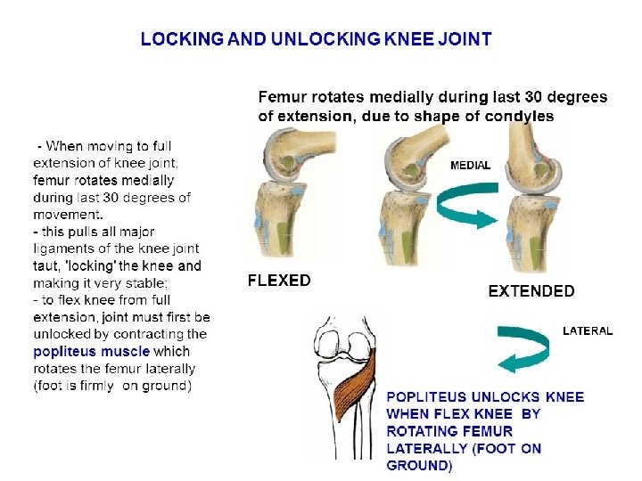
Functionally, the knee joint is a condylar & modified hinge joint. 1. Transverse axis of movement is not fixed, & moves forward during extension & translates backward in flexion; 2. Along with extension & flexion, there is a conjunct rotation of femur on tibia(or vice versa) around a more or less vertical axis.
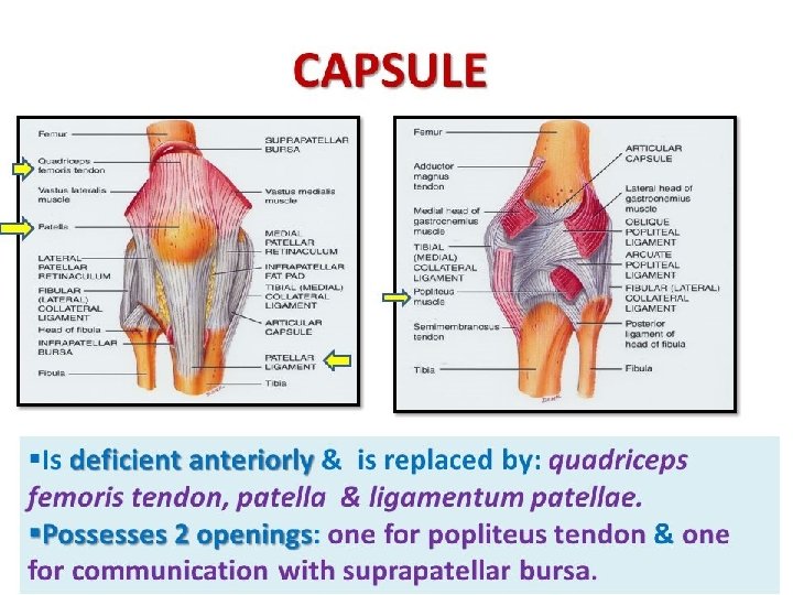
BONES FORMING THE JOINT: 1. CONDYLES OF FEMUR 2. CONDYLES OF TIBIA 3. ARTICULAR SURFACE OF PATELLA
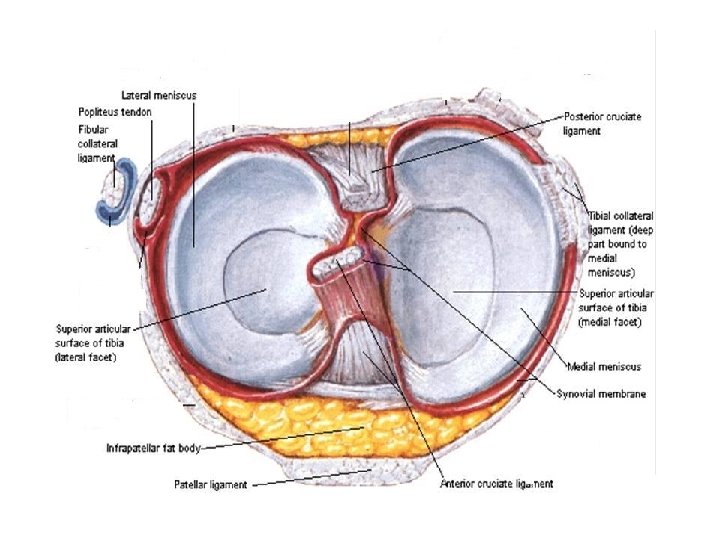
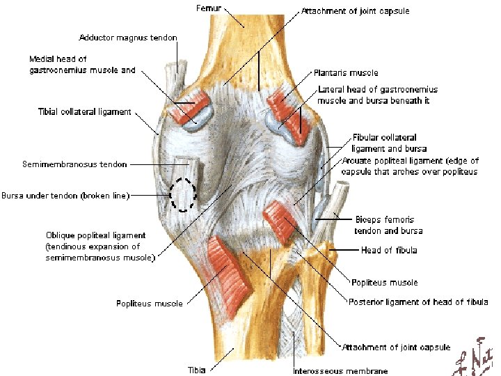
LIGAMENTS of KNEE JOINT 1. Capsular ligament 2. Synovial membrane 3. Ligamentum patellae 4. Tibial collateral ligament 5. Fibular collateral ligament 6. Oblique popliteal ligament 7. Arcuate popliteal ligament 8. Medial & lateral menisci
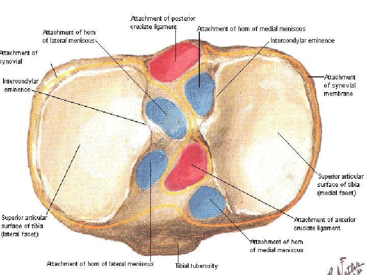
9. Anterior & posterior cruciate ligments. 10. Coronary ligament 11. Transverse ligament. 12. Menisco - femoral ligaments.
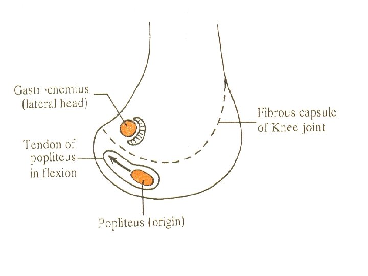
Proximal attachment of capsule
Posteriorly (1 gap ) for passage of TENDON OF POPLITEUS
Distal attachment of capsule Anteriorly (1 gap)
REFLECTION OF SYNOVIAL MEMBRANE
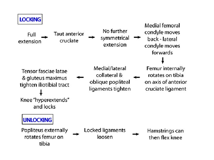
LIGAMENTUM PATELLAE It is derived from the tendon of quadriceps femoris & extends from the apex of patella to the upper part of the tubercle of tibia. When the knee joint is locked at the end of extension , all ligaments are taut except the ligamentum patellae.
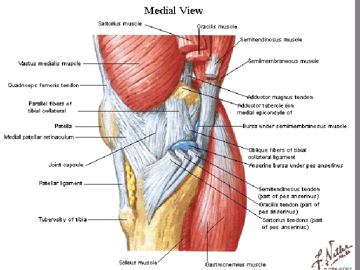
TIBIAL COLLATERAL LIGAMENT The ligament consist of superficial & deep part. Both part are attached above to the medial epicondyle of femur. The superficial part extends downward & forward as a flattened band & is attached to the medial condyle & upper part of medial border of shaft of tibia along a rough strip of bone.
FIBULAR COLLATERAL LIGAMENT It extends from lateral epicondyle of femur to the head of fibula close to its styloid process.
OBLIQUE POPLITEAL LIGAMENT It is an expansion derived from the insertion of semimembranosus & extend from the posterior surface of medial condyle of tibia to the lateral part of intercondylar line of femur.
ARCUATE LIGAMENT It forms a Y-shaped band. The stem is fixed to the styloid process of head of fibula. The posterior band is attached to the lateral condyle of tibia & anterior band is attached to the lateral condyle of femur.
CRUCIATE LIGAMENT The anterior & posterior cruciate ligaments are intracapsular but extrasynovial. They are named as anterior & posterior according to their tibial attachment.
CORONARY LIGAMENT: These are parts of capsule that provide attachment to peripheral margins of medial and lateral menisci. TRANSVERSE LIGAMENT: It connect the anterior horn of medial meniscus to the anterior margin of lateral meniscus.
MEDIAL & LATERAL MENISCI They are composed of fibro-cartilage. Each menisci divides the condylar part of the joint into meniscofemoral & menisco-tibial compartments. Function of the menisci 1. They increase the concavity of tibial condyles for the better adaptation with femoral condyles. 2. They maintain a potential joint space for flushing of synovial fluid to provide nutrition to the articular cartilage. 3. Menisci act as shock absorber. 4. Menisci help in the complex mechanism of gliding & angular movements. 5. Peripheral attached part of the menisci is vascular & gets nutrition from the capsular arteries.
MENISCO-FEMORAL LIGAMENT The posterior horn of lateral meniscus is connected to the medial condyle of femur by anterior & posterior menisco-femoral ligaments.
BURSAE AROUND KNEE JOINT Anteriorly 1. Subcutaneous pre - patellar bursa. (inflammation causes House maid knee). 2. Subcutaneous infra- patellar bursa ( inflammation causes Clergy-mans knee) 3. Deep infra-patellar bursa 4. Supra-patellar bursa (Communicates with the joint cavity. )
BURSA AROUND KNEE JOINT
Laterally : • Between lateral head of gastrocnemius & the capsule • Between tendon of biceps femoris & fibular collateral ligament. • Between fibular collateral ligament & tendon of popliteus muscle • Between tendon of popliteus & lateral condyle of tibia (communicates with joint cavity).
Medially 1. Medial head of gastrocnemius & the capsule (communicates with joint cavity) 2. Superficial part of tibial collateral igament sartorius, gracilis &semitendinosus. 3. Superficial & deep part of tibial collateral ligament. 4. Semi-membranosus & medial condyle of tibia (may communicate with joint cavity)
RELATIONS OF KNEE JOINT Anteriorly- Quadriceps femoris Antero-medially- medial patellar retinaculum Antero-laterally- lateral patellar retinaculam & iliotibial tract Postero-medially- Sartorius, gracilis, semimembranosus & semitendinosus Postero-laterally- Tendon of biceps femoris & common peroneal nerve Posteriorly- popliteal vessels, tibial nerve, both head of gastrocnemius & plantaris
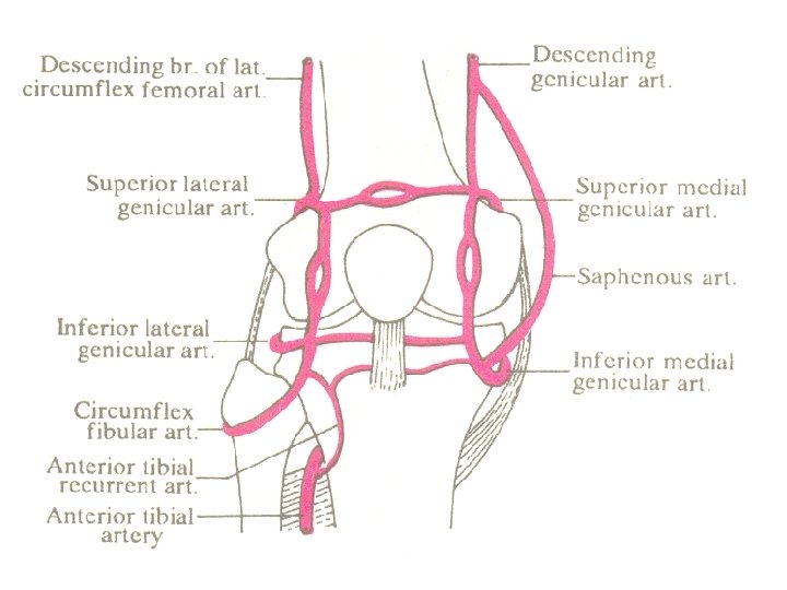
ARTERIAL SUPPLY 1. Descending genicular branch of femoral artery. 2. Descending branch of lateral circumflex femoral artery 3. Genicular branches of popliteal artery. 4. Recurrent branches of anterior tibial artery. 5. Circumflex fibular branch of posterior tibial artery.
NERVE SUPPLY • From Femoral nerve – • nerves to the vasti. • 3 from the Tibial nerve – • superior medial Genicular • inferior medial genicular • middle genicular nerve. • 3 from the Common peroneal nerve • superior lateral Genicular • inferior lateral genicular • recurrent genicular nerve
MUSCLES PRODUCING THE MOVEMENTS: Active movements performed at the knee joint areq. Extension or straightening continues until leg & thigh are in the same vertical lines. Extension is produced by Quadriceps femoris & assisted by tensor fascia latae. q. Flexion –prime movers: semimembranosus, semitendinosus, biceps femoris; initiated by, popliteus Assisted by, sartorius, gracilis, both head of gastrocnemius & plantaris. q Medial rotation- by semimembranosus, semitendinosus, popliteus, sartorius & gracilis. q Lateral rotation-biceps femoris.
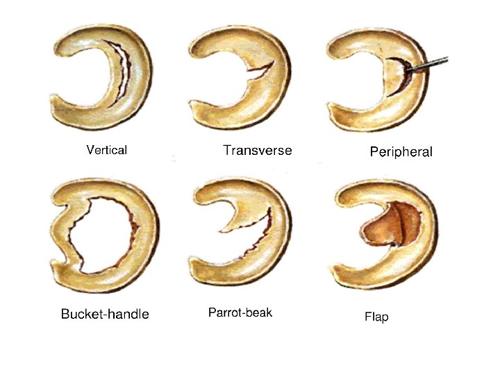
Applied Anatomy Injuries to menisci q. Most injured structures of the knee are Tibial collateral ligament, the medial menisci & ACL o A blow to lateral aspect of the knee when the foot is on the ground may sprain the tibial collateral ligament, the attached medial menisci may also be torn. o. ACL tears may occur when the tibial collateral ligament & medial menisci are injured, a blow to the anterior aspect of the flexed knee may tear only the ACL. q Injuries to the menisci commonly occur in the flexed knee as a result of forcible rotation or abduction. The medial meniscus suffers more frequently than the lateral.
• Rupture of cruciate ligaments is less common. Anterior ligament is more commonly affected. • If both cruciate ligaments are injured , excessive forward & backward gliding of tibia , abduction & adduction of joint occur. • Fractureof patella is a frequent problem. Does not heal as it is a sesamoid bone. Wiring has to be done. • Acute traumatic synovitis. Joint cavity is distended with fluid. swelling is above and on the sides of patella.
. Inflammation of bursa. Housemaids knee - Subcutaneous pre-patellar bursa. Clergyman's knee - Subcutaneous infra patellarbursa. . Osteoarthritis of knee joint.
Genu valgum (Knock knees) Angle between long axis of thigh and leg is reduced laterally Genu varus ( bow legs) Angle between long axis of thigh and leg is increased laterally.
• Total knee replacement or knee joint arthroplasty done in severe destructive arthritis of the joint.