Anatomy and histology of the skin Skin the


; § stratum spinosum (Squamous")

")


, squamous (black), granulous (red), lucidum")


: §")


,")

 § is a form of terminal differentiation of the")
 § degenerative phenomena: of nucleus, cellular organelles, cell envelope,")
 § each of the keratins is the product of")


 α, β, γ:")
 radiation § the spectrum of UV radiation: § UVC (200 -290")


 Cellular components of SIS § lymphocytes: T lymphocytes (T helper/")
 Molecular components of SIS § antigens and haptens; § antibodies")


: brown in color; ex.")

 or plaque (10")


, by")


- Slides: 116
Anatomy and histology of the skin
Skin – the more large organ in the body § surface – 1. 5 -1. 8 sqm; § thickness: § 0. 2 -0. 5 mm (eyelid, prepuce); § 3 -5 mm (palms and soles). § weight: § 4 -5 kg; § 20 kg with hypodermis. § glabrous skin – palms and soles; § hair bearing skin. Organization of the human skin: § the skin is composed of four distinct layers: § the epidermis; § the dermo-epidermal junction; § the dermis; § the hypodermis.
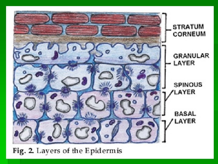
Histology of the epidermis § stratum germinativum (Basal cell layer); § stratum spinosum (Squamous cell layer); viable epidermis; § stratum granulosum (Granular cell layer); § stratum lucidum; § stratum corneum (Horny layer) – nonviable epidermis. Keratinocytes Dendritic cells
Basal Cell Layer § a single range of columnar cells, with basophilic cytoplasm, dark-staining oval or elongated nucleus, many ribosomes, tonofibrils (keratin filaments) into fine bundles, melanin granules (transferred from melanocytes); § the cells are connected with each other by desmosomes (symmetrical, laminated structures) and with basement membrane by hemidesmosomes.
Basal cell layer (glutaraldehide- osmium fixation)
Squamous Cell Layer § 5 -20 ranges of polygonal cells with long axis parallel to the skin surface, eosinophilic cytoplasm, clear rounded nucleus, large bundles of keratin filaments, attached to desmosomes; § the new organelles in the upper spinous cells → lamellar granules (keratinosomes, Odland bodies) – they contain polysaccharides, free sterols, lipids, hydrolytic enzymes; their contents are discharged into the intercellular space at interface with granular layer.
Squamous cell layer
Granular Cell Layer § 3 -10 ranges of diamond-shaped or flattened cells, with horizontal long axis, cytoplasm filled with basophilic kertohyaline granules (irregular in size and shape, with particulate substructure); § the dissolution of the nucleus and cell organelles is initiated; keratin filaments in large bundles; § keratinosomes migrate towards the periphery of the cells and discharge their lipid components.
Horny Layer § 4 -10 ranges of eosinophilic, flattened cells, arranged in vertical stacks, that have lost nuclei and cytoplasmic organelles; § keratin filaments are aggregated in macrofibres under the influence of filaggrin; § highly insoluble cornified envelope within the plasma membrane, that contain involucrin, loricrin, keratolinin; § the loss of desmosomes.
Stratum lucidum § an additional homogenous eosinophilic zone between granullar cell layer and horny layer, in palmo-plantar skin; § contains cells with dense cytoplasm, opaque membranes, degenerated nucleus, without organelles; § keratin filaments are immersed in a matrix of eleidine.
Acral skin – layers of the epidermis: basal (green), squamous (black), granulous (red), lucidum (blue), horny (yellow). Blood vessels (arrows from dermis).
Normal skin
Acral skin
Epidermis Langerhans Cells Keratinocyte Melanin granules Melanocyte Dermis
Dendritic Cells of the Epidermis - Melanocytes § dendritic cells localized between the basal cells; § in section stained with hematoxylin-eosin-clear cells, with small, dark-staining nucleus, clear cytoplasm; § the dendritic processes recognized with dopa reaction, staining with silver nitrate; § in electron microscopy differ from keratinocytes by lack of tonofilaments or desmosomes; § melanocytes contain melanosomes, specialized organelles that synthesize melanin, that is transferred to keratinocytes (apocopation); § the evolution of melanosomes in 4 stages: round, ellipsoid, partially melanized, completely melanized.
Melanocyte
Langerhans Cells § dendritic cells of mesenchimal origin, localized in suprabasal epidermis, dopa negative; § in sections stained with hematoxylin-eosin-clear cells; § histochemical stains (adenosine phosphatase, aminopeptidase) – for identifying and differentiating from melanocytes alfa D manositase; staining with gold chloride; § demonstrated with monoclonal antibody OKT 6; by immunoperoxidase technique or immunofluorescence;
Langerhans Cell
Langerhans Cells § in electron microscopy – relatively clear cytoplasm, lobulated nucleus, well developed endoplasmic reticulum, Golgi complex and lysososmes; lack of tonofilaments and desmosomes, presence of Bierbeck granules; recognized by monoclonal antibody anti-Lag (Langerhans-associated granule); § Langerhans cells express on their surface HLA-DR, DP and DQ antigens, T 6 antigen CD 1 (CD 1 a, CD 1 c), CD 4 (HIV receptor), CD 14, CD 33, VLA (very late antigen), adhesion molecules et al. and have receptors for Fc (Fcγ and Fcε) of Ig. G and Ig. E and C 3 b, C 4 d, CD 23 (Ig. E receptor in atopic dermatitis), DEC 205 multilectine etc. A new identified lectine (Langerin) is a endocitic receptor.
Merkel Cells § dendritic cells localised between the basal cells, directly above the basement membrane; § in light microscopy can be recognized in silver impregnated sections; § in electron microscopy – lobulated nucleus, clear cytoplasm, electron-dense granules, strands of filaments, occasional desmosomes; unmyelinated nerve endings are associated with Merkel cells.
Indeterminate Cells § have ultrastructural features of Langerhans cells but lack Birbeck granule; § react specifically with the monoclonal antibody OKT 6.
Histology of the dermo-epidermal junction § the indeterminate filaments of basal keratinocytes; § the basal plasma membrane of keratinocytes, melanocytes and Merkel cells; § hemidesmosomes: focal thickenings of the basal plasma membrane of keratinocytes, with an intercellular component (the attachement plaque) and an extracellular component (sub-basal dense plate); § the basement membrane with 3 layers: lamina lucida, lamina densa and lamina fibroreticularis; § anchoring filaments: very fine, vertically orientated structures that connect hemidesmosomes at lamina densa; § anchoring fibrils: short curved structures that insert into the lamina densa and extend into the dermis and insert into anchoring plaques or curve back in lamina densa; § anchoring plaques: amorphous bodies in the superficial dermis; § the elastic microfibrils, that extends into the dermis and may enmesh with dermal elastic fibres.
Histology of the dermis § Layers of the dermis: § papillary dermis (the uppermost part); § reticular dermis (subpapillary layer). § Components of the dermis: § cells; § fibres; § ground substance.
Cells of the dermis: § § § fibroblast - a cell at an early stage of differentiation; fibrocyte – a cell fully differentiated; mesenchymal origin; § small and spindle shaped (S-cell) or larger, flatter, amoeboid (A-cell); § produce collagen and elastine.
Cells of the dermis: § mast cell: originate from haematopoietic bone marrow § ovoid or spindle shape, mono or binuclear, with round acidophilic cytoplasmic granules, which stain metachromatically with toluidine blue or Giemsa; § localized around blood vessels, nerves and appendages, in subpapillary dermis;
Cells of the dermis: § macrophage: originate from bone marrow, is larger than monocyte, with clear cytoplasm, lighty staining elongated nucleus, well developed rough endoplasmic reticulum and Golgi apparatus, intermediate filaments; § is a general scavenger implicated in the ingestion and killing of micro-organisms and the degradation of foreign substances; § secrete several enzymes; § possess numerous receptors.
Fibers of the dermis: § collagen fibers: § in light microscopy: bands with faint longitudinal striations; in dark field, each fiber is a bundle of parallel fibrils; § in E. M. : fibrils with cross striations; § structure of collagen: 3 polypeptide chains, coiled in a triple helix (helical domain) and a globular domain at each end (amino-N-terminal domain and carboxyterminal domain). § reticulin fibers: § a special type of thin collagen fibers; § in light microscopy, with silver nitrate stains black; § in E. M. : fibrils separated by an interfibrilar substance.
Fibers of the dermis: § elastic fibers: § in light microscopy, with orcein or resorcin-fuchsin, thin bundles, that in the papillary dermis form an intermediate plexus of elaunin fibers running parallel to the dermal-epidermal junction; oxytalan fibers, perpendicular to the dermal-epidermal junction;
Ground substance § an amorphous substance: contains proteoglycans, glycosaminoglycans, electrolytes, water; § the staining methods of the ground substance: § the production of metachromasia; § colloidal iron; § Alcian blue, ruthenium red; § in electron microscopy: flocculent and filamentous material.
Histology of the hypodermis § adipocytes, organized into lobules defined by septa of fibrous connective tissue with blood vessels and nerves.
Skin and its appendages
Histology of the skin appendages Eccrine sweat gland § 2 -4 million, distributed over nearly entire body surface; most numerous on the palms, soles and in the axillae; § two segments: § the secretory coil; the duct. § the secretory coil: 3 cell types: § clear (secretory) – pyramidal, with abundent mitochondria, a lipofuscin cell granule; § dark (mucoid) – cuboidal or pyramidal, with dark granules; § myoepithelial cells; § the duct: § in the dermis: 2 layers: basal cells (outer ring), luminal cells (inner ring); membrane; § in the epidermis: 1 layer – luminal cells; § opens directly onto the skin surface.
Eccrine sweat gland
Histology of the skin appendages Apocrine sweat gland § limited to the axillae, nipples, perineum and genitalia, groin, periumbilical area; § larger than eccrine gland; § opens in the folliculary duct. Apoeccrine sweat gland § 50% of axillary glands in the adults with axillary hyperhydrosis; § a long duct which opens directly into skin surface.
Apocrine sweat gland
Histology of the skin appendages Sebaceous gland § distributed in large numbers in the face, scalp, chest, upper back; § associated with hair follicles; § microscopically: multilobed, with cells with small dark nucleus and a foamy cytoplasm; § in E. M. : peripheral cells with tonofilaments and few lipid droplets; central cells full of lipids; excretory duct with stratified squamous epithelium.
Sebaceous gland
Sebaceous gland
Histology of the skin appendages Hair: § distributed on all areas of the skin, except the palms, soles and portions of the genitalia; § types of hair: § lanugo hairs: fine, long hairs covering the fetus; § vellus hairs: fine, short, unmedullated; § indeterminate scalp hairs: under 1 cm in length; fine, hypopigmented (between 11 and 16 years); § terminal hairs: longer than 1 cm, coarse medullated, pigmented (in adults on the scalp, eyebrows, eyelashes, beard, axillae, pubic region);
Skin appendages
Histology of the skin appendages Hair: § in a longitudinal section: § the lower portion (hair bulb): § dermal hair papilla with capillary loops; § hair matrix; § hair; § inner root sheath and outer root sheat. § the middle portion (isthmus) – from the attachment of the arrector pili muscle to the entrance of sebaceous duct; § the upper portion (infundibulum) – above the entrance of the sebaceous duct;
Histology of the skin appendages Hair: § the hair shaft (in horizontal section): § medulla; § cortex; inward to outward. § hair cuticle. § the inner root sheath (3 concentric layers): § inner root sheath cuticle; § Huxley layer; § Henle layer. § the outer root sheath – a downward extension of the epidermis; § vitreous layer; § fibrous root sheath.
Histology of the skin appendages Nail: § the matrix: underlies the proximal nail fold but some extends under the nail plate; produce nail plate; § the nail bed: an epithelium that produces a minimal amount of keratin, adherent to the bottom of the nail plate; § the nail plate: grow out from the nail matrix; rests on the nail bed; contain hard keratin; normally – thin and translucent; § the nail folds: 1 proximal, 2 laterals; § the hyponychium: extends from the nail bed to the distal groove; § cuticle: extension of dorsal epidermal stratum corneum on the nail plate; seals proximal nail fold.
Cutaneous vasculature Blood vessels: § vascular network: vertically distributing and collecting vessels and horizontal plexuses; § the subcutaneous plexus: small branches from musculocutaneous arteries that penetrate the subcutaneous fat; § the subpapillary plexus: arterioles that ascend into the dermis and form an horizontal arteriolar plexus; § the superficial plexus (papillary plexus): arterioles became capillary loops in the dermal papillae; § the layers of the vessels: intima (endothelial cells), internal elastic lamina, media (muscle cells); adventitia (connective tissue); § the glomus: a special arterio-venous shunt located within the reticular dermis in certain areas (the pads and nail beds of the fingers and toes, the ears, in the center of the face).
Cutaneous vasculature Cutaneous lymphatics: § begin with a blind-ended capillary in the dermal papilla; § a superficial lymphatic plexus in the papillary dermis; postcapillary lymph vessels; § two deeper horizontal plexuses.
Nerves and receptors of the skin § the nerve network of the skin contain somatic sensory and sympathetic autonomic fibers; § the sensory fibers alone (free nerve endings) or in association with specialized structures (corpuscular receptors) are receptors of touch, pressure, itch, pain, temperature; § sympathetic motor fibers innervate the sweat glands, vascular smooth muscle, the arrector pili muscle, sebaceous glands; § the free nerve endings (including those associated with Merkel cells) – the most widespread in the skin;
Nerves and receptors of the skin § the corpuscular receptors (capsule and inner core), contain neural and nonneural components: § Meissner corpuscle: elongated or ovoid mechanoreceptor located in the dermal papillae of digital skin; § mucocutaneous end organs: located in the lips, eyelid, perianal canal, glans, clitoris; § Vater-Pacini corpuscle: ovoid or flattened sphere; lies in the deep dermis and hypodermis in weight-bearing surfaces, nipple, anogenital region; § Krause corpuscle: irregular round; capsule, fibrillar structure; § Ruffini corpuscle: amielinic fibers, thin capsule.
Physiology of the skin
Process of keratinization (keratin synthesis) § is a form of terminal differentiation of the keratinocytes; § the cytoskeleton of all mammalian cells is composed of three filament systems: § microfilaments (6 nm); § intermediate filaments (8 -10 nm); § microtubules (25 nm); § the keratins are members of the intermediate filaments; § the process of keratinization: § degenerative phenomena; § synthetic processes;
Process of keratinization (keratin synthesis) § degenerative phenomena: of nucleus, cellular organelles, cell envelope, desmosomes; § synthetic processes: § keratin intermediate filament formation; § synthesis of keratinosomes; § cell envelope (CE) formation – cornified envelope; § keratohyaline granules proteins synthesis; § keratins family: 20 epithelial keratins, 10 hair keratins; type I (acidic), type II (neutral-basic); molecular masse between 40 and 68 k. Da);
Process of keratinization (keratin synthesis) § each of the keratins is the product of a unique gene; § the keratins are expressed as pairs with one member of each subfamily; § soft keratinization (skin); § hard keratinization (hair, nail, lingual papillae). The control of keratinization: § factors of activation: oestrogens, corticosteroids, timic extracts, mechanical pression, ultraviolet radiation; § factors of inhibition: vitamin A, thyroid hormones.
Melanogenesis Cells implicated – melanocytes - Epidermal melanin unit: a melanocyte and 36 keratinocytes to which the pigment is transferred; - the keratinocytes phagocytize the melanosome – laden dendritic tips of the melanocyte (apocopation); - there is no differences in the number of melanocytes per square millimeter of the skin on the same anatomic region of black and white patients; - a difference in melanocyte density for various regions of the body: the lowest number on the trunk and arm, the highest on the face; - the major difference is in the activity of individual melanocytes; in black skin, larger, more dendritic, with greater quantity of melanin, than in white skin.
Melanogenesis Melanosomes: § highly organized organelles that contain: § structural matrix proteins; § tyrosinase; § several proteins of unknown structure; § 4 stages (I-IV) in melanosome development: § spherical: contains filaments with a distinct periodicity; § ellipsoidal: oval in shape, with numerous parallel longitudinal fillaments, no melanin depostion; § partially melanized: the internal structure partially obscured by the deposition of melanin pigment; § completely melanized: melanin deposited throughout the structure;
Melanogenesis § the melanosomes pass from the perinuclear area of the melanocyte to the dendrites and progress from stage I to stage IV; § the relationship between skin color and melanosomes: § in black skin they are larger, individually dispersed in the keratinocytes; § in white skin they are smaller, aggregated into groups of two or three; § the degradation of melanosomes in keratinocytes by lisosomal enzymes with a faster rate in white skin and slowly in black skin.
Melanogenesis Melanin synthesis The classic Mason-Raper pathway: tyrosinase tyrosine → → dopa quinone → leucodopachrome conversion factor tyrosinase → dopachrome → → 5, 6 – dihydroxyindole → → indole-5, 6 - quinone → → melanochrome → melanin § Dopachome oxyreductase (dopachrome tautomerase) – other regulatory factor. § Forms of melanin: § eumelanin – brown to black in color, with Mason-Raper pathway of synthesis; § phaeomelanin – yelow-red in color, with another pathway of synthesis; § mixed type of melanins.
Melanogenesis The control of melanogenesis Melanocyte-stimulating hormones (MSH, melanotropins) α, β, γ:
Melanogenesis Ultraviolet (UV) radiation § the spectrum of UV radiation: § UVC (200 -290 nm); § UVB (290 -320 nm); § UVA (320 -400 nm). § immediate pigmentary darkening: within minutes of sun exposure, fades within 6 -8 hours; produced by UVA and visible light, as a result of an oxidation of persisting melanin or its precursors; § delayed tanning: is apparent within 48 -72 hours, produced by UVB and UVA, as a result of new melanin production. Other factors of stimulation: estrogens, estrogens and progesterone, prostaglandins E 2 and D 2, copper, nitrogen mustard, ACTH, PUVA; Factors that inhibit melanogenesis: vitamins B 1 and C, silver, hydroquinone.
Physiology of skin appendages Hair § Hair growth rate: 0. 35 -0. 37 mm/ day § Hair cycle: regular cycles of growth and shedding; 3 phases of follicular activity: § anagen: the growth stage (3 -5 years on the scalp); § catagen: the degenerative stage, with conversion from active growth to the resting phase (few days on the scalp); § telogen: the resting stage (3 -4 month on the scalp); § 100 hairs may be shed from the scalp every day
Physiology of skin appendages Hair: § Regulation of the hair growth: § genetic influences for normal growth of hairs on different anatomic locations; § hormonal influences: § androgens “upregulate” the pubic hair, axillary hair, beard hair and “downregulate” genetically predisposed scalp hair follicles in andro-genetic alopecia; § corticosteroids, the ACTH, thyroid hormones, somatothrope hormone stimulate the hair growth; § estrogens stimulate indirectly the axillary and pubic hair; § other factors: UV and infrared radiation stimulate the hair growth; Bi, cytostatics, inhibits the hair growth.
Physiology of skin appendages Nail § the average rate of growth of the thumbnail: 0. 10 -0. 12 mm/ day; finger nails: 0. 5 -1. 2 mm/ week; § fingernails grow faster than toenails; § the hard keratin of the nail plate is formed in the nail matrix; § functions of the nail: § strength and protection for the terminal phalanx; § help with fine touch; § handling of small objects; § the esthetic role.
Physiology of skin appendages Eccrine sweat gland § type of secretion: merocrine (the integrity of the secretory cell after secretion); § the responsiveness of the sweat gland to cholinergic agents, α and β adrenergic stimulants and other neurotransmiters (VIP, ATP); § periglandular acetylcholine – the major stimulant of eccrine sweat secretion; § sweat formation in 2 steps: § secretion of primary fluid with nearly isotonic Na. Cl concentrations by the secretory coil; § reabsorption of Na. Cl from the primary fluid by the duct under the influence of aldosterone and antidiuretic hormone; sweat becomes hypotonic.
Physiology of skin appendages Eccrine sweat gland § composition of eccrine sweat: § inorganic ions: Na, Cl, K, Ca, P, Mg, I, HCO 3, Fe, Zn et al. ; § organic compounds: urea, ammonia, amino acids, proteins, lactate, glucose, piruvate, histamine, bradikinine, immunoglobulins, prostaglandins, vitamine C, B 2, B 6, K, amphetamine-like substances, proteolytic enzymes, et. al. ; § p. H between 4. 0 and 6. 8; § control of eccrine sweat secretion: § thermal factors: sweating all over the body, especially the chest, back, forehead, scalp and axillae; § physic exercise: sweating mainly on the palms, soles and axillae.
Physiology of skin appendages Apocrine sweat gland § type of secretion: holomerocrine or decapitation secretion (the luminal part of the cell is lost); § composition of secretion: apocrine sweat is milky, viscid; the chemical composition is little known because of difficulties in collecting a pure secretion (mixed with sebum and eccrine sweat); § control of secretion: § emotional factors (only after puberty) the secretion can be stimulated by epinephrine or norepinephrine; adrenergic-neuron blocking agents and adrenergicreceptor blocking drugs inhibit apocrine sweating; § hormonal factors: the development of the apocrine glands is dependent on the sex hormones and not the maintenance of their functional activity.
Physiology of skin appendages Sebaceous gland § type of secretion: holocrine (entire sebaceous cell break down to form the secretion); § composition of sebum: squalene, wax esters, triglycerides, fatty acids, cholesterol esters; § control of sebum secretion: § androgenic hormones stimulate sebaceous gland, especially dihydrotestosterone; § exogenous estrogens lower the sebum secretion when they are administrated in a great quantity; § fasting decrease the sebum secretion; § exogenous retinoids supress the sebum secretion by inhibiting sebaceous cells; § damage to the epidermis inhibits the sebaceous glands underneath the wound.
Protective functions of the skin § protection against the loss of essential body fluids: the role of epidermis and horny layer; § protection against the entrance of toxic agents: stratum corneum restricts the absorption of selected classes of molecules and serves as a reservoir for exogenous toxic and nontoxic agents; § protection against the entrance of microorganisms: § the structural integrity of horny layer; § the desquamation: remove adherent microorganisms; § sebaceous lipids: antibacterial effects; § biochemical defenses: the acid environment (p. H of 5 to 6. 5), cytokines, prostaglandins, antioxidants, enzyme systems; § Langerhans cells and T lymphocytes;
Protective functions of the skin § protection against damage from UV radiation: § the stratum corneum; § melanin barrier; § protection against damage from mechanical agents: § stratum corneum; § dermis and hypodermis; § protection against environmental temperature: the role of the skin in the maintenance of a constant body temperature: § the effector role of the skin in thermoregulation: skin thermoreceptors, skin vasculature, sweat gland responses with heat transfer from the skin to the environment by evaporation of the sweat; § protection against damage from low-voltage electric current: the stratum corneum.
Skin immune system (SIS) Cellular components of SIS § lymphocytes: T lymphocytes (T helper/ inducer cells, T supressor cells, T cytotoxic cells) B lymphocytes; § null cells (non. T, non. B): NK (natural killer) and K (killer) cells; § Langerhans cells and other dendritic cells; § keratinocytes; § cells of the monocyte/ macrophage system: monocytes, macrophages; § granulocytes (neutrophils, eosinophils, basophils); § mast cells.
Skin immune system (SIS) Molecular components of SIS § antigens and haptens; § antibodies (immunoglobulins); § complement; § cytokines; § adhesion molecules; § histocompatibility antigens (HLA).
Sensory function § the capacity of the skin to detect changes in the surrounding environment and transmit these informations to central nervous system for processing; § sensory fibers may contain one or various neuropeptides that may act as mediators of inflammation.
Basic pathologic reactions of the skin
Pathologic reactions in the epidermis Disturbances of epidermal cell kinetics Acanthosis: a broadening of the stratum spinosum by an increase of cell population. Granulosis: a thickening of the granular cell layer. Hyperkeratosis: a thickening of the horny layer by an increased production or a reduced desquamation of corneocytes. Atrophy of the epidermis: a diminution of the germinative cell volume and a flattening of the rete ridges.
Pathologic reactions in the epidermis Disturbances of epidermal cell differentiation Parakeratosis: a falty and accelerated cornification leads to a retention of pyknotic nuclei of epidermal cells; the stratum granulosum is rudimentary or may not be present and the horny layer contain pyknotic nuclei and other organelles in a dense cytoplasmic net of keratin fibrils. Diskeratosis: a premature cornification of individual cells within the viable layers of the epidermis. Dyskeratotic cells: an eosinophillic cytoplasm, a pyknotic nucleus, keratin filaments arranged in perinuclear aggregates → a breakdown of the cytoplasmic skeleton → a loss of the ability of the cell to adjust its shape and form → the cell tend to round up and lose its attachements to the surrounding cells. Dyskeratosis – associated with acantholysis.
Hiperkeratosis, acanthosis, agranulosis, parakeratosis
Hiperkeratosis, acanthosis, agranulosis, parakeratosis
Parakeratoza, agranuloza
Diskeratoza
Disturbances of epidermal coherence Acantholysis: a primary loss of cohesion of epidermal cells; initially a separation of the interdesmosomal regions of the cell membranes of keratinocytes followed by splitting and disappearance of desmosomes; the cells round up and intercellular gaps and slits result; the influx of a serous exudate from the dermis leads to a cavity; the cells remain metabollicaly active for some time; degeneration and cell death → a secondary phenomena; ex. pemphigus vulgaris. Spongiosis: a secondary loss of cohesion between epidermal cells due to the influx of a serous exudate from the dermis into the intercellular compartment of the epidermis; epidermal cells remain in contact with each other only at the sites of desmosomes and acquire a stellate appearance. Spongiform vesicle: individual cells rupture and lyse - microcavities; ex. eczema. Spongiform pustule: the migration of leukocytes within the epidermis v separation of epidermal cells and subsequent destruction; ex. psoriasis.
Acantholysis
Acantholysis
Spongiosis
Spongiosis
Disturbance of dermal-epidermal cohesion Epidermolytic blistering: a cytolysis of basal cells; ex. epidermolysis bullosa simplex; Junctional blistering: a cleft formation through the lamina lucida, accompaned by un influx of a serous exudate from the dermal vessels; ex. bullous pemphigoid. Dermolytic blistering: a dermal-epidermal separation below the basal lamina; ex. recessive epidermolysis bullosa, dermatitis herpetiformis, porphyria cutanea tarda.
Pathologic reactions in the dermis Inflammatory reactions § vasodilatation; § increased permeability and edema; § cellular infiltration: § acute: polymorphonuclear leukocytic infiltrate; § chronic nonspecific: lymphohistiocytic; § chronic specific: tuberculosis (giant cells, epithelioid cells, lymphocytes, caseation), leprosy (foamy histiocytes), syphilis (plasmocytes) et al. Hemorrhage Hyperplasia (proliferative processes) Atrophy (atrophic processes) Necrosis (a death of the dermis) Degenerative processes: fibrinoid, amiloid, mucin accumulation et al.
Pathologic reactions in the hypodermis § Inflammatory processes § Necrosis § Lipogranuloma § Hyperplasia
Basic skin lesions
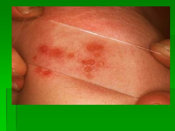
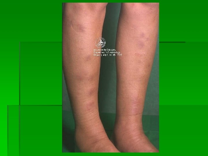
Macule § a circumscribed, flat lesion that differ from the surrounding normal skin by its color; the size and shape are variable. Types of macules: § by vascular abnormalities: § erythematous (by capillary dilatation): red in color, reduced by diascopy; ex. psoriasis, eczema, lupus erythematosus et. al; § purpuric (by extravasation of the red blood cells): redviolet → yellow-green → brown; nonreduced by diascopy; petechiae (small), ecchymoses (larger) sugillation (“black and blue”); vibices (linear); ex. purpura.
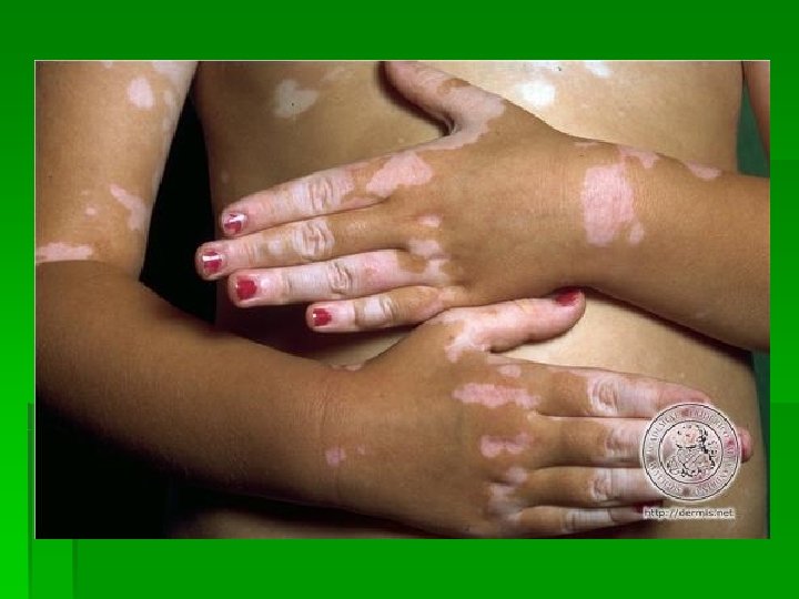
Macule § by melanin abnormalities: § hiperpigmentation (excess of melanin): brown in color; ex. freckles, naevi, chloasma. § hypopigmentation – depigmentation (lack of pigment production): white in color; ex. albinism, vitiligo, pityriasis versicolor; § artificial: by others pigmentary substances; ex. tatoos, carotenaemia, jaundice, licopenaemia. Patch: a large macule with some surface change – slight scale or fine wrinkling; ex. psoriasis, eczema.
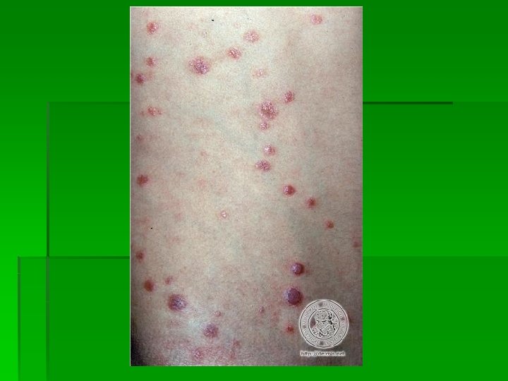
Papule § a small, solid, elevated skin lesion less than 0. 5 cm in diameter; oblique lighting with a flashlight in a darkened room detect a slight elevation; The shape of the papule: § acuminate (pointed): miliaria rubra; § dome-shaped: secondary syphilis; § flat-topped: lichen planus. The color of the papule: red, copper, violaceous, yellow et. al. Hystological types: § epidermal (the result of an increase in the number of epidermal cells): verruca plana (plane warts); § dermal (the result of an infiltrate in the dermis): secondary syphilis; § dermal-epidermal ( a hyperplasia of cellular components of the epidermis and an infiltrate in the dermis): lichen planus.
Wheal § a rounded or flat-topped elevated papule (3 -4 mm) or plaque (10 -12 cm in diameter), characteristically evanescent, desappearing within hours. The shape: round, oval, serpiginous, annular. The color: pale red or white (especially in the center). The borders: sharp, but not stable (move). Histology: an edema in the upper portion of the dermis. Exemples: urticaria, ocasionally, dermatitis herpetiformis, bullous pemphigoid. Dermographism: a wheal produced by stroking of the skin in persons with urticaria, or in normal persons.
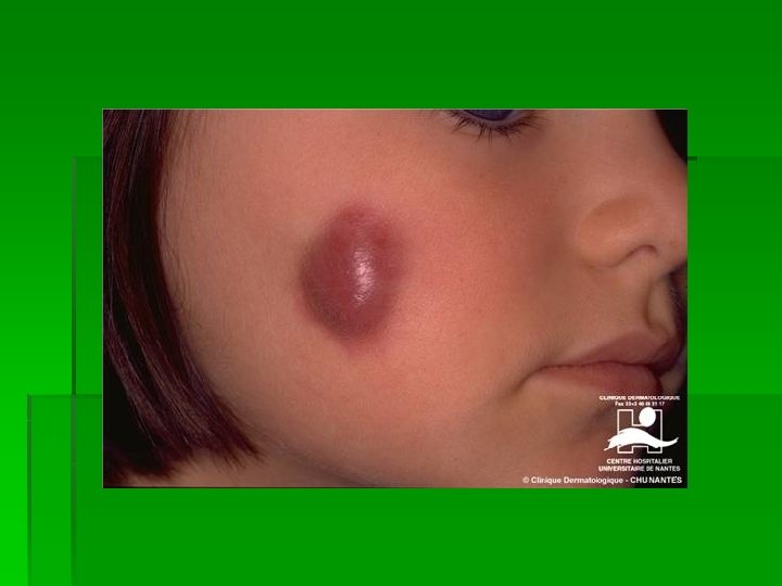
Nodule § a palpable, round or oval lesion, greater and deep than a papule; the consistence is hard, firm, soft or fleshy; the surface is smooth, keratotic, ulcerated or fungatic; is warm, painless or painful. Histological types: § epidermal: keratoacanthoma; § epidermal-dermal: mycosis fungoides, lupus vulgaris; § dermal: granuloma annulare; § dermal-subdermal: erythema nodosum, hypodermitis; § subcutaneous: lipoma.
Gumma: a granulomatous nodular lesion with stadial evolution. ex. late syphilis, tuberculosis, the deep mycoses. Vegetation: a small, soft and smooth, pediculated elevation, red in color; can be covered with thick dry scales (keratotic); vegetations are aglomerated in the cauliflowerlike masses, with a moist, macerated surface. ex. condylomata acuminata (anogenital warts), common warts, pemphigus vegetans, warty tuberculosis. Lichenification: a thickened plaque with accentuated skin margins, produced after repeated rubbing. Histologically: a proliferation of keratinocytes and stratum corneum in combination with changes in the collagen of the underlying dermis. § ex. chronic eczema, prurigo, neurodermatitis.
Tumor § a benign or malignant proliferation of the skin, variable in color, fleshy, soft, firm or hard. § exemples: papilloma, keratoachantoma, lipoma, naevi, carcinoma, melanoma, sarcoma, et al.
Vesicle § an elevated lesion filled with clear fluid, with a diameter less than 0. 5 cm. § Spongiform vesicle – by an intraepidermal interstitial vesication (intercellular edema and individual cells rupture): eczema. § Parenchimatous vesicle – by a ballon degeneration of the epidermal cells: herpes simplex, herpes zoster (shingles), varicella.
Bulla § a blister filled with clear fluid greater than 0. 5 cm in diameter; tense or flaccid; Histological types: § superficial bulla (subcorneal): impetigo; § medium (acantholytic) bulla: pemphigus vulgaris; § profound (subepidermal) bulla: bullous pemphigoid, dermatitis herpetiformis, porphyria cutanea tarda, bullous erythema multiforme.
Pustule § a circumscripted, raised lesion filled with a purulent exudate, white, yellow or greenish-yellow in color; may vary in size and shape. Types: § follicular or nonfollicular; § sterile or bacterian; § primary or secondary. Exemples: rosacea, acne vulgaris, pustular psoriasis, folliculitis.
Erosion § a moist, usually depressed lesion that results from loss of all or a portion of the viable epidermis; usually do not scar. § primary or secondary (after the rupture of vesicles, bullae, the denudation of papules). § Exemples: primary chancre, eczema, herpes simplex, pemphigus vulgaris, et al.
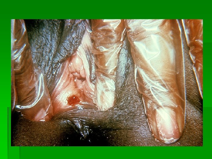
Ulceration § a destruction of the epidermis and papillary dermis; may vary in size and shape; the base is soft or firm; heels with scarring; § ulcer – a chronic ulceration. § exemples: stasis ulcer, tuberculosis, neoplasms.
Fissure § a thin linear tear in then skin, superficial or profound (painful), by the loss of the extensibility of the skin by hiperkeratosis or inflamation. § exemples: chronic eczema of the hands and feet, perleche.
Excoriation § a linear or punctate cleavage in the skin as a result of scrathing in all types of pruritus. § exemples: pediculosis, scabies, eczema et al.
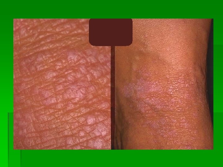
Scale § abnormal shedding or accumulation of horny layer in perceptible flakes; dry, usually whitish. Types of scale: § pityriasiform (little and thin): pityriasis simplex, pityriasis versicolor; § psoriasiform (brittle platelets of several loose layers): psoriasis; § seborrheic (yellow-to-brown, greasy): seborrheic dermatitis; § ichtyosiform (like fish scales): ichthyosis vulgaris; § cuticular and lamellar (thin, relatively large flakes): lamellar ichtyosis; § exfoliative (large sheets): erythroderma; § follicular (keratotic plugs, spines, fillaments): keratosis pilaris; § hystrix-like (little horns): ichthyosis hystrix.
Crust § liquid debris that has dried on the surface of the skin; result from breakage of vesicles, bullae or pustules; the color: yellow (dried serum), green or yellow-green (dried purulent exudate), brown or black (dried blood). § exemples: impetigo, eczema, herpes simplex, herpes zoster, ecthyma et al.
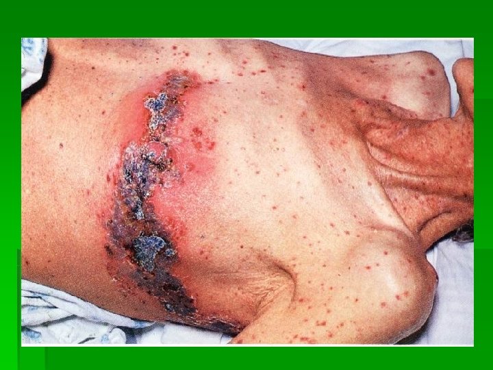
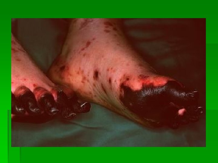
Gangrene and sphacelus § a lesion resulting from a necrose § gangrene: sharply demarcated, blue-black in color; § sphacelus: adherent, dry, necrotic membrane or necrotic core. § exemples: gangrene from arterial occlusion, decubitus ulcer, furuncle.
Scar § a residual lesion, replacing an ulceration by healing; dissapearance of normal skin lines and appendages. Types: § hypertrophic: prominent, hard; § atrophic: thin, depressed; Exemples: herpes zoster, varicella, acne, porphyria cutanea tarda, ecthyma, syphilis, tuberculosis, leprosy, lupus erythematosus et al.
Atrophy § thining of the skin § epidermal atrophy: the surface is thin and wrinkled, often associated with dermal alteration. § dermal atrophy: a depression of the skin; may be or not associated with epidermal atrophy. § exemples: lupus erythematosus, striae of pregnancy, scleroderma et al.