Urinary system The urinary system also known as


, potassium (K+) and chloride (Cl−)")


 are cells in the Bowman's capsule in the kidneys")


 , this is an insoluble substance mainly made up from")


- Slides: 35
Urinary system
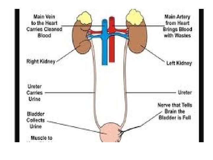
The urinary system, also known as the renal system, consists of the two kidneys, ureters, the bladder, and the urethra. Each kidney consists of millions of functional units called nephrons. The purpose of the renal system is to eliminate wastes from the body, regulate blood volume and pressure, control levels of electrolytes and metabolites, and regulate blood p. H.
The kidneys have extensive blood supply via the renal arteries which leave the kidneys via the renal vein. Following filtration of blood and further processing, wastes (in the form of urine) exit the kidney via the ureters, tubes made of smooth muscle fibers that propel urine towards the urinary bladder, where it is stored and subsequently expelled from the body by urination (voiding). The female and male urinary system are very similar, differing only in the length of the urethra.
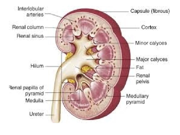
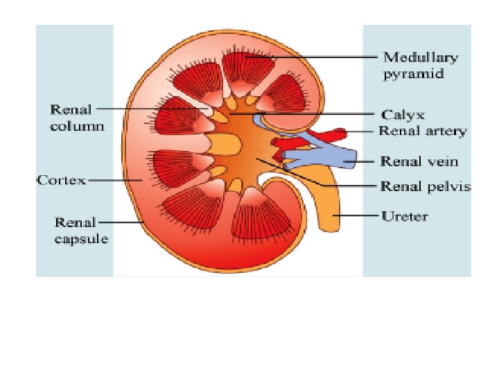
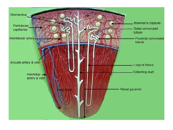
The kidneys are bean shaped organs. The kidney has a bean-shaped structure having a convex and a concave border. A recessed area on the concave border is the renal hilum, where the renal artery enters the kidney and the renal vein and ureter leave. The kidney is surrounded by tough fibrous tissue, the renal capsule, The kidney is organised into many lobes, organised in a pyramidal structure, where the outer portion is made up of cortex, and the inner portion is made up of the medulla
kidney contains about 1 million functional units called nephrons, which are continuous with a system of collecting tubules. Each kidney is held in place by connective tissue, called renal fascia, and isis surrounded by a thick layer of adipose tissue, called perirenal fat, which helps to protect it. A tough, fibrous, connective tissue renal capsule closely envelopes each kidney and provides support for the soft tissue that is inside. The renal cortex, renal medulla, and renal pelvis are three main internal regionsfound in a kidney. Nephrons, masses of tiny tubules, are largely located in the medulla and receive fluid from the blood vessels in the renal cortex.
Function of kidney 1. controlling ACID-base balance. 2. controlling WATER balance. 3. maintaining ELECTROLYTE balance. 4. removing TOXINS and waste products from the body. 5. controlling BLOOD PRESSURE. 6. producing the hormone ERYTHROPOIETIN. 7. activating vitamin D
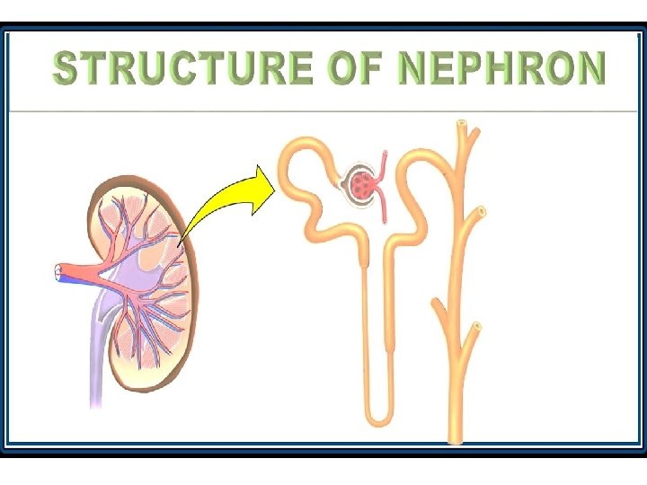
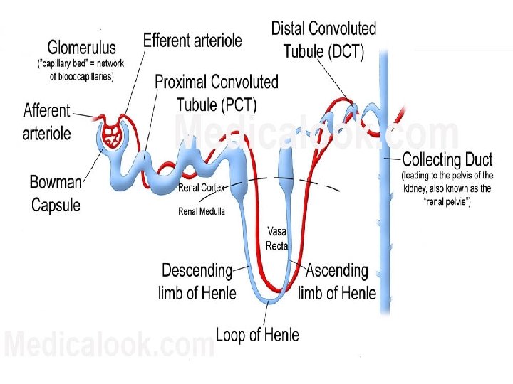
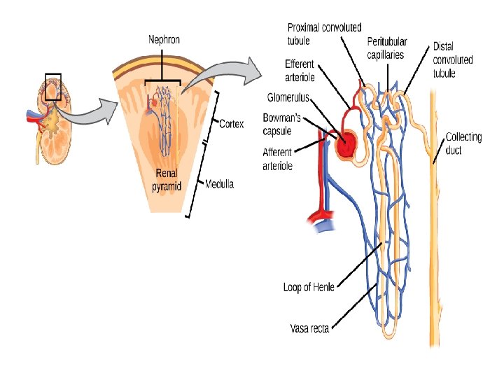
Uriniferous tubule: The kidney composed of many Uriniferous tubule, each one consist of 1 -Nephron 2 -Collecting duct Nephron Basic structural and functional unit of the kidney. In humans, a normal kidney contains 800, 000 to 1. 5 million nephrons. All nephrons wall simple epithelial tissue, each nephron consist of ; 1 - Renal corpuscles. 2 - Proximal convoluted tubule. 3 - Loop of Henle. 4 - Distal convoluted tubule
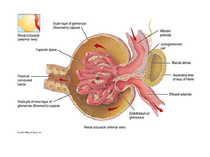
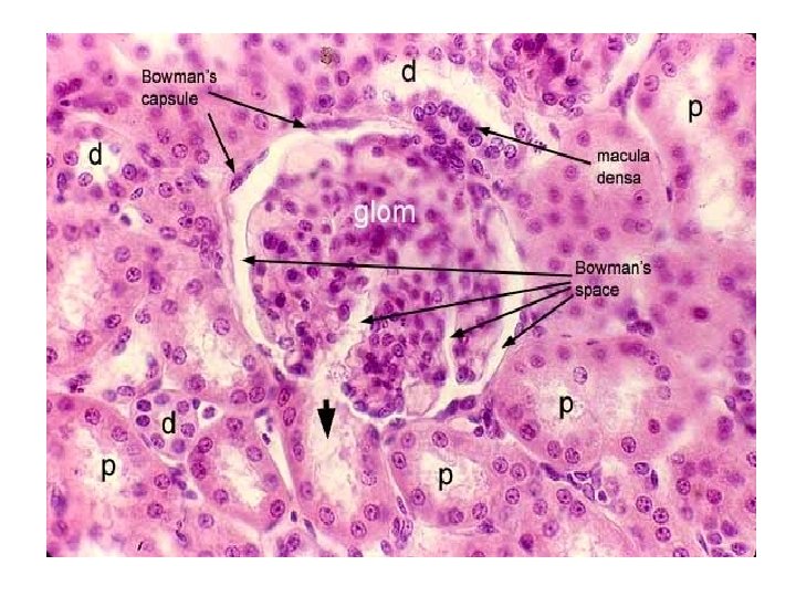
Renal corpuscle In the kidney, a renal corpuscle is the initial blood-filtering component of a nephron. It consists of two structures: a glomerulus and a Bowman's capsule. The glomerulus is a small tuft of capillaries containing two cell types. Endothelial cells, which have large fenestrae, are not covered by diaphragms. Mesangial cells are modified smooth muscle cells that lie between the capillaries. They regulate blood flow by their contractile activity and secrete extracellular matrix, prostaglandins, and cytokines. Mesangial cells also have phagocytic activity, removing proteins and other molecules trapped in the glomerular basement membrane or filtration barrier.
The Bowman's capsule has an outer parietal layer composed of simple squamous epithelium. The visceral layer, composed of modified simple squamous epithelium,
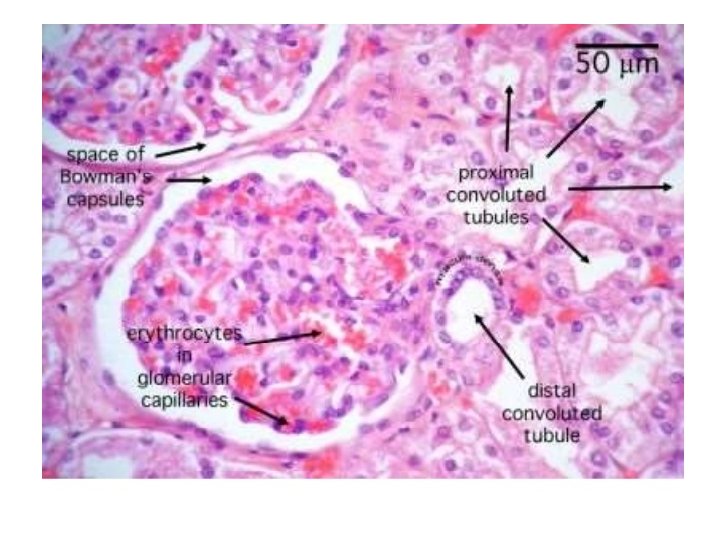
The renal corpuscle filtration barrier permits passage of water, ions, and small molecules from the bloodstream into Bowman's space (the space between the visceral and parietal layers). Large and/or negatively charged proteins are prevented from passing into Bowman's space, thus retaining these proteins in the circulation. The basal lamina is composed of 3 layers: lamina externa, lamina densa, and lamina interna.
Fluid from blood in the glomerulus is collected in the Bowman's capsule to form "glomerular filtrate", which is then further processed along the nephron to form urine. The three types of filtration carried out in the Bowman's capsule are: 1) Basement Filtration 2) Visceral Filtration 3) Endothermic Filtration.
Proximal convoluted tubule The proximal tubule is the portion of the duct system of the nephron of the kidney which leads from Bowman's capsule to the loop of Henle. The most distinctive characteristic of the proximal tubule is its brush border (or "striated border"), Cuboidal epithelial cells lining the proximal tubule have extensive lateral interdigitations between neighboring cells
Loop of Henle. The loop of Henle can be divided into four parts: Thin descending limb of loop of Henle The thin descending limb has low permeability to ions and urea, while being highly permeable to water. The loop has a sharp bend in the renal medulla going from descending to ascending thin limb. Thin ascending limb of loop of Henle The thin ascending limb is impermeable to water, but it is permeable to ions.
Thick ascending limb of loop of Henle Sodium (Na+), potassium (K+) and chloride (Cl−) ions are reabsorbed from the urine by secondary active transport by a Na-K-Cl cotransporter (NKCC 2). The electrical and concentration gradient drives more reabsorption of Na+, as well as other cations such as magnesium (Mg 2+) and calcium (Ca 2+). Cortical thick ascending limb The cortical thick ascending limb drains urine into the distal convoluted tubule. The tissue type of the loop is simple squamous epithelium. The "thick" and "thin" terminology does not refer to the size of the lumen, but to the size of the epithelial. The loop is also sometimes called the Nephron loop.
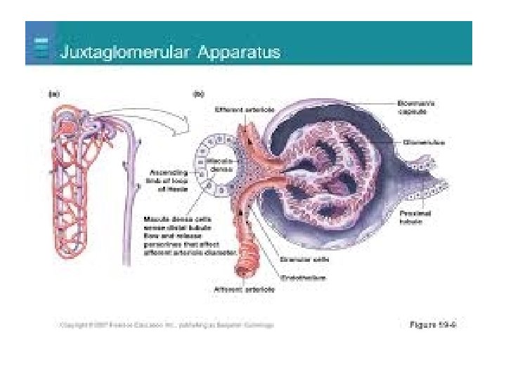
Distal convoluted tubule The DCT is lined with simple cuboidal cells that are shorter than those of the proximal convoluted tubule (PCT). The lumen appears larger in DCT than the PCT lumen because the PCT has a brush border (microvilli). DCT can be recognized by its numerous mitochondria, basal inholdings and lateral membrane interdigitations with neighboring cells. The point where DCT contacts afferent arteriole of renal corpuscle is called macula densa. It has tightly packed columnar cells which display reversed polarity and may monitor the osmolarity of blood.
Collecting duct The collecting duct system of the kidney consists of a series of tubules and ducts that connect the nephrons to the ureter. It participates in electrolyte and fluid balance through reabsorption and excretion, processes regulated by the hormonesaldosterone and vasopressin (antidiuretic hormone).
Podocytes (or visceral epithelial cells) are cells in the Bowman's capsule in the kidneys that wrap around the capillaries of the glomerulus. [1] The Bowman's capsule filters blood, holding back large molecules such as proteins, and passing through small molecules such as water, salts, and sugar, as the first step in forming urine.
Ureter: Ureters are tubes made of smooth muscle fibers that propel urine from the kidneys to the urinary bladder. In the adult, the ureters are usually 25– 30 cm (10– 12 in) long and ~3– 4 mm in diameter Histology The ureter is surrounded by urothelium, a type of transitional epithelium that is capable of responding to stretches in the ureters. The transitional epithelium may appear as a columnar epithelia when relaxed, and squamous epithelia when distended. Below the epithelium, a sub mucosa exists. The sub mucosa is made up of connective tissue interspersed with blood vessels, veins and lymphatic. The ureter is surrounded by two muscular layers, an inner longitudinal layer of muscle, and an outer layer of muscle arranged circularly. There is also growing evidence of a large number of multivesicular bodies and other lysosomal components seen in these cells, indicating a high endocytic activity. Pedicels" (or "foot processes") extend from the podocyte and increase the surface area which is crucial for the efficiency of ultrafiltration.
Urinary bladder is the organ that collects urine excreted by the kidneys before disposal by urination. A hollow muscular, and distensible (or elastic) organ, the bladder sits on the pelvic floor. Urine enters the bladder via the ureters and exits via the urethra. Histology of the Urinary Bladder Upon examining the histology, it can be seen that the bladder wall has the same tissue layers as the renal pelvis and ureter. The layers are: mucosa, muscularis, . and serosa/adventita
Histology of the Mucosa; The innermost portion of the urinary bladder is the mucosa The histology of the mucosa is that composed of transitional epithelium and connective tissue. When looking at a histology slide of the bladder mucosa, the transitional epithelium has domed shaped cells on the apical surface. The epithelial layer contains no blood vessels or lymphatics. The basement membrane is a single layer of cells. The basement membrane separates the epithelium from the connective tissue. .
The epithelium lies upon connective tissue called the lamina propria. The lamina propria is composed of areolar connective tissue. It contains blood vessels, nerves, and, in some regions, glands. The urinary sites have no muscularis mucosae, and therefore, the lamina propria and the submucosa tend to merge. When examinging a histology slide of the bladder, a well delineated submucosa is not visible. Sometimes the connective tissue closest to the muscularis is called the submucosa
Calculus (kidney stone) , this is an insoluble substance mainly made up from calcium salts of uric acid and phosphate. They can found in the central part of the kidney, the renal pelvis. They can move and lodge themselves in the ureter or bladder. They may need to be removed surgically if they interfere with the passage of urine.
kidney transplantation: This is an individual has damaged kidneys he may need to have a kidney transplant . This means surgically removing a living kidney from a donor (living or recently deceased) and putting into likely to be successful if the donor and recipient are closely related. The problem with organ transplants is the rejection of foreign.
Cystitis : This is an inflammation of the urinary bladder usually caused by bacteria but it can be caused by injury or chemicals. This condition is usually accompanied by painful passage of urine