Histology of the Digestive System Basic Histological Layers

Histology of the Digestive System Basic Histological Layers 1. Mucosa a. Epithelium b. Lamina Propria c. Muscularis Mucosae 2. Submucosa a. Submucosal plexus “Plexus of Meissner” 3. Muscularis a. Myenteric plexus “Plexus of Auerbach 4. Serosa

Histology of the Mucosa Organ Epithelium Mouth Nonkeratinized Stratified Squamous Pharynx Nonkeratinized Stratified Squamous Esophagus Nonkeratinized Stratified Squamous Stomach Simple Columnar Small Intestine Simple Columnar Large Intestine Simple Columnar Anus Nonkeratinized Stratified Squamous

Histology of the Mucosa Organ Folds of the epithelium Esophagus none Stomach L: Rugae, S: gastric pits Small Intestine L: Plicae circulares, Villi S: Crypts of Lieberkuhn, microvilli L: Haustra S: Intestinal glands Large Intestine

Histology of the Submucosa Organ Specialized structures Esophagus Submucosal mucous glands Stomach None Duodenum Brunner’s glands Ileum Peyer’s Patches Large Intestine None

Histology of the Muscularis Organ Smooth muscle layers Esophagus 2, circular and longitudinal Stomach 3, oblique, circular, and longitudinal Small Intestine 2, circular and longitudinal Large Intestine 2, circular and longitudinal

Histology of the Serosa Organ Serosa Esophagus Stomach Adventitia due to the fact that the esophagus is not in a cavity Visceral Peritoneum Small Intestine Visceral Peritoneum Large Intestine Visceral Peritoneum Anus Adventitia

Microscopic View of the Esophagus

Endoscopic View of the Esophagus

Low power view of the Stomach

Low and High power view of the Stomach Mucosa

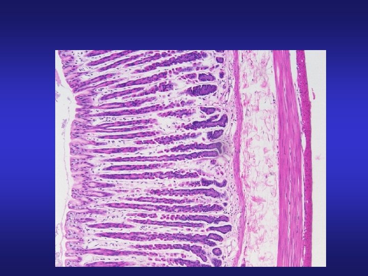
Scanning view of the Small Intestine demonstrating Plicae Circularis

High power view of the duodenal Mucosa

High Power View of Villi

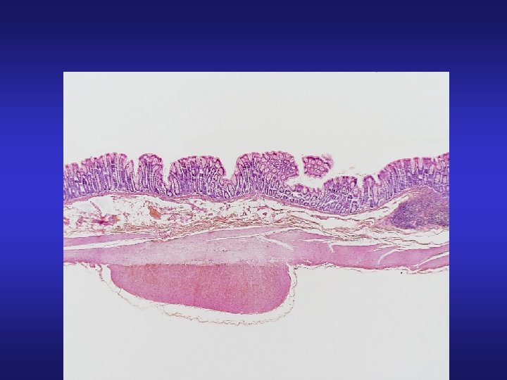
X-sectional view of the duodenum

Gross view and low-power view of the ileum

High-power view of the Ileum demonstrating Peyer’s patches

High power view of the colon demonstrating intestinal glands

Large Intestine

Large Intestine

Ano-Rectal Junction
- Slides: 27