Stomach Histology Dr Zahid Sarfaraz Khan AP Anatomy


 gland in submucosa. Penetrate the m. mucousa")

- Slides: 30
Stomach Histology Dr. Zahid Sarfaraz Khan A/P Anatomy Kgmc
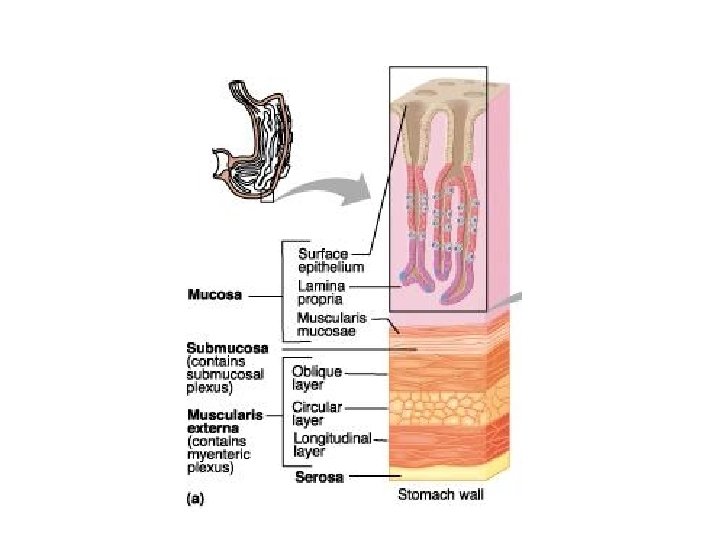
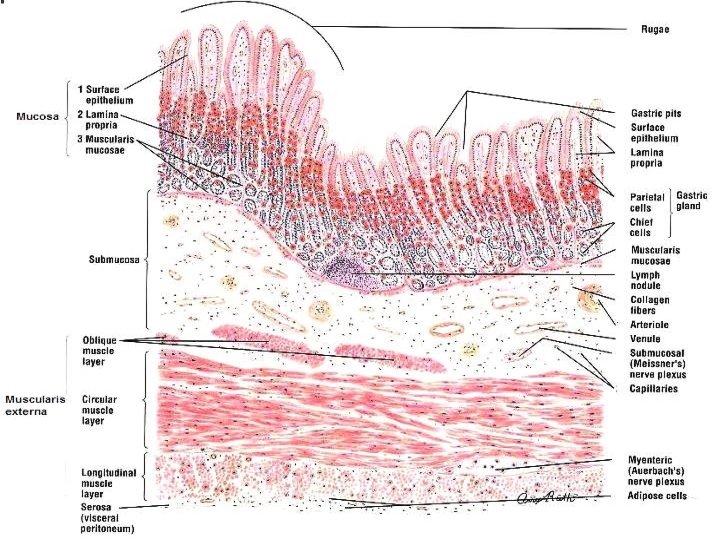
STOMACH • Stomach is a dilated segment of the digestive tract, that digest food and secrets hormone • There are three histological regions: Ø Cardia Ø Fundus and body Ø Pylorus • The fundus and body are identical in microscopic structure • The mucosa and submucosa of the undistended stomach lie in longitudinally directed folds known as rugae • When the stomach is filled with food, folds flatten out
MUSCULARIS EXTERNA: Inner: Oblique Middle: circular Outer: Longitudinal SEROSA: Outermost layers of the stomach which consists of loose connective tissue covered by mesothelium
Cellular Composition of the stomach
Surface mucous cells of stomach • Cover luminal surface of stomach • Partly cover gastric pit • Cytoplasm contain mucigen granules • Stain poorly • Have short surface microvilli • Secrete protective bicarbonate in deeper layer of surface mucous coat.
Mucous neck cells q Located just below gastric pit. q Columnar in shape q but Less columnar than the surface mucous cells lining the gastric pits q Contain mucinogen granules in apical cytoplasm( Vacuolated foamy) q While nuclei are situated basally. q Produces soluble mucus q But their mucus secretion is less alkaline (Acidic Mucus) than that of the surface Epithelial mucous cells.
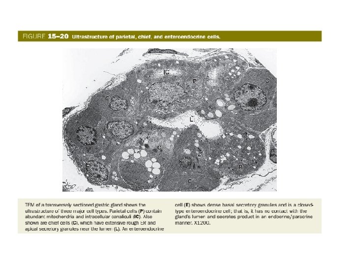
Parietal cell of stomach They are large, ovoid or polyhedral cells with a large central nucleus. - More numerous in the upper half of the gland than in the lower half q. Eosinophilic cytoplasm q. Fried egg like appearance. q. Plasma membrane form deep branching canaculi - Secretes HCL and intrinsic factor. v Intrinsic factor combines with vitamin B 12 to form a complex necessary for erythrocytes formation.
Chief, peptic or zymogenic cells • Located at base of the gland • Basally located nuclei • Strongly basophilic granular cytoplasm • Pepsin secreting cells
Neuroendocrined OR DNES Diffused Neuroendocrine system • Also found in based of the gastric gland Secrete: • Serotonin • Peptide hormone gastrin called G cell. • Somatostatin
Regerative Stem Cells • Relatively thin and columnar • Few in no. and interspersed among neck of fundic gland. • Rich supply of ribosome • BUT Less orgenelles • Nuclei basally located • Have little heterochromatine and display a large nucleous • These cells have regenerative ability and replace all those cell where need occur.
Types of cells:
Stomach • Stomach is divided into three histological regions on the basis of nature of glands: Ø Cardiac region Ø Fundic region (fundus & body) Ø Pyloric region
Cardiac region of the stomach Mucosa: • Epithelial lining at the cardio-oesophageal junction changes from stratified sq. to simple columnar epithelium • Presence of Mucous surface cells and cardiac glands and gastric pit. Submucosa: Consists of Meissner’s plexus and blood vessels
Stomach - fundus • Mucosa: - Lining Epithelium – Simple columnar epithelium that invaginates to various extents into the lamina propria, forming gastric pits. - These cells are involved in mucus secretion. The mucus protects the epithelial lining from damage due to the presence of acid in the stomach. - HCL - PEPSINOGEN
MUSCULARIS MUCOSAE: q. It consists of two thin layer of smooth muscles. i. e. , Outer longitudinal and inner circular
PYLORIC GLANDS
STOMACH - PYLORUS • MUCOSA: - Epithelium: Simple columnar as in fundic part - Pyloric glands occupy the lamina propria - Gastric pits are deeper - Glands are short, tortuous and branched - Produce mucus contain lysoenzyme and Gastrin G cells Presence of amino acid + distention of stomach stimulate G cell to release gastrin---parietal cell --release acid secretion.
Pyloris have Muscularis mucosa and Submucosa are similar to fundic part
• MUSCULARIS EXTERNA: Inner: Oblique Middle: circular Outer: Longitudinal Similar to Fundic part, but the circular fibres are much thickened to form pyloric sphincter
DIFFERENCE BETWEEN CARDIA, FUNDUS & BODY, AND PYLORUS CARDIA FUNDUS & BODY PYLORUS Contain cardiac gland Contain gastric gland Contain pyloric gland Gastric pit less deeper than pyloric gland Gastric pit more deeper than gastric or cardiac gland Parietal cells absent Parietal cells more or very few Parietal cells few 23 23
MEDICAL APPLICATION • For various reasons; • including autoimmunity, parietal cells may be damaged to the extent that insufficient quantities of intrinsic factor are secreted and vitamin B 12 is not absorbed adequately. • low levels of vitamin B 12 can reduce proliferation of erythroblasts, producing pernicious anemia
Gastric and duodenal ulcers Ø Are painful erosive lesions of the mucosa. Ø That may extend to deeper layers. Ø Such ulcers can occur anywhere between the lower esophagus and the jejunum, and Their causes include: A- Bacterial infections with Helicobacter pylori, B- Effects of nonsteroidal anti-inflammatory drugs, C- Overproduction of HCl or pepsin, D- Lowered production or secretion of mucus or bicarbonate.
ATROPIC GASTRITIS • Both parietal cell and chief cells are became decrease. • Little gastric juice, no acid or pepsin activity. • Intrinsic factor produce by parietal cell decrease.
MEDICAL APPLICATION • Tumors called carcinoids, which arise from enteroendocrine • EC cells, are responsible for the clinical symptoms caused by overproduction of serotonin. • Serotonin increases gut motility.
Small intestine ØThree subdivisions: üDuodenum Jejunum Ileum
Duodenum ØContain Four layer ØDuodenal (brunner ) gland in submucosa. Penetrate the m. mucousa and secretion of hormone called urogastrone. ØVilli ØMicrovilli forming brush border ØGoblet cell. §Light stain interspead among absortive cell of intestinal epithelium. §More in distal portion. ØIntestinal gland (crypt of lieberkuhn) in lumina propria. open into intervillous space ØIntervillous space. ØLacteal ØMyenteric plexus
Thank you