Preparation of tissues for study v HISTOLOGY It


 • Buffered gluteraldehyde (electron microscope preparation)")


 Dehydration b) Clearing c) Impregnation (infiltration)")


 It is the process in which the water content in")


: Ø In this the tissue is kept in a wax bath containing")

 Xylene (II) for 1. 5 hours Infiltration: 1. 2.")


 Forceps")

 (reservoir) Heated chambers Wax flow adjuster Mould")


 Ø It is the procedure in which the blocks which have")
 microtome • Ultramicrotome")

")

")

 1 -30µm Paraffin Block Steel knife")
 and the block holder is moved up")


 microtome • Frozen tissue embedded in a freezing medium is cut on")
 microtome")
 microtome")


 Hydration (alcohol) Deparaffinization (xylene) Clearing (xylene)")


- Slides: 108
Preparation of tissues for study
v HISTOLOGY : It is the branch of science which deals with the microscopic study of normal tissue v HISTOPATHOLOGY : It is the branch of science which deals with the microscopic study of tissue affected by disease Tissue for study can be obtained from: Ø Ø Biopsies Autopsies
Microtechnique • Microtechnique: is tissue preparation for microscopic examination • There are different methods used, however the basic principles are similar • It usually involves hardening of the tissue followed by sectioning (cutting) - Paraffin technique - Freezing technique
Histological Techniques: 1. Paraffin Tissues are hardened by replacing water with paraffin. The tissue is then cut in the microtome at thicknesses varying from 1 to 30 µm thick. From there the tissue can be mounted on a microscope slide, stained and examined using a light microscope.
2. Freezing technique: • Water-rich tissues are hardened by freezing and cut frozen; sections are stained and examined with a light microscope • This technique is much faster than traditional histology (20 minutes vs. 16 hours) and are used in operations to achieve a quick diagnosis • Cryosections can also be used in immunohistochemistry as freezing tissue does not alter or mask its chemical composition
Protocols followed in Histotechniques 1. 2. 3. 4. 5. 6. 7. 8. 9. 10. Receipt & Identification Labeling of the specimen with numbering Fixation Dehydration Clearing Impregnation (infiltration) Embedding Section cutting Staining Mounting
Specimen Dissection - the stage
Fixation
Fixation • This is the process by which the constituents of cells and tissue are fixed in a physical and chemical state so that they will withstand subsequent treatment with various reagents with minimum loss of architecture • This is achieved by exposing the tissue to chemical compounds: fixatives • Fixatives prevent autolysis and bacterial decomposition and preserves tissue in their natural state and fix all components
Tissue fixatives • Buffered formalin (light microscope preparation) • Buffered gluteraldehyde (electron microscope preparation) • Osmium tetraoxide (electron microscope preparation, preserve and stain) • Zenker’s formal saline • Bowen’s fluid
• No fixative will penetrate a piece of tissue thicker than 1 cm.
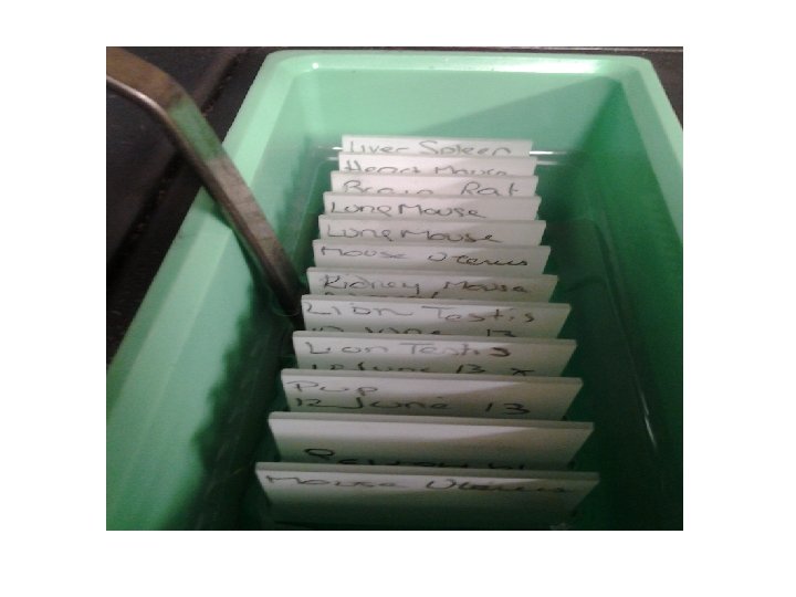
Cassette
Specimen is placed in porous cassettes
Labeling
Cassettes are collected in fixatives 10% formalin 1 mm/hour fixation
Processing
Tissue Processing ü In order to cut thin sections of the tissues, it should have suitable hardness and consistency when presented to the knife edge. ü These properties can be imparted by infiltrating and surrounding the tissue with paraffin wax, various types of resins or by freezing. ü This process is called tissue processing.
Tissue Processing It can be subdivided into: a) Dehydration b) Clearing c) Impregnation (infiltration)
Types of tissue processing • There are two types : 1. Manual Tissue Processing 2. Mechanical Tissue Processing
Mechanical Tissue Processing • In this the tissue is moved from one jar to another by mechanical device • Timings are controlled by a timer which can be adjusted in respect to hours and minutes • Temperature is maintained around 60 ºC • Automatic tissue processor: Ø Overnight Ø 12 Baths Ø 16 hours
Tissue basket
Tissues processor
Manual Tissue Processing • In this process the tissue is changed from one container of reagent to another by hand Note: The processing, whether manually or mechanically, involves the same steps and reagents in same sequence
Dehydration (removal of water) It is the process in which the water content in the tissue to be processed is completely removed by passing the tissue through increasing concentrations of dehydrating agents • Tissues are dehydrated by using increasing strength of alcohol; e. g. 70%, 90% and 100% • Water is replaced by diffusion
• During dehydration water in tissue has been replaced by alcohol • The next step alcohol should be replaced by paraffin wax • As paraffin wax is not alcohol soluble, we replace alcohol with a substance in which wax is soluble. This step is called clearing.
Clearing • Replacing the dehydrating fluid with a fluid that is totally miscible with both the dehydrating fluid and the embedding medium • Some clearing agents: - Xylene - Toluene - Chloroform - Benzene
Impregnation (infiltration): Ø In this the tissue is kept in a wax bath containing molten paraffin wax
The duration of the procedure can be noted down as: Fixation 1. 2. 10 % Formalin saline (I) 10 % Formalin saline (II) for 1. 5 hours Dehydration 1. 2. 3. 4. 5. 6. 80 % alcohol 95 % alcohol (I) 95 % alcohol (II) Absolute alcohol (100%) (III) for for for 1 hour 1 hour
Clearing: 1. 2. Xylene (I) Xylene (II) for 1. 5 hours Infiltration: 1. 2. Paraffin wax (I) Paraffin wax (II) for 1. 5 hours
Embedding
Embedding: g is the process by which impregnated tissues are surrounded by a medium such as agar, gelatin, or wax which when solidified will provide sufficient external support during sectioning
Embedding: Ø It is done by transferring the tissue to a mould filled with molten wax & is allowed to cool & solidify Ø After solidification, a wax block is obtained which is then sectioned to obtain ribbons
Embedding tools Moulds Paraffin wax
Metal moulds
Disposable plastic moulds
Embedding tools Paraffin wax: is a polycrystalline mixture of solid hydrocarbons. Paraffin wax is traditionally marketed by its melting points which range from 39°C to 68°C
Embedding tools Scalpel (blade) Forceps
Embedding Centre
Embedding Centre Used lids Molten wax (60°C) (reservoir) Heated chambers Wax flow adjuster Mould Hot surface Cold plates (5°C) Cassette Wax dispenser
General Embedding Procedure
Fill the mould with paraffin wax
Using warm forceps select the tissue, take care that it does not cool in the air
Orienting the tissue in the mould
Orientation Of Tissue In The Block ü Correct orientation of tissue in a mould is the most important step in embedding 1. cross section 2. longitudinal section ü Incorrect placement of tissues may result in diagnostically important tissue elements being missed or damaged during microtomy
Cool the block on the cold plate
Blocks on the cold plate
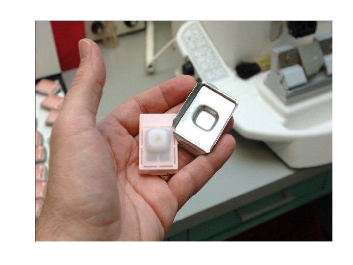
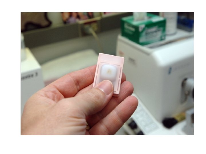
Remove the block from the mould
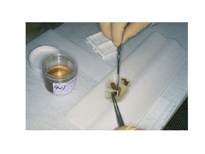
Blocks of embedded tissue are usually trimmed to remove the excess wax on the surface
Sectioning
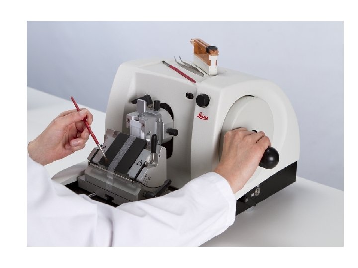
Sectioning (Section Cutting) Ø It is the procedure in which the blocks which have been prepared are cut or sectioned and thin strips of uniform thickness are prepared Ø The instrument by which this is done is called as a Microtome
TYPES OF MICROTOMES: • Rotary microtome • Freezing (cryostat) microtome • Ultramicrotome
Microtome knives • • • Steel Knives Non-corrosive Knives For Cryostats Disposable Blades Glass Knives Diamond Knives • Microtome blades are extremely sharp, and should be handled with great care. Safety precautions should be taken in order to avoid any contact with the cutting edge of the blade
Steel Knives (for rotary microtome)
Disposable Blades
Glass Knives (for ultramicrotome)
Rotary Microtome Block holder Knife holder Drive wheel
Micron adjustment (section thickness) 1 -30µm Paraffin Block Steel knife
Rotary Microtome The knife is stationary (fixed) and the block holder is moved up and down in a vertical plane by the rotary action of the hand wheel Ø It is the most commonly used Ø Suitable for paraffin embedded sections Ø It is suitable for cutting of small tissues Ø Ideal for cutting serial sections
Ultra Microtome
Ultra Microtome ü Used for very thin section ü The typical thickness of tissue cut is between 20 -100 nm for TEM ü Knife: Diamond or Glass
Ultra Microtome
Ultra Microtome
Freezing (Cryostat) microtome • Frozen tissue embedded in a freezing medium is cut on a microtome in a cooled machine called a cryostat
Freezing (Cryostat) microtome
Freezing (Cryostat) microtome
Sectioning procedure
Ribbon of sections
Tissue floatation bath § It is a thermostatically controlled water bath • It is maintained at a temperature 5 – 6 degrees below the melting point of paraffin wax
Flattened paraffin sections
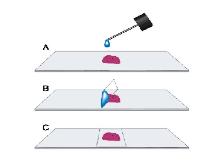
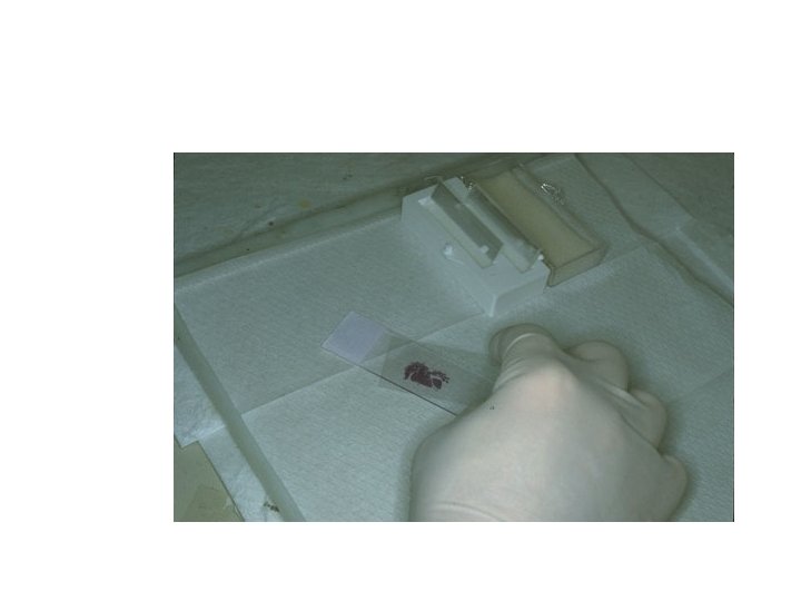
Taking the floating sections onto slide Adhesives used for fixing the sections on the slides Albumin solution ( Mayor’s egg albumin)
Taking the section onto slide Flat, no air bubbles, no stretch or breaks
Staining
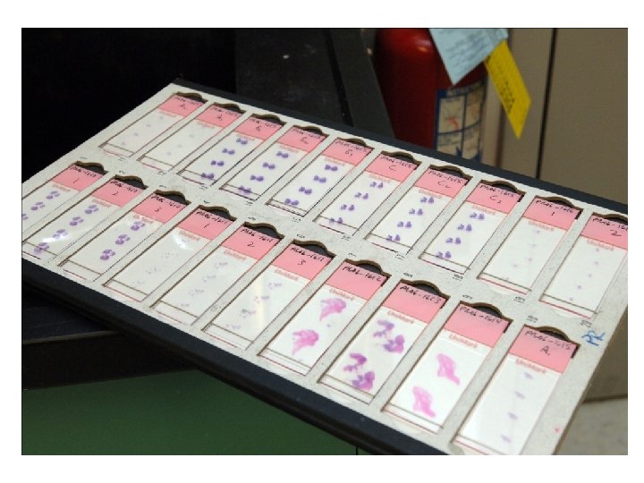
Staining • Staining is a process by which we give colour to a section. Staining of the section is done to bring out the particular details in the tissue under study • There are hundreds of stains available • The most commonly used stain in routine practice is Hematoxylin & Eosin stain
Staining • Classification of Stains: • Acid stains • Basic stains • Staining steps: a. rehydration b. stain c. dehydration
Acid Dyes In an acidic dye: • The basic component is colored and the acid component is colorless • Acid dyes stain basic components e. g. eosin stains cytoplasm • The color imparted is shade of red
Basic Dyes In a basic dye: • The acid component is colored and the basic component is colorless • Basic dyes stain acidic components e. g. Hematoxylin stains nucleus • The color imparted is shade of blue
H & E stains are universally used for routine histological examination of tissue sections
• Result : The nucleus stains Blue The cytoplasm stains pink
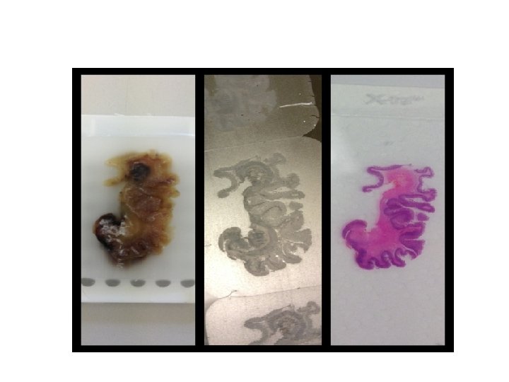
Special stains • When a specific component of tissue e. g. fibrous tissue, elastic tissue, nuclear material is to be stained, certain special stains are used which specifically stain that component tissue Examples: 1. PAS 2. Masson’s trichrome 3. Silver
Normal glomerulus of kidney is stained with H&E
Normal glomerulus of kidney is stained with PAS detects glycogen, glycoproteins, glycolipids and mucins in tissues
Masson Trichrome Stain § Nuclei stain black or blue-black • Muscles stain red • Collagen and mucus stain green or blue • Cytoplasm of most cells stains pink Normal glomerulus of kidney is stained with Masson_trichrome
Normal glomerulus of kidney is stained with methenamine silver Silver stains reticular fibers (type III collagen)
Staining Tools
Slide rack
Slide rack
Solutions
Procedure of staining: • There are two types of staining: • Manual Staining • Automatic staining • Slides stained either manually or by automatic stainer, pass through same sequences of reagents.
Manual Staining • Different reagent containers are placed in a special sequence and the slides are removed from one container to another manually.
Manual Staining
Manual Staining
Automatic stainer
Automatic stainer
Haematoxylin Eosin Dehydration (alcohol) Hydration (alcohol) Deparaffinization (xylene) Clearing (xylene)
Mounting • Stained section on microscope slide is mounted using mounting medium dissolved in xylene • Examples of Mountants : DPX ( Distrene Dibutyl phthalate Xylene ) ü A coverslip is placed on top, to protect the sample
Light Microscopic Examination
Microscopic Examination
Concering Electron Microscopy u Electron beam instead of light u Gluteraldehyde fixative u Embedding is in hard epoxy plastic u Ultramicrotome • Diamond or Glass knives • Thin section (0. 02 -0. 1 µm). u Specimen is mounted on a metal grid
Ultra Microtome