Development of teeth malformations Dr Gallatz Katalin DEVELOPMENT


")
")


 – dental")


 mineralization zománc szekréció AMELOGENESIS:")


 1. phase: granular process appears on the apical")


")


")


- Slides: 58
Development of teeth, malformations Dr Gallatz Katalin
DEVELOPMENT OF THE CROWN DEVELOPMENT OF THE ROOT ERUPTION MALFORMATIONS
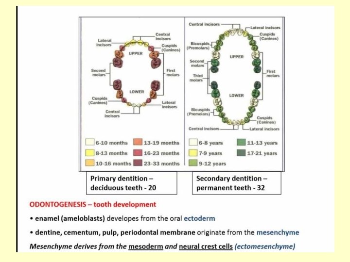
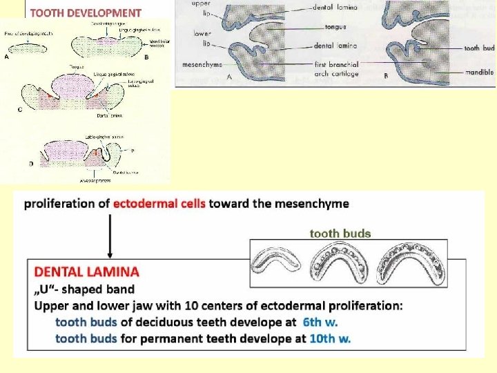
The development of the teeth starts at 6 th week with the proliferation of the ectoderm of the stomodeum
-The tooth development starts in the 6 th week, with the formation of the dental lamina from the oral epithelium. -The dental laminaes (U-shaped bands) follow the curves of the primitive jaws. Dental lamina
Dental lamina - Each dental lamina develops ten centers of proliferation, the tooth buds of the decidous teeth. -The tooth buds of the permanent teeth appear later on the palatine or lingual aspect of the decidous teeth
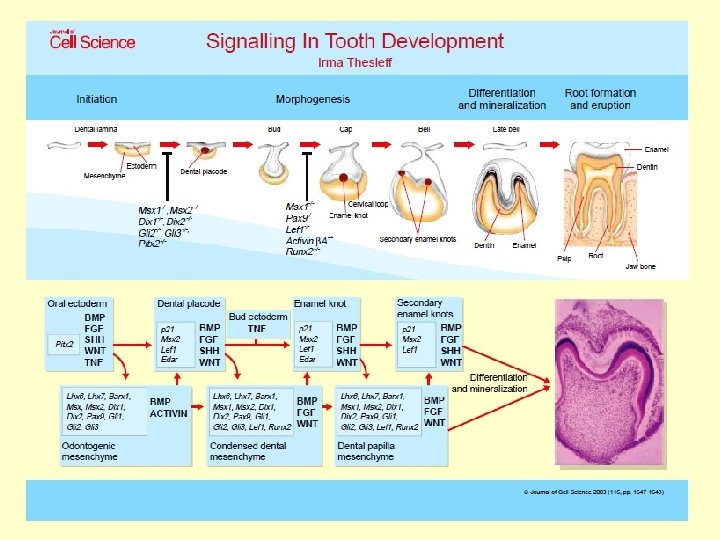
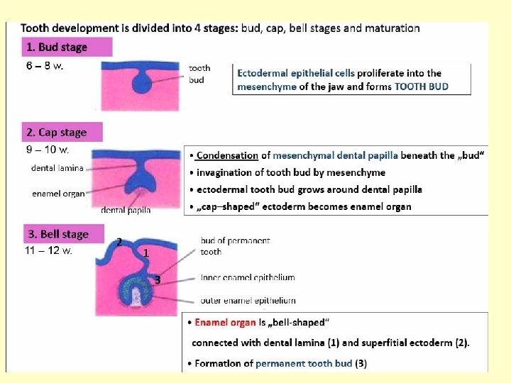
1 TOOTH DEVELOPMENT • • 2 initiation stage – 6 th to 7 th week bud stage – 8 th week cap stage – 9 th to 10 th weeks EO bell stage – 11 th to 12 th weeks 3 EO – enamel organ 4 • apposition stage • maturation stage
CAP STAGE BELL STAGE ENAMEL ORGAN DENTAL SAC The mesenchyme invaginates into the tooth buds resulting the cap stage than the bell stage
CAP STAGE • Enamel organ • Dental papilla • Dental sac (follicle)
PARTS OF THE DEVELOPING TEETH ECTOMESENCHYME ECTODERM Enamel organ ENAMEL (from the neural crest) Dental papilla Dental sac DENTIN CEMENT PULP PERIODONTAL LIG. ALVEOLUS
DS DP
DENTAL SAC ENAMEL ORGAN DENTAL PAPILLA
Parts of the developing tooth enamel organ dental papilla dental sac ameloblasts – enamel producing cells differentiate from the inner enamel epithelium odontoblasts – dentin producing cells differentiate from the ectomesenchymal cells of the dental papilla (neural crest origin) enamel knot
ENAMEL KNOT – SIGNAL CENTER * Non-dividing cells from the inner enamel epithelium, • produce different signal molecules, which transport the information between the ectodermal and ectomesenchymal cells BMP, FGF, Sh. H, activin enamel knot
Tooth formation: Initial stages - development of the dental lamina - neural crest cells migrate into the developing mesenchyme - a basement membrane separates the developing oral epithelium and mesenchyme The initiation of tooth formation starts around the 37 th day of gestation.
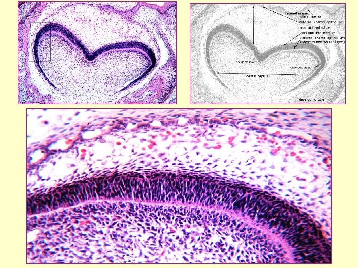
CAP STAGE • Tooth bud proliferates and forms a cap shaped organ • this stage marks the beginning of histodifferentiation (differentiation of tissues) • the tooth germ also begins to take on form – start of morphodifferentiation • enamel organ is formed – produces the future enamel (ectodermal origin)
CAP STAGE Below this cap is a condensing mass of mesenchyme (ectomesenchyme) – dental papilla – produces the future dentin and pulp tissue. The basement membrane separating the enamel organ and the dental papilla becomes the future site for the dentinoenamel junction (DEJ) • The mesenchyme surrounding the enamel organ is the dental sac
• enamel organ + dental papilla + dental sac is considered the developing tooth germ • tooth germs are found in the developing dental arches primary dentition • Enamel organ • Dental papilla • Dental sac (follicle) CAP STAGE
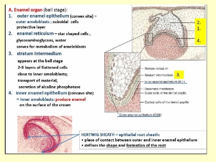
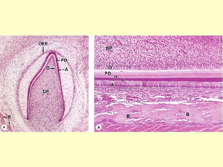
Bell Stage • Continuation of histodifferentiation and morphodifferentiation • differentiation produces 4 cell layer within the ENAMEL ORGAN – 1. inner enamel epithelium – 2. outer enamel epithelium – 3. stellate reticulum – 4. stratum intermedium
LAYERS OF THE ENAMEL ORGAN
• DENTAL PAPILLA undergoes differentiation and produces two types of cells 1. outer cells of the DP –ectomesenchymal cells differentiate into dentin-secreting cells odontoblasts 2. central cells of the DP form the connective tissue of the pulp Dental sac/follicle increases its collagen content and differentiates at a later stage than the EO and DP Bell Stage
APPOSITION AND MATURATION
APPOSITION MATURATION DENTINOGENESIS: first the odontoblasts produce predentin (organic material) mineralization zománc szekréció AMELOGENESIS: the first layer of predentin induces the inner enamel epithelial cells, they differentiate into ameloblasts and secret the organic matrix of the enamel, mineralization ECTO-MESENCHYMAL INTERACTIONS dentin szekréció
APPOSITIONAL STAGE 1 1. oral ep. 2. outer enamel ep. 3. stellate reticulum 4. inner enamel ep. 5. dental papilla 6. cervical loop 2 3 4 5 blood vessels 6 http: //www. iob. uio. no/studier/undervisning/histologi/index. php
APPOSITIONAL STAGE 8 1. dental papilla 2. preameloblasts 9 3. Preodontoblasts 4. odontoblasts 6 7 5. predentin 6. ameloblasts 7. dentin 5 2 4 8. stratum intermedium 9. enamel 3 1 http: //www. iob. uio. no/studier/undervisning/histologi/index. php
DIFFERENTIATION OF THE AMELOBLASTS
DIFFERENTIATION OF THE AMELOBLASTS
Amelogenesis – Ameloblasts (enamel producing cells) 1. phase: granular process appears on the apical surface of the ameloblasts the Tomes-process, it comes off and forms the organic matrix 2. phase: calcification (mineralisation and maturation) 4 micrometer thick enamel layer/day Retzius lines.
Stages of the amelogenesis 1. stage of the secretion 2. stage of the preabsorption 3. stage of the maturation
Summary of the development of the crown 1. Formation of the dental lamina 2. Formation of the enamel organ and the preameloblasts 3. Formation of the dental papilla and odontoblasts 4. Secretion of the predentin 5. Differentiation of the ameloblasts amelogenesis 6. Mineralization of the dentin mineralization of the enamel 7. End of the amelogenesis secretion of the Nasmyth’s membrane
TOOTH DEVELOPMENT Development of the crown
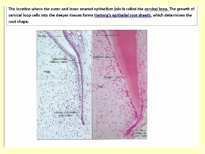
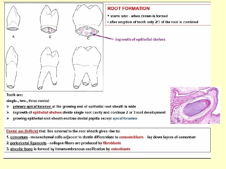
DEVELOPMENT OF THE ROOT HERTWIG’S ROOT SHEATH
HERTWIG-root sheath Composed by the inner and outer enamel epithelium
DEVELOPMENT OF THE ROOT 1. Formation of the Hertwig’s root sheath 2. Formation of the odontoblasts
DEVELOPMENT OF THE ROOT 3. dentinogenesis 4. desintegration of the Hertwig’s sheath 5. formation of the epithelial islands of Malassez
DEVELOPMENT OF THE ROOT 6. cementogenesis
Development of the root
ERUPTION • Axial movement toward oral epithelium starts when the root develops. http: //www. anat. ucl. ac. uk/research/arnett_lab/ 1 mm
Changes in epithelium during eruption enamel cuticle oral ep. junctional ep. reduced enamel ep.
ERUPTION Alveolar bone and connective tissue are resorbed as teeth erupt. osteoclasts http: //www. anat. ucl. ac. uk/research/arnett_lab/
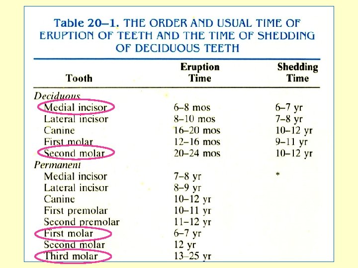
Timetable for tooth development
Development of permanent dentition Embryonic age 0 1 Primary incisor Permanent incisor 2 bud 3 4 5 Cap bell mineralization of crown bud Cap 6 bell tooth germs of primary teeth dental lamina tooth germs of permanent teeth 7 8 months mineralization
Abnormalitis of the teeth 1. Abnormal number of the teeth total anodontia (anodontia totalis) supernumerary teeth (dentes supernumerarii) 2. Abnormal shape of the teeth fused teeth twinning of teeth 3. Abnormal size of the teeth microdontia (small teeth) macrodontia (large teeth)
Molecular mechanism of tooth development • Many genes control tooth development - shape, number of cusp (incisor, molar) – size – number (2 vs 3 molars…. . ) – location (mesio-distal, maxillo-mandibular…. ) – timing of formation and eruption
Abnormalitis of the development of the enamel 4. Amelogenesis imperfecta mutation of the amelogenin or enamelin genes amelogenesis imperfecta
Abnormalitis of the development of the dentin 5. Dentinogenesis imperfecta DSPP GENE MUTATION (dentinsialo-phosphoprotein) Dentinogenesis imperfecta
Summary of the tooth development
Thank you for your attention! Tooth development - You. Tube