ANATOMY OF UTERUS Dr K Bharathi Professor Head

ANATOMY OF UTERUS Dr. K. Bharathi Professor & Head Dept. of Prasutitantra & Striroga National Institute of Ayurveda Jaipur

Three avarthas of Yoni

Description of Uterus in Ayurvedic Classics

THE UTERUS �Synonyms: metra, womb, matrix �A pear shaped hollow muscular organ �Measuring around 7. 5 x 4. 0 x 2. 5 cm in the longitudinal, transverse, and anteroposterior diameters. �It is slightly larger in the multipara than in the nullipara.

�Anteversion = a tipping forward of an organ �Anteflexion = bending forward of an organ �Pyriform = pear – shaped �Hollow = having a space or cavity inside; not solid; empty

Divisions 1. The corpus uteri: � Body that lies above the internal os � Cornu = the area of insertion of the fallopian tubes � Fundus lies above the insertion of the tubes. � Three structures are attached to the cornu �round ligament anteriorly, �Fallopian tube centrally, �ovarian ligament posteriorly.

Divisions 2. The isthmus: �an area 4 -5 mm in length that lies between the anatomical internal os above, and the histological internal os below. It is lined by low columnar epithelium and few glands. �The isthmus expands during pregnancy forming the lower uterine segment (10 cm) during the last trimester.

Divisions 3. The cervix: � The elongated lower part of the uterus � Measuring 2. 5 -3. 0 cm. � Divided by the vaginal attachment into � supravaginal portion above � vaginal portion (portio-vaginalis) below. � The cervical canal is the cavity that communicates above with the uterine cavity at the internal os and below with the vagina at the external os. � The external os is round in nulliparas and slit shaped in multiparas. � The cervical mucosa has two ridges (anterior and posterior) from which transverse ridges radiate to form the arbor vitae uteri.

Dissection showing the cephalic aspect of the female genitalia and its relations

Ventral view of a deep dissection of the urinary bladder and the blood supply to the left side of the internal genitalia, showing the relation of the uterine vessels to the ureter
, with the")
Position � The uterus is kept in an anteverted anteflexed position (AVF), with the external os lying at the level of the ischial spines, by the support of the cervical ligaments, endopelvic fascia and pelvic floor muscles (levator ani). � Anteversion: The uterus is inclined anteriorly to axis of the vagina. � Anteflexion: The body of the uterus is bent forwards upon the cervix.

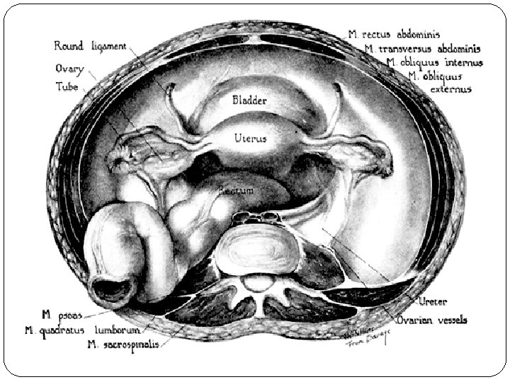
Relations of the Body of the Uterus �Anteriorly: �The bladder and vesicouterine pouch. �Posteriorly: �The pouch of Douglas. �Laterally: �The broad ligament on each side.

Relations of the Supravaginal cervix �Anteriorly: �Urinary bladder. �Posteriorly: �Forms the anterior wall of Douglas pouch. �Laterally: � 1/2 an inch lateral to the internal os the ureter is crossed by the uterine artery (i. e. ureter below the uterine artery). �The uterosacral, cardinal, and pubocervical ligaments are attached to its posterior, lateral, and anterior surfaces respectively.

Ligaments of the Uterus �The uterus is partially supported by three pairs of ligaments. �The paired round ligaments extend from the anterosuperior surface of the uterus through the internal inguinal rings and through the inguinal canals to end in the labia majors. �They are composed of muscle fibers, connective tissue, blood vessels, nerves, and lymphatics.

�The round ligaments stretch with relative ease, particularly in pregnancy. �The uterosacral ligaments are condensations of endopelvic fascia that arise from the posterior wall of the uterus at the level of the internal cervical os. �They fan out in the retroperitoneal layer and attach broadly at the second, third, and fourth segments of the sacrum. �They are predominately composed of smooth muscle but also contain connective tissue, blood vessels, lymphatics, and parasympathetic nerve fibers.
 or transverse cervical ligaments arise from the anterior and posterior")
�The paired cardinal (Mackenrodt's) or transverse cervical ligaments arise from the anterior and posterior marginal walls of the cervix and fan out laterally to insert into the fascia overlying the obturator muscles and the levator ani muscles. �The cardinal ligaments form the base of the broad ligament. They are composed of perivascular connective tissue and nerves that surround the uterine artery and veins. �The cardinal and uterosacral ligament complex is collectively called the parametrium.
 � 2. Myometrium (musculosa)")
Histology of the Uterus �Three layers: � 1. Endometrium: (mucosa) � 2. Myometrium (musculosa) � 3. The peritoneal covering or perimetrium

Histology of the Uterus Endometrium: � Lined by simple cubical or columnar epithelium � Contains tubular glands. � Shows cyclic changes with the menstrual cycle under the influence of ovarian hormones

Histology of the Uterus Myometrium �Three layers �outer longitudinal muscle layer �middle layer of interlacing crisscross muscle fibres surrounding the blood vessels �inner circular muscle layer

Histology of the Uterus Perimetrium: �Anteriorly: �firmly attached to the fundus and body till the isthmus where it becomes loose and is reflected on the superior surface of the urinary bladder forming the vesicouterine pouch. �Posteriorly: �firmly attached to the fundus, body, cervix, and posterior vaginal fornix then is reflected on the pelvic colon forming the Douglas pouch. �Laterally: �the anterior and posterior peritoneal coverings blend as the anterior and posterior layers of the broad ligaments.

Histology of the Cervix � Endocervix: Lined by simple columnar epithelium with compound racemose glands or crypts that are liable to chronic infection. It secretes alkaline cervical mucus. � Muscle layer: Outer longitudinal and inner circular muscles. (2 layers only) � Ectocervix: Formed of stratified squamous epithelium covering the outer portion of the cervix. The junction between squamous and columnar epithelium at the external os is either abrupt or it may form a transitional zone 1 -3 mm known as the transformation zone.

Blood Supply Arterial supply: � THE UTERINE ARTERIES �Arise from the anterior division of internal iliac artery. �in the base of the broad ligament, crossing above the ureter 1/2 an inch lateral to the supravaginal cervix. � 2 branches: �An ascending �A descending branch

Blood Supply � The ascending branches pass upwards in a tortuous manner parallel to the lateral border of the uterus between the 2 layers of the broad ligament to end by anastomosing with branches of the ovarian arteries near the uterine cornu. � The descending cervical branch supplies the lower cervix.

Blood Supply � Venous drainage: � Starts as a plexus between the 2 layers of the broad ligament (Pampiniform plexus) that communicate with the vesical plexus and drains into the uterine and ovarian veins. � Lymphatic drainage: � Fundus: To the para-aortic lymph nodes via ovarian vessels. � Cornu: To the superficial inguinal lymph nodes via lymphatics of the round ligament. � Body: To the internal then external iliac lymph nodes via the uterine vessels. � Isthmus: As that of the cervix. � Cervix: Two groups of lymphatics: � Primary groups: Paracervical, parametrial, obturator, internal and external iliac nodes. � Secondary groups: Common iliac, para-aortic, and lateral sacral lymph nodes.

Nerve supply of the Uterus �The cervix and body are relatively insensitive to touch, cutting and burning. �The cervix is sensitive to dilatation and the body is sensitive to distension. �Innervations �Parasympathetic form S 2, 3, 4 �Sympathetic from: �T 5 and T 6 (motor) �T 10, T 11, T 12, and L 1 (sensory). �Both reach the uterus through branches of inferior hypogastric plexus.

THE VAGINA �A fibromuscular tube from the vulva to the uterus forming an angle of 60° with the horizontal plane. �Vagina = Sheath �Length: �anterior wall is 8 -9 cm �posterior wall is 10 -11 cm �Vaginal Fornices: The cervix projects in the upper blind end of the vagina that forms a pouch (vaginal pouch) around the cervix and is divided into four fornices : two lateral,

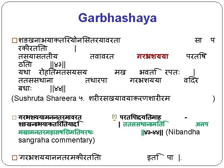
YONI � Definition: ‘Yujyate yoni’ � Synonyms: गहय पसथमपतय धव य न परजनन भगम || (Paryayaratnamala – 520) other synonyms – garbhavartma, raktapatha, manobhavagara mukha Shape: Shankhanabhi – hollow of the conch shell शङखन भय कत रय न सतरय वरत स परक रत त | तसय सतत य तव वरत गरभशयय परत षठ त || (Su. Sha. 5/43) Importance – Surgical anatomy: Is considered as one of the important parts, and hence advised to avoid injury to this

Anatomical Relations of the Vagina � Anteriorly: �Upper 1/3: trigone of urinary bladder �Lower 2/3: urethra. � Posteriorly: �Upper 1/3: peritoneum of Douglas pouch. �Middle 1/3: ampulla of rectum. �Lower 1/3: the perineal body. � Laterally: �Lower end: Bulbocavernosus muscle, vestibular bulb, and Bartholin gland. � 1 cm above orifice: urogenital diaphragm � 2½ cm above the orifice: levator ani muscle with the pelvic fascia above it. �The lateral fornix gives attachment, to the lower part of the cardinal ligaments. �The ureters pass through the cardinal ligaments 1 cm lateral to the vagina.

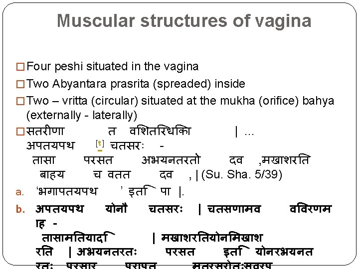
Relations of the Vagina according to Ayurveda �Superiorly – Uterus �तसय सतत य परत षठ त तव वरत गरभशयय �Laterally – extra peshi of female like – shukra artava praveshini, �Superiorly – smaratapatra (clitoris) �Inferiorly – bahirmukha srotas - external orifice opening outside �Around/ encircled by three avartas (envelops)

Structures of the Vagina: 3 Nadis – Sameerana, Chandramukhi, Gauri

Functions of vagina/Apana vayu �Artava vahana/ rakta vahana – carrying down of menstrual blood through the external orifice (bahirmukha srotas) �Garbha nishkrama – during the time of labour

Nadis of the vagina �Madanata patra �Kandarpageha �Upasthagarbha

Vaginal Supports �Ligaments attached to the upper vagina: �Pubocervical ligament anteriorly �Mackenrodt’s ligament laterally �Uterosacral ligament posteriorly �Levator ani muscles : pubo-vaginalis part �Triangular ligament, and the Perineal membrane. �Vaginal fascia: Connective tissue fascia that condenses anteriorly forming the vesico-vaginal fascia and posteriorly forming the recto-vaginal fascia.

Histology of the Vagina � The cut section of the vagina is “H” shaped with approximation of the anterior to the posterior vaginal walls. It is formed of � Three layers; �mucosa, formed of squamous epithelium without glands, the �musculosa, which is fibromuscular with some fibres from the levator ani inserted into it, and the �adventitia, which is connective tissue continuous with the paracolpos.
 �Additional branches from:")
Arterial supply: Blood Supply �The vaginal artery (from internal iliac artery) �Additional branches from: �Middle rectal artery (from internal iliac artery) �Inferior rectal artery (from the internal pudendal artery, of the internal iliac artery) Venous drainage: n A plexus around the vagina (the vaginal plexus), drain into the internal iliac vein by veins that accompany their corresponding arteries.

Lymphatic drainage and Nerve Supply �Lymphatic drainage of the vagina �lower 1/3 drains to the inguinal lymph nodes, �upper 1/3 follows lymphatic drainage of the cervix, �middle 1/3, drains in both upper and lower directions. �Nerve supply of the vagina: �The pudendal nerve gives sensory fibres to the lower vagina.
")
Applied Anatomy � Vaginal Prolapse: Weakness of the vaginal supports (ligaments, fascia and muscles) may lead to: �descent of anterior vaginal wall (cystocele or urethrocele), �descent of posterior vaginal wall (rectocele or enterocele), or �descent of the vaginal vault after hysterectomy (vault prolapse).

Applied Anatomy � The posterior fornix: �offers a passage to the pouch of Douglas for performing culdoscopy, culdocentesis and for drainage of a pelvic abscess. � The lateral fornix: �The ureter lies 1 -2 cm lateral to it so that it may be injured during clamping the angle of the vagina in hysterectomy operation.

Applied Anatomy �Pudendal nerve block: �Transvaginal injection of a local anaesthetic solution around the pudendal nerve as it passes around the ischial spine gives a local anaesthesia sufficient for minor operations on the vulva and vagina, and has been used for low forceps operations in obstetrics.

Applied anatomy - Ayurveda �Yonisamvarana – complications of Mudhagarbha �Yonibhramsha – bad prognosis of Mudhagarbha �Yonivyapad treatments – Veshavarabndha, Yonipurana etc �Uttaravasti Procedures – Insertion of vastinetra
- Slides: 43