Diseases of Skin and Eyes Anatomy of Skin


- Slides: 29

Diseases of Skin and Eyes

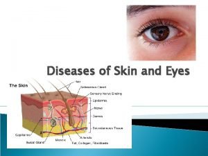
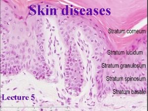
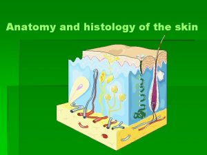
Anatomy of Skin Layers of skin (3) Epidermis Dermis Subcutaneous ◦ Gives rise to hair follicles and sebaceous glands (oil and sweat)

Function of the skin Protection from environmental factors Barrier to infection Sensitive to pressure ◦ Pacinian corpuscles Nerve endings for light touch/light sensation – hairs standing on end when excited/nervous

Modes of infections (Manifestations of Infections in Skin) Break or breach of the skin Associated with systematic infection Toxin mediated by microorganism that is actually infecting some other site of body and producing toxins that effect the skin

Modes of infections (Manifestations of Infections in Skin) Break or breach of the skin ◦ Enter the body through carbuncles etc. . . usually associated with puss

Furnucle

Modes of infections (Manifestations of Infections in Skin) Associated with systematic infection – see symptoms on the skin ◦ ◦ Chicken pox Herpes Syphilis Small pox

Modes of infections (Manifestations of Infections in Skin) Toxin mediated by microorganism that is actually infecting some other site of body and producing toxins that effect the skin ◦ Scarlet fever Streptococcus pyogenes is causative agent ◦ Scalded skin syndrome – like third degree burns Staphylococcus aureus is causative agent

Scalded Skin Syndrome

Normal Flora on Our Skin Normal flora on our skin- we want to have these microorganisms because they outcompete most other invading microorganisms for space and nutrients. ◦ Staphylococci ◦ Yeast ◦ Gram negative enterics ◦ Diptheroids

Skin Bioload (Microbiota) Variety and amount of microorganisms on our skin is affected by temperature moisture p. H amount of sweat produced ◦ chemicals excreted ◦ ◦ Oleic acid Urea Sebum

Bacterial Pyoderma (skin w/ puss) aka Cellulitis Most common causes of skin infections are Staphylococcus aureus and Streptococcus pyogenes. Clinically Presented as: ◦ ◦ Carbuncles Boils Pimples Furnucles

Bacterial Diseases - Impetigo Most common skin infection in children. Signs and symptoms: local infections, characterized by isolated pustules that become crusted and rupture Transmission: mostly through contact, bacteria penetrate skin through minor abrasion or insect bite Causative agent: S. pyogenes Tx: Topical antibiotic-Bactroban, Systemic antibiotics-Cephalexin, Erythromycin

Bacterial Infection - Folliculitis Infection of hair follicle Signs and symptoms: rash, pimples surrounding hair follicle, itching, reddened skin area Transmission: result of injury or damage to hair follicle Causative agent: S. aureus Tx: Topical antibiotic-Bactroban, Systemic-Erythomycin

Necrotizing Fasciitis Severe type of tissue infection that can involve the skin, subcutaneous fat, the muscle sheath (fascia), and the muscle. It causes gangrenous changes, tissue death, systemic disease, and frequently death. Signs and Symptoms: Severe pain in the area swelling in the area Discoloration in the area May appear reddened, bronzed, bruised, or purple (purpuric) Progresses to dusky, dark color Bleeding into the skin Visible dead (necrotic) tissue Skin color, patchy Skin breaks (open wound) Skin around the wound feels hot and looks reddened, raised, or discolored (inflammed) Oozing fluid ranging from yellowish clear or yellowish bloody to pus-like fever General ill feeling (malaise)

Necrotizing Fasciitis Causative Agent: Streptococcus pyogenes Mycobacterium ulcerans Vibrio vulnificus) Tx: Powerful broad-spectrum antibiotics must be administered immediately. They are given intravenously (in a vein) to attain high blood levels of the antibiotic in an attempt to control the infection. Surgery is required to open and drain infected areas and remove (debride) dead tissue. Skin grafts may be required after the infection is cleared. If the infection is in a limb and cannot be contained or controlled, amputation of the limb may be considered. Sometimes pooled immunoglobulins (antibodies) are given by vein to help fight the infection. Fatalities are high

Erysipelas A type of (cellulitis) skin infection ◦ Signs and Symptoms: An erysipelas skin lesion typically has a raised border that is sharply demarcated from normal skin. The underlying skin is painful, intensely red, hardened (indurated), swollen, and warm. Facial erysipelas classically involve the cheeks and the bridge of the nose. Blisters may develop over the skin lesion. Fever and shaking chills are common ◦ Causative Agent: S. pyogenes

Anthrax Etiology Bacillus anthrax Gram-positive Endospore forming 3 clinical manifestations ◦ Cutaneous ◦ Pulmonary ◦ GI

Cutaneous Anthrax Flu like symptoms Malignant pustule called eschar – surrounded by swelling (edema) redness black in the middle becomes a scab. Not common – associated with an occupation or hobby that involves livestock or ranching

Pulmonary Anthrax – Woolsorter’s Disease ◦ Spores can be inhaled in the lungs ◦ Very pathogenic ◦ Known as Wool Sorters disease – associated with livestock ◦ Organism is beta hemolytic with a double zone of hemolysis ◦ Organism has a polyglutamic acid capsule ◦ Organism produces at least three toxins Destroy tissues and cells Promotes growth of organism in tissues If it becomes systemic it can spread through lymph ◦ Vaccination available but it is usually not give to humans (just animals) ◦ PCN is drug of choice

Leprosy Etiology Mycobacterium leprae Causative agent related to TB Development of multiple lesions on skin Loss of sensory perception in areas of skin that have been infected ◦ Areas that are cooler than body temperature ◦ Fingers ◦ Toes ◦ Nose ◦ Elbows ◦ Ears Organism doesn’t actually damage but the body’s cell mediated response destroys the nerve endings (pacinian corpuscles) Organism is acid fast

Leprosy The average generation period is about 12 -14 days. 1 cell becomes 2 during this period. Pretty slow growing bug. Incubation period is 12 weeks to 40 years! With a mean of two years BCG – vaccine for TB, preventative for leprosy Not very common Tx - topical steroids can be used to control swelling. Dapsone used for treatment.

Toxic Shock Syndrome Severe disease caused by a toxin made by S. aureus or S. pyogenes, characterized by shock and multiple organ dysfunction. ◦ Signs and Symptoms: High fever, sometimes accompanied by chills Profound malaise Nausea, vomiting and/or diarrhea Diffuse red rash resembling a sunburn Rash followed in 1 or 2 weeks by peeling of the skin, particularly the skin of the palms or soles Redness of eyes, mouth, throat Confusion, seizures, headaches Myalgias (muscle aches) Hypotension (low blood pressure) Organ failure (usually kidneys and liver)

Scarlet Fever Causative agent: S. pyogenes Childhood disease famous in 1800 s Begins as pharyngitis, organism begins to produce toxin Symptoms: ◦ Pinkinsh-red rash covers the whole body except palms of hands and soles of feet ◦ Rash is the body’s rxn to the circulating toxin ◦ Tongue has spotted, strawberry appearance and the upper membrane is lost. Red and large

Scarlet Fever Signs

Conjunctivitis – Pink eye Bacterial conjunctivitis due to the common pyogenic (pus-producing) bacteria causes marked grittiness/irritation and a stringy, opaque, grey or yellowish mucopurulent discharge (gowl, goop, "gunk", "eye crust") that may cause the lids to stick together (matting), especially after sleeping. Common etiologies: S. aureus and C. trachomatis

Diseases of Eyes Bacterial Neonatal Gonorrheal Opthalmia: serious form of conjunctivitis Symptoms: Acute infection with much pus formation At more advanced stages ulcers form on cornea infection carries high risk of blindness Causative Agent: Neisseria gonorrhoeae Transmission: acquired as infant passes through the birth canal Rx: oral Tetracycline or Erythromycin drops for prevention

Viral Disease of Eye Keratitis is a condition in which the eye's cornea, the front part of the eye, becomes inflamed. The condition is often marked by moderate to intense pain and usually involves impaired eyesight. Herpetic Keratitis: Herpes simplex keratitis is a serious viral infection. It may have recurrences that are triggered by stress, exposure to sunlight, or any condition, disease or treatment which impairs the immune system. ◦ Symptoms: eye pain Impaired vision Eye redness White patch on the cornea Sensitivity to light Increased tearing Causative Agent: Herpes simplex 1 (cold sores) Transmission: problem for contact lens wearers especially

Protozoan Disease Acanthameoba keratitis Frequently results in severe eye damage Symptoms: eye pain Impaired vision Eye redness White patch on the cornea Sensitivity to light Increased tearing Transmission: contact lenses Tx: Topical ointment (propamidine or miconazole), corneal transplant or eye removal may be required
 Define a primary skin lesion and list three types
 Chapter 8 skin disorders and diseases
 Milady chapter 8 skin disorders and diseases
 Blue eye phenotype
 Brown eyes are dominant to blue eyes
 Anemia eyes vs normal
 Eyes punnett square
 Eye color hazel
 4 eyes in 4 hours
 Organum germinativum
 Thin skin vs thick skin
 Milady chapter 23 facial review questions
 Modern lifestyle and hypokinetic diseases
 Is athlete's foot communicable or noncommunicable
 Section 19-3 diseases caused by bacteria and viruses
 Chapter 6 musculoskeletal system diseases and disorders
 Chapter 24 sexually transmitted diseases and hiv/aids
 Chapter 22 genetics and genetically linked diseases
 Chapter 21 mental health diseases and disorders
 Chapter 17 reproductive system diseases and disorders
 Chapter 15 nervous system diseases and disorders
 Milady nail diseases and disorders
 Nail disease
 Icd 10 morbus hansen
 Chapter 8 cardiovascular system
 Pulpitis classification
 Venn diagram of communicable and non-communicable diseases
 Purulent diseases of lungs and pleura
 Tronsmo plant pathology and plant diseases download
 Tronsmo plant pathology and plant diseases download
