Histology and Embryology Department Human Histology n Introduction

Histology and Embryology Department Human Histology 孔 力

n Introduction 1. Goal of Histology is the study of the tissue of the body and of how these tissues are arranged to constitute organs. Cell: Tissue: made of cells and extracellular matrix Basic tissue: Epithelium Connective tissue Muscle tissue Nerve tissue

n cells and extracellular matrix n Epithelium Tissue: Connective tissue Muscle tissue Nerve tissue n n n Organ: systems : 2. Relationship between histology and other medicine courses
 resolution— 0. 2")
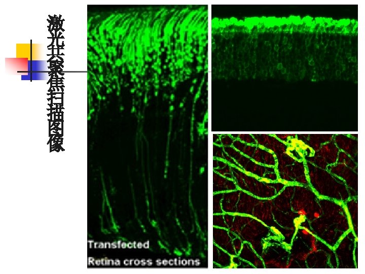
3. Histologic methods 3 -1. microscopy/显微镜 Fine structure light microscopy: (LM) resolution— 0. 2 um 1 um= 10-3 mm= 10-6 m H. E. Staining ( H: Hematoxylin E : Eosin ) basophilic, acidophilic 激光共聚焦扫描显微镜 (confocal laser scanning microscope)
 : ultra structure resolution---0. 2 nm scanning EM / 1 um=1000 nm")
Electronmicroscopy (EM) : ultra structure resolution---0. 2 nm scanning EM / 1 um=1000 nm transmission EM

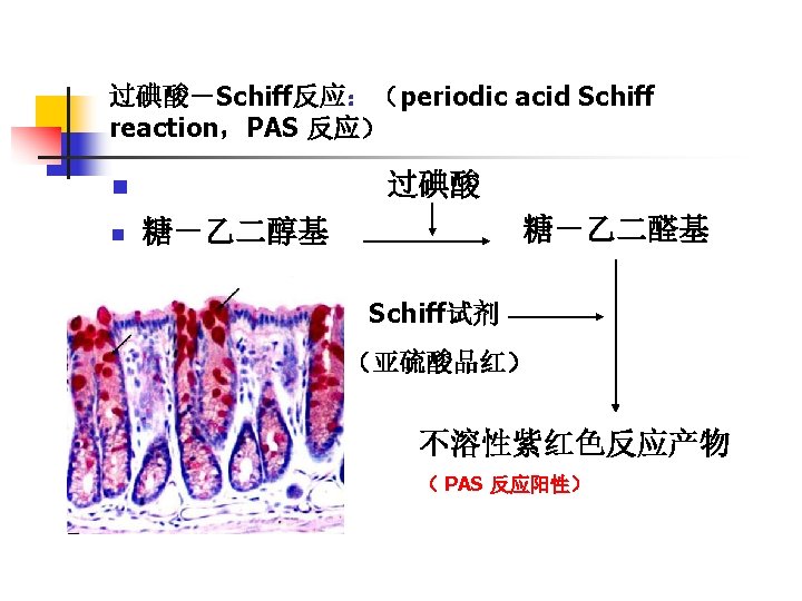
3 -2 Specific Localisation Methods for LM and EM n Histochemistry and Cytochemistry Photomicrograph of an intestinal villus stained by PAS. Staining is intense in the cell surface brush border (arrows) and in the secretory product of goblet cells (G) because of their high content of polysaccharides. The counterstain was hematoxylin.
 Molecule A has a high and specific affinity toward a portion")
Immunohistochemistry, Immunocytochemistry (1) Molecule A has a high and specific affinity toward a portion of molecule B. (2) When A and B are mixed, A binds to the portion of B it recognizes. (3) Molecule A may be tagged with a label that can be visualized with a light or electron microscope. The label can be a fluorescent compound, an enzyme such as peroxidase, a gold particle, or a radioactive atom. (4) If molecule B is present in a cell or extracellular matrix that is incubated with labeled molecule A, molecule B can be detected.

3 -3 Cell and Tissue Cultured neural crest cells seen with different optical techniques. The cells are unstained, and the same cells appear in all photographs. Use the two pigmented cells for orientation in each image. A: Conventional light microscopy. B: Phase contrast microscopy. C: Nomarski differential interference microscopy.

3 -4 Hybridization Techniques In situ Hybridization 原位杂交 Tissue section of a benign epithelial tumor (condyloma) submitted to in situ hybridization. The brown areas are places where DNA of human papillomavirus type 2 is present. The counterstain was hematoxylin.

4. Problems in the interpretation of tissue section How different 3 dimensional structures may appear when thinsectioned. A: Different sections through a hollow ball and a hollow tube. B: A section through a single coiled tube may appear as sections of many separate tubes. C: Sections through a solid ball (above) and sections through a solid cylinder (below).
- Slides: 14