A joint conference with MAPA and PANE 2009


- Slides: 33
A joint conference with MAPA and PANE 2009 Ophthalmic Pathology Emiko Furusato, M. D. Department of Neuropathology and Ophthalmic Pathology Armed Forces Institute of Pathology
Contents • Grossing the globe • Ocular histology • 10 Representative cases
Grossing the ocular specimen • Four basic steps 1. 2. 3. 4. Orient the specimen and determine laterality Measurements Transillumination (TI) Sectioning the globe
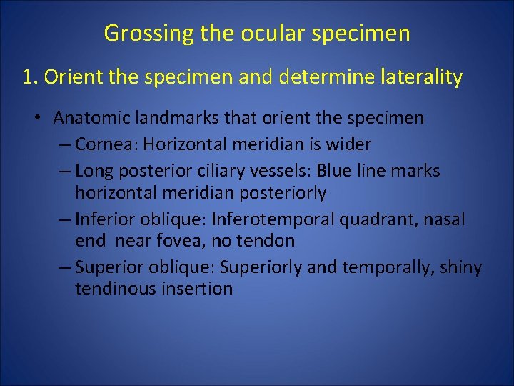
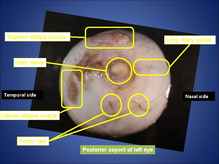
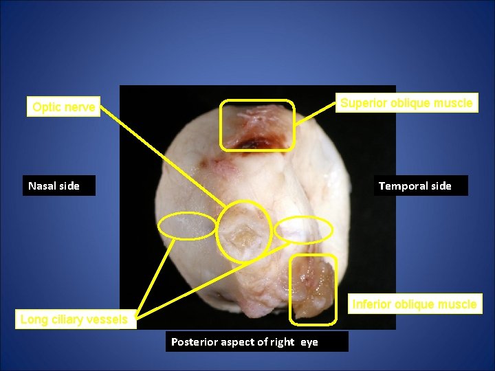
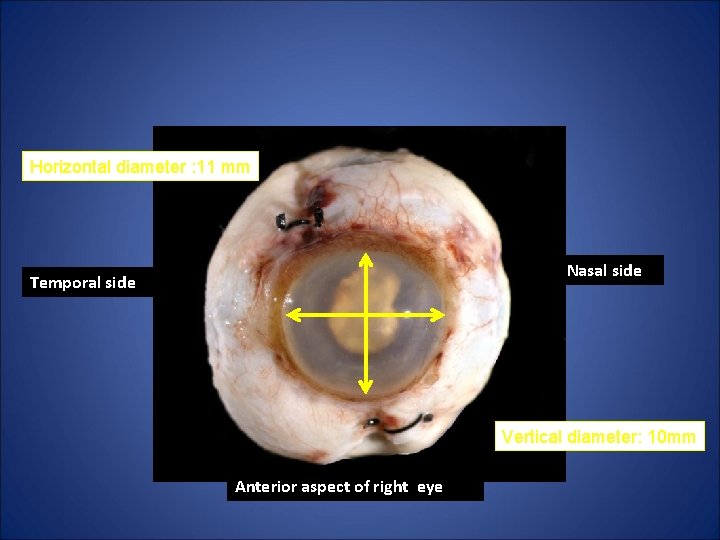
Grossing the ocular specimen 1. Orient the specimen and determine laterality • Anatomic landmarks that orient the specimen – Cornea: Horizontal meridian is wider – Long posterior ciliary vessels: Blue line marks horizontal meridian posteriorly – Inferior oblique: Inferotemporal quadrant, nasal end near fovea, no tendon – Superior oblique: Superiorly and temporally, shiny tendinous insertion
Superior oblique muscle Long ciliary vessel Optic nerve Temporal side Nasal side Inferior oblique muscle Vortex vein Posterior aspect of left eye
Superior oblique muscle Optic nerve Nasal side Temporal side Inferior oblique muscle Long ciliary vessels Posterior aspect of right eye
Horizontal diameter : 11 mm Nasal side Temporal side Vertical diameter: 10 mm Anterior aspect of right eye
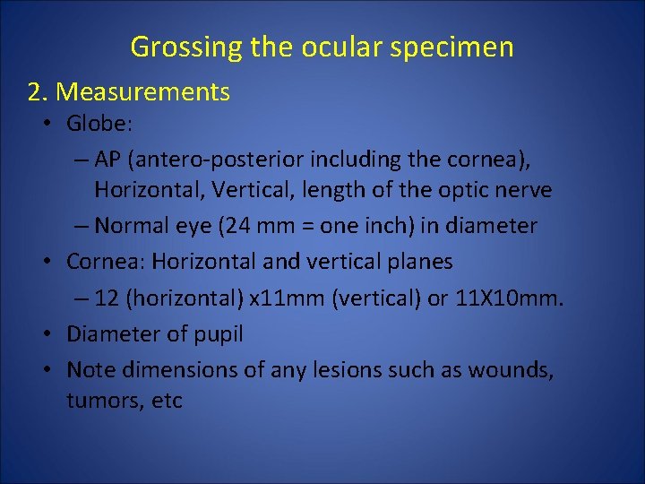
Grossing the ocular specimen 2. Measurements • Globe: – AP (antero-posterior including the cornea), Horizontal, Vertical, length of the optic nerve – Normal eye (24 mm = one inch) in diameter • Cornea: Horizontal and vertical planes – 12 (horizontal) x 11 mm (vertical) or 11 X 10 mm. • Diameter of pupil • Note dimensions of any lesions such as wounds, tumors, etc
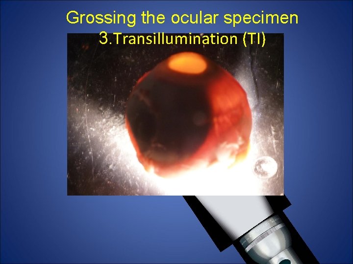
Grossing the ocular specimen 3. Transillumination (TI)
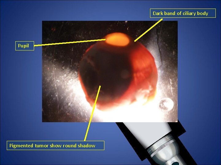
Grossing the ocular specimen 3. Transillumination (TI) • Transilluminated prior to dissection. • Use dissecting microscope illuminator in a darkened room. • Applications – Uveal malignant melanoma – Dark shadow on the sclera
Dark band of ciliary body Pupil Pigmented tumor show round shadow
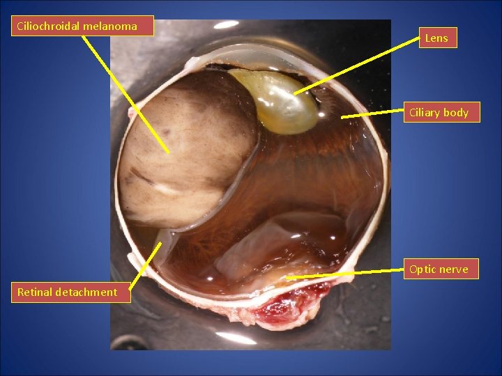
Ciliochroidal melanoma Lens Ciliary body Optic nerve Retinal detachment
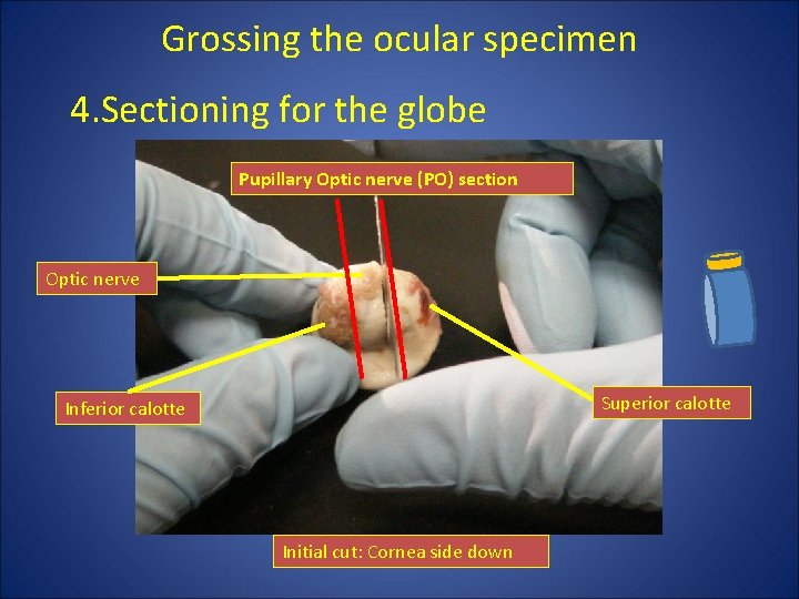
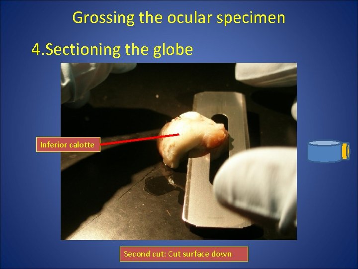
Grossing the ocular specimen 4. Sectioning for the globe Pupillary Optic nerve (PO) section Optic nerve Superior calotte Inferior calotte Initial cut: Cornea side down
Grossing the ocular specimen 4. Sectioning the globe Inferior calotte Second cut: Cut surface down
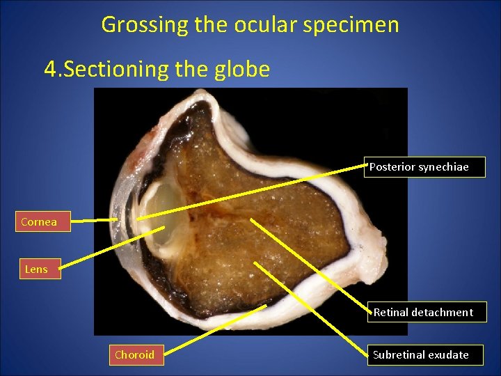
Grossing the ocular specimen 4. Sectioning the globe Posterior synechiae Cornea Lens Retinal detachment Choroid Subretinal exudate
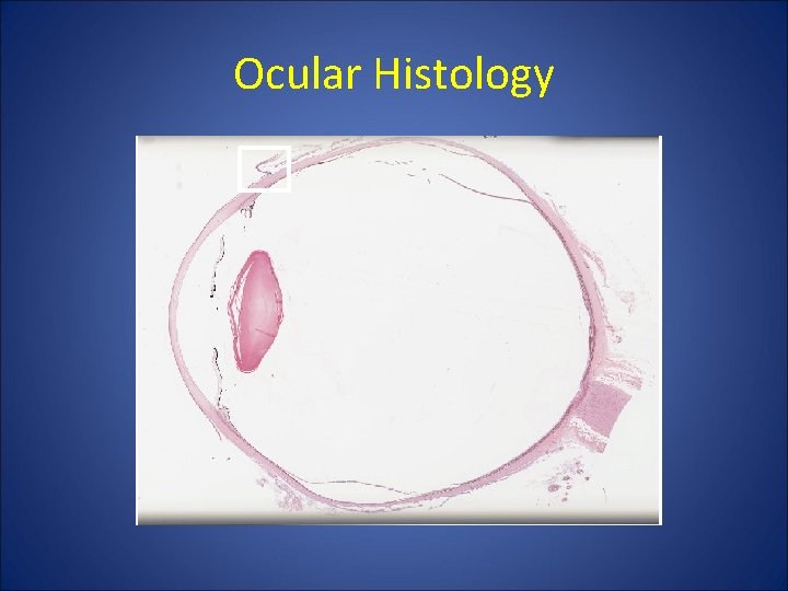
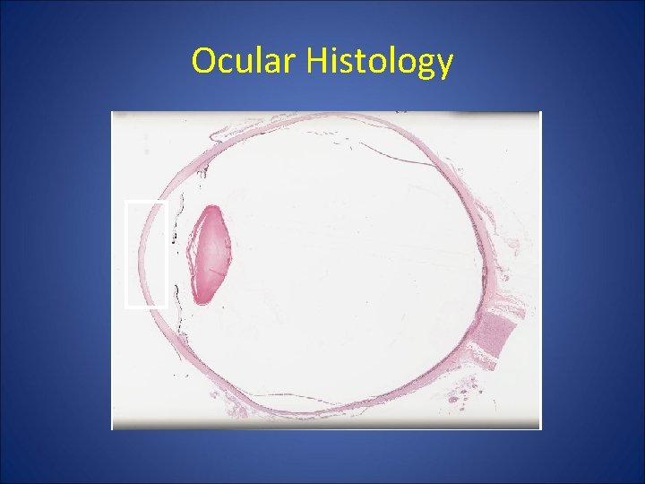
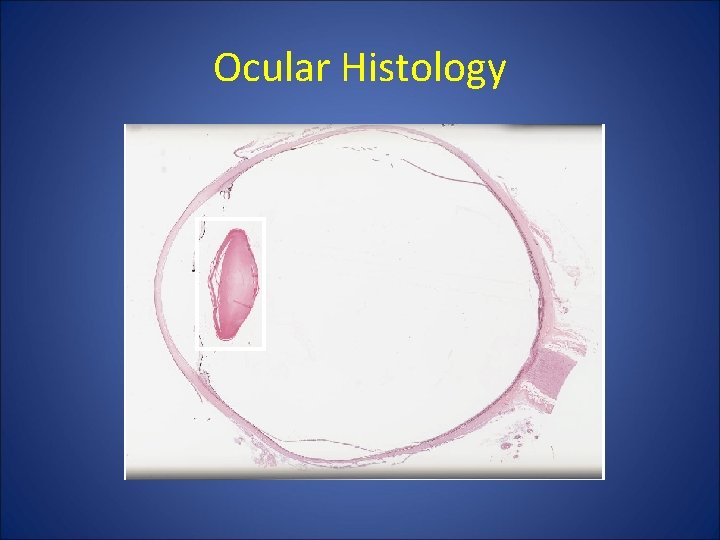
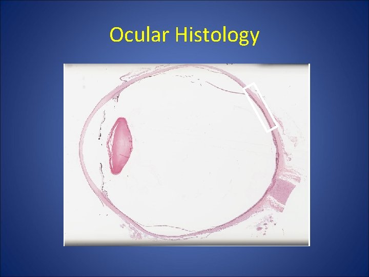
Ocular Histology • • • Conjunctiva Cornea Iris / Ciliary body Lens Retina Choroid
Ocular Histology
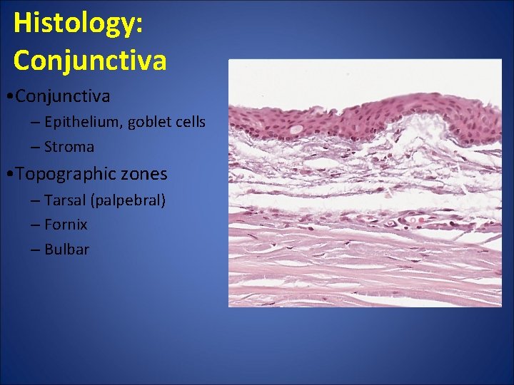
Histology: Conjunctiva • Conjunctiva ‒ Epithelium, goblet cells ‒ Stroma • Topographic zones ‒ Tarsal (palpebral) ‒ Fornix ‒ Bulbar
Ocular Histology
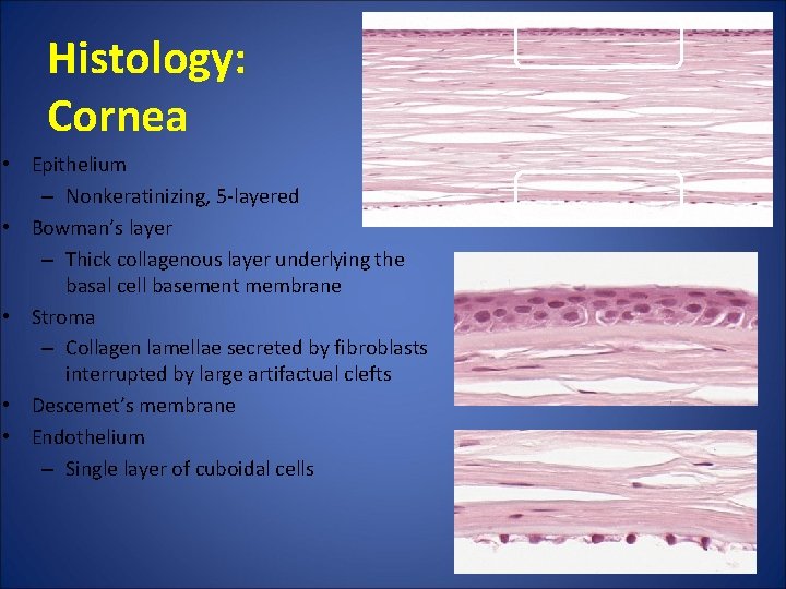
Histology: Cornea • Epithelium – Nonkeratinizing, 5 -layered • Bowman’s layer – Thick collagenous layer underlying the basal cell basement membrane • Stroma – Collagen lamellae secreted by fibroblasts interrupted by large artifactual clefts • Descemet’s membrane • Endothelium – Single layer of cuboidal cells
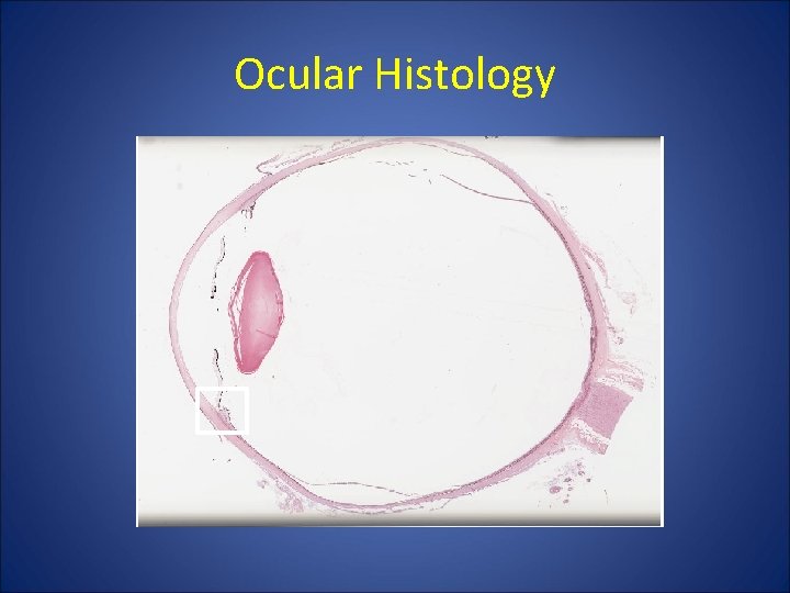
Ocular Histology
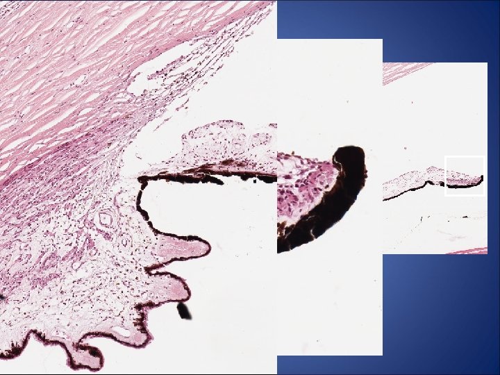
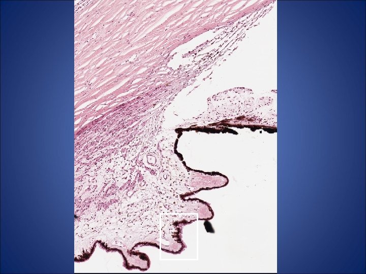
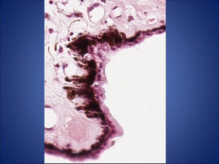
Histology: Iris & Ciliary body • Iris (anterior to posterior 4 zone) – Anterior border layer – Stroma – Partially pigmented epithelium – Completely pigmented posterior pigment epithelium Ø Sphinctor muscle appears as a ring in the pupillary stroma • Ciliary body - Nonpigmented ciliary epithelium - Pigmented ciliary epithelium - Stroma, ciliary muscle
Histology : Iris/ ciliary body • Iris – Anterior limiting layer’-Stroma • Sphincter pupillae • Pigment epithelium( double layer) • Ciliary body • Nonpigmented epithelium-Pigmented epithelium’-Stroma, ciliary muscle
Ocular Histology
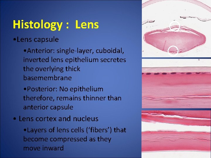
Histology : Lens • Lens capsule • Anterior: single-layer, cuboidal, inverted lens epithelium secretes the overlying thick basemembrane • Posterior: No epithelium therefore, remains thinner than anterior capsule • Lens cortex and nucleus • Layers of lens cells (‘fibers’) that become compressed as they move inward
Ocular Histology
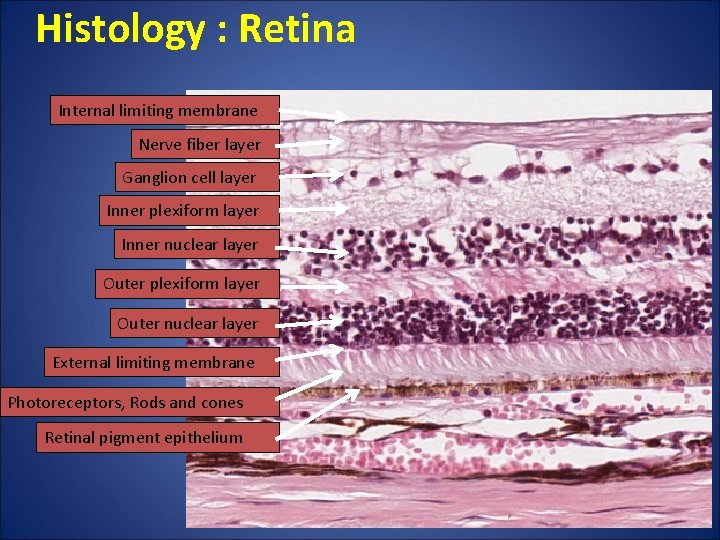
Histology : Retina Internal limiting membrane Nerve fiber layer Ganglion cell layer Inner plexiform layer Inner nuclear layer Outer plexiform layer Outer nuclear layer External limiting membrane Photoreceptors, Rods and cones Retinal pigment epithelium
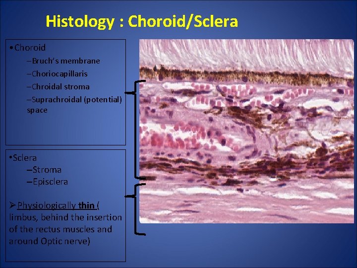
Histology : Choroid/Sclera • Choroid –Bruch’s membrane –Choriocapillaris –Chroidal stroma –Suprachroidal (potential) space • Sclera –Stroma –Episclera ØPhysiologically thin ( limbus, behind the insertion of the rectus muscles and around Optic nerve)
10 Representative cases
Conjunctival Cases
Case 1