Histology Lecture 6 Blood Composition of Blood Blood

Histology Lecture 6 Blood

Composition of Blood Ø Blood sample separates into 2 parts – plasma - straw colored liquid on top • ~55% of the volume – formed elements • ~45% of the volume – red blood cells – buffy coat: white blood cells and platelets
 Ø Packed Cell Volume is the % of the blood which is")
Hematocrit (Hct) Ø Packed Cell Volume is the % of the blood which is RBC’s ØMales: 40 -54% (47%) ØFemales: 38 -46% (42%) Ø Hct indicates the status of RBC production, the state of hydration, or various disease states

Hematocrit Procedure
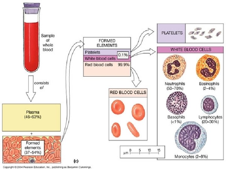

The Formed Elements of the Blood: Ø Leukocytes = White Blood Cells – Granular leukocytes (granulocytes) • neutrophils • eosinophils • basophils – Agranular leukocytes (agranulocytes) • lymphocytes - T cells, B cells • monocytes tissue macrophages

Granular Leukocytes Eosinophil 2 -4% Neutrophil 60 -70% Basophil 0. 5 -1%

Agranular Leukocytes Lymphocyte 20 -25% Monocyte 3 -8%

Leukocyte Life Span and Number Ø 5, 000 - 10, 000 WBC’s/mm 3 blood ØRBC/WBC ratio 700/1 Ø Differential WBC count (a standard clinical lab report) ØNeutrophils 60 -70% ØLymphocytes 20 -25% ØMonocytes 3 -8% ØEosinophils 2 -4% ØBasophils 0. 5 -1% Ø Abnormal proportions are correlated with different types of disease processes

Differential WBC Count Lymphocyte 20 -25% Monocyte 3 -8% Eosinophil 2 -4% Neutrophil 60 -70% Basophil 0. 5 -1%

Leukocyte Identification Agranular Granular Dark Hidden nuc. Small Spherical nucleus All have many large granules in cytoplasm & multilobed nuclei Basophil Lymphocyte Large 2+ lobes Red gran. Eosinophil no large granules in cytoplasm Monocyte Faint gran. Neutrophil
 and formed elements cells (cells) include erythrocytes,")
Blood ÏBlood consists of fluid element (plasma) and formed elements cells (cells) include erythrocytes, platelets and leukocytes ÏAdult has about 5. 5 Liters ÏHematocrit is volume of packed RBC § normal male = 40 -50%; female = 35 -45% ÏBlood usually studied in stained smear § Wright’s stain: eosin Y w/ azure A & B and methylene violet (made from base-treated methylene blue) ÏBlood cells produced in bone marrow from stem cells

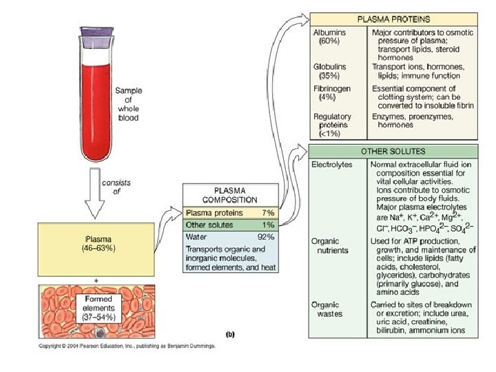
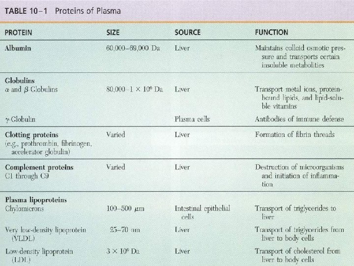
Plasma A yellowish fluid in which cells , platelets, organic compounds, and electrolytes suspended or dissolved Major component is water(90% volume) Proteins(9%) Inorganic salts ions, nitrogenous compounds, nutrients and gases constitute the remaining 1% Serum After coagulation, some of organic and inorganic components leave the plasma forming clot remaining fluid is called Serum

Erythrocyte (Biconcave disk which has no nucleus (There is about 7. 8 µm diameter in fresh state and 7. 2 - 7. 4 µm in stained smears (Large surface area for gas exchange (4 -6 million per mm³ (Flexible; squeeze when pass through capillaries (Survive about 120 days in circulation (Old RBC removed by macrophages in spleen and bone marrow (Reticulocytes are about 1% of total population

Hemoglobin is a large protein composed of four H polypeptide chains, each of which is covalently bound to a iron-containing heme group

Erythrocyte Cell Membrane ÚErythrocyte cell membrane of the RBC and the underlying cytoskeleton are highly pliable and can withstand great shear forces Most of proteins are transmembrane, principally glycophorin A, ion channels, and anion transporter band 3 protein, which act as an anchoring site for ankryin Band 4. 1 protein act as an anchoring site for glycophorins ankryin and band 4. 1 protein anchor the cytoskeleton composed of spectrin, actin and adducin
 have specific")
Leukocytes NThere is about 6 -10, 000 per mm³ NGranulocytes (polymorphonuclear leukocytes) have specific granule o neutrophils 60 -70% o acidophils 2 -4% o basophils 0. 5 -1% NAgranulocytes lack specific granule o lymphocytes 20 -30% o monocytes 3 -8% NPlatelets 200, 000 -400, 000 per mm³

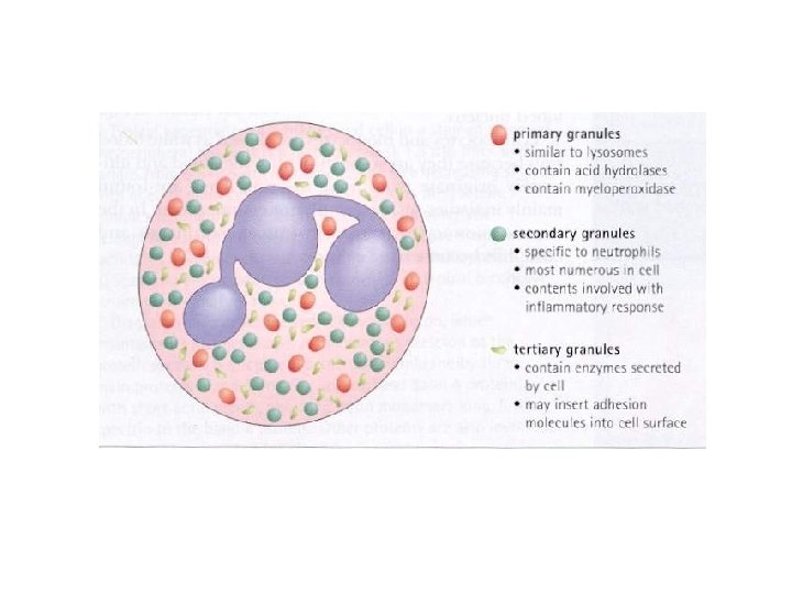
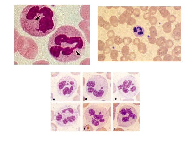
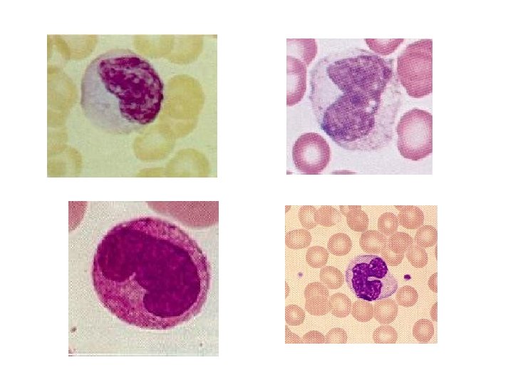
Neutrophils f 12 -15 µm in diameter f. Multi-lobed nucleus f. Human females may have inactivated second X chromosome (drumstick) f. Specific granules: salmon pink, Alkaline PO 4 ase, collagenase, lactoferrin, lysozyme and phagocytins f. Azurophilic granules: deep red or purple, primary lysosomes containing acid hydrolase myeloperoxidase, lysozyme f. Tertiatry granules: gelatinase, cathepsin

Neutrophils ¶ Half-life 6 -7 hours ¶ lifespan 1 -4 days in tissue ¶ Active phagocytes which is an important defense against infection ¶ Form H 2 O 2 and powerful cytotoxin ¶ Lysozyme breaks down bacterial cell wall ¶ Lactoferrin binds Fe which is needed by some bacteria

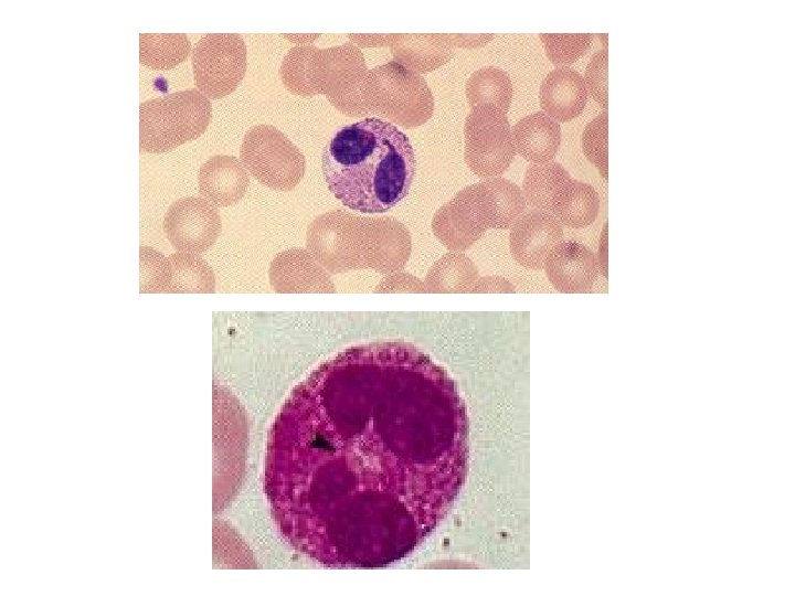
Eosinophils © About 12 -15 µm in diameter © Bi-lobed nucleus © Many granules (primary lysosomes) © Kill parasites, especially worms © Phagocytose Ag-Ab complexes formed in allergy © Specific granules of eosinophil possess an internum surrounded by externum © Internum contain: major basic protein, eosinophilic cationic protein (parasites) © Nonspecific granules are lysosomes © Corticosteroids decrease eosinophils in blood

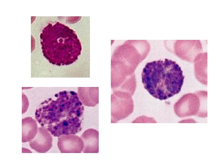
Basophils Scarce, hard to find in smears S-shaped nucleus Ig E receptors on their plasmalemma Many blue (basophilic) specific granules with heparin and histamine Content of specific granules cause vasodilatation Nonspecific granules are lysosomes Function as initiator of the inflammatory process May act as supplement of mast cell function

Lymphocytes § 20 -25 % of circulating leukocytes § 6 -8 µm dia most common; large ones up to 18 µm dia also found in blood § Dark, heterochromatic nucleus § Thin layer of blue cytoplasm, many ribosomes § Three types: T-cells(80%), B-cells (15%) and Null cells(remainder) § Differentiation occurs in bone marrow (B cells) and thymus (T cells) § T-cells may live many years, B-cells live a few months

Lymphocytes ÌAfter stimulation T-cells and B-cells become : Memory cells and Effector cells ÌB cells form plasma cells, function in humoral immunity via immunoglobulins ÌT cells function in cell-mediated immunity ÌEffector T-cells: T helper cells, T suppressor cells, cytotoxic T cells ÌSome T cells with “memory” of antigen exposure survive long periods; immunization ÌNull Cells are composed of: Stem cells and Natural killer cells ÌNK cells kill some foreign and virally alerted cells


Monocytes 812 -20 µm in diameter 8 Oval, eccentric, horseshoe or kidney shaped nucleus 8 Lighter stained nucleus than large lymphocytes 8 Cytoplasm light blue due to azurophilic granules (lysosomes) and ribosomes 8 In tissues, differentiate into macrophages 8 Lifespan 12 -100 hours 8 Do not re-enter into circulation

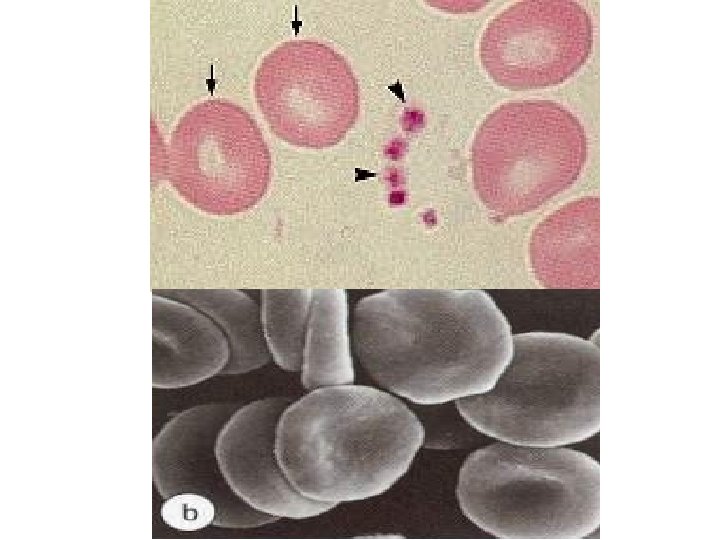
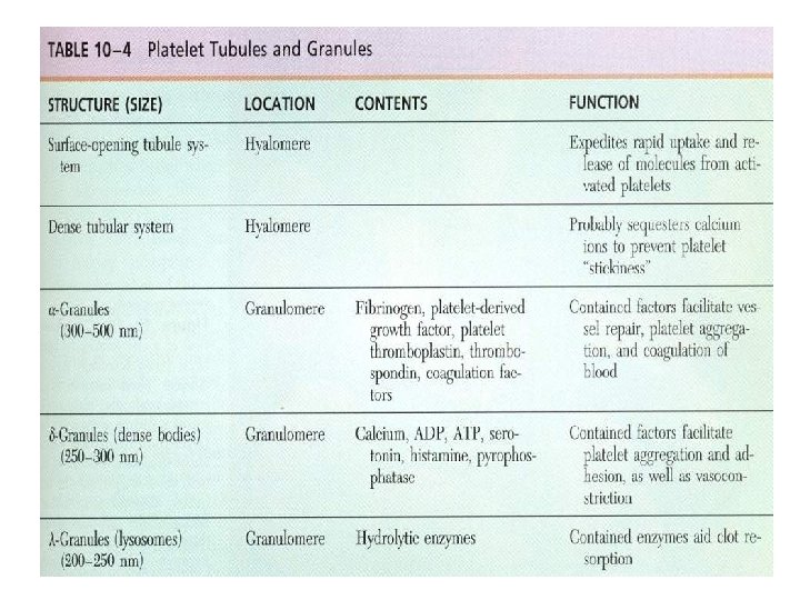
Platelets • • Non-nucleated disk-shaped cell fragments 2 -5 µm in diameter, derived from megakaryocyte cytoplasm Life span less than 14 days in blood Hyalomere is the peripheral clear region Granulomere is the central darker region Light blue cytoplasm, dark blue or purple granules Microtubule bundles at periphery Dense bodies (delta granules) contain calcium, pyrophosphate, and serotonin • Alpha granules contain fibrinogen, PDGF and other proteins • Lambda granules contain lysosomal enzymes
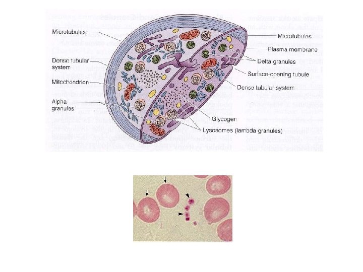

Platelet Function ©Blood clotting; aggregation of platelets ©Endothelium plasminogen activators ©Clot retraction due to actin/myosin ©Clot removal due to plasmin which is a proteolytic enzyme

Department of Histology F. Rajaei
- Slides: 38