HISTOLOGY OF TONGUE TONGUE HISTOLOGY Defn Tongue is


 part: Location- floor of oral cavity Apex- touching incisor teeth Margin-")
part: Location: forms base of tongue. Mucosa: Reflected laterally onto palatine tonsil")


- Slides: 32
HISTOLOGY OF TONGUE
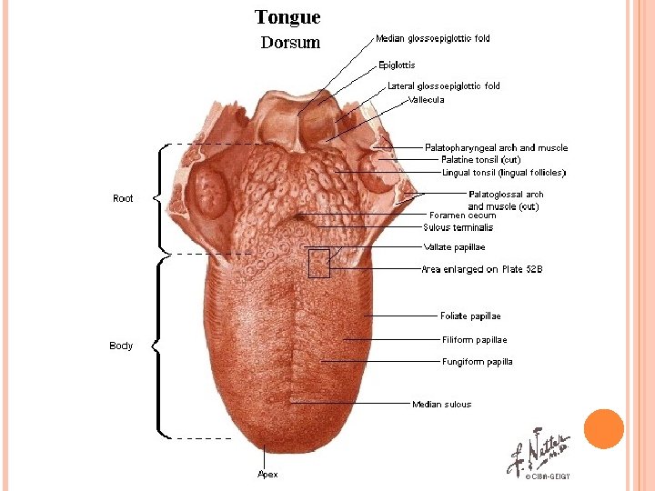
TONGUE HISTOLOGY Defn: Tongue is a highly muscular organ of deglutition, taste and speech. Position: partly oral and partly pharyngeal. Parts: - it has root apex curved dorsum and inferior surface.
Dorsum: - convex at rest divided by a v-shaped sulcus terminalis into - Anterior or oral or pre-sulcal part- anterior 2/3 - Posterior pharyngeal or post-sulcal part- posterior 1/3 Sulcus terminalis – two limbs run from -a median depression-foramen caecum (the site of the upper end of embryonic thyroid diverticulum) -anterolaterally to palatoglossal arch.
Oral (presulcal) part: Location- floor of oral cavity Apex- touching incisor teeth Margin- in contact with gums and teeth Dorsum- related to hard and soft palate.
Pharyngeal (postsulcal)part: Location: forms base of tongue. Mucosa: Reflected laterally onto palatine tonsil and pharyngeal wall.
Lingual tonsil: Has low elevation due to lymphoid nodules embedded in the submucosa collectively known as lingual tonsil. Ducts of seromucous glands open on apices of these elevations.
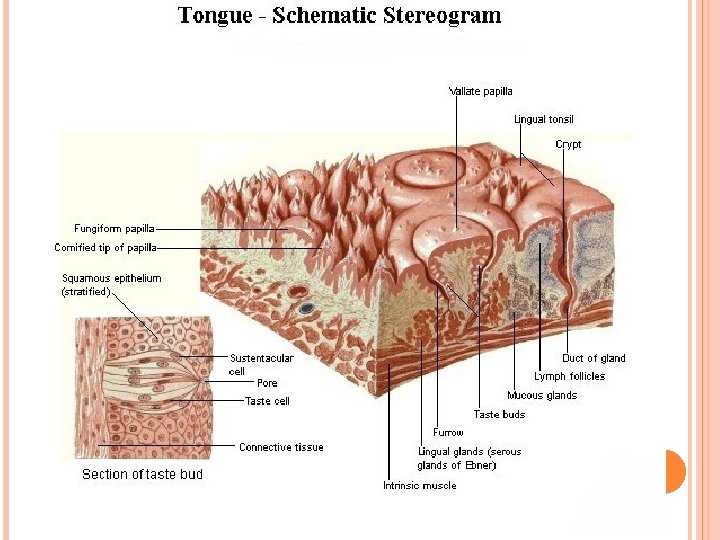
LINGUAL PAPILLAE: Defn: Projection of mucosa from dorsum of tongue. Papillae are indented by lamina propria, underlying connective tissue. Location – dorsum of tontue in the presulcal part. Types: - 4 types.
Filiform papillae – minute, conical or cylindrical They have irregular cores of connective tissue Epithelium is keratinised
papillae increase friction between tongue and food. Facilitate movement of particles by tongue with in oral cavity. In gastrointerstinal problems normal shedding is delayed and unshed cells with bacteria forms a greyish coating on the papilla.
Fungiform palillae: -Numerous than vallate but less numerous than filiform papillae. - occurs mainly on lingual margin but also irregularly on dorsum, interspersed among filiform papillae. - taller, longer and broader than filiform papilla. - Mushroom like rounder shape, lower part narrower than upper. - deep red in colour. bears one or more taste buds on its apical surface.
Foliate papillae: - 4 or 5 vertical folds bilaterally on side of tongue near sulcus terminalis. - red and leaf like. - they bear numerous taste buds.
Vallate papillae: - 8 to 12 in number. - from v-shaped row immediately in front of sulcus terminalis - 1 to 2 mm in diameter. - large cylindrical structure encircled by deep furrow. Narrower at base than apex. - covered by stratified squamous epithelium. - taste buds abound in both walls of the sulcus. - Numorous excretory ducts from underlying serous (von ebner’s) glands, located in connective tissue empty into the base of furrow.
Taste buds: -Total about 5000 taste buds. - found on fungiform, vallate and foliate papillae, absent in filiform papillae. -
Tip of the tongue most sensitive to weet and salt. Posterior part – bitter Lateral edges – sour - Posterior region of tongue Exhibits large mucosal ridges and elevations or folds resembling fungiform papilla. Stratified squamous epithelium comvers the surface. Lamina propria is wider.
- - Under epithelium aggregations of diffuse lymphatic tissue is observed. Occumulation of adipose tssue, nerve fibres and blood vessels is also observed. Deep to the lamina propria between interlacing skeletal muscle fibres are mucous acini of posterior lingual glands. Excretory ducts of posterior lingual glands open into dorsal surface of tongue between bases of mucosal ridges and folds. These glands extend through root of tongue.
TONGUE MAGNIFICATION: X 40 STAIN: H&E
FILLIFORM PAPILLAE MAGNIFICATION: X 640 STAIN: H&E
FOLIATE PAPILLAE FROM RABBIT
CIRCUMVALLATE PAPILLAE
Taste buds
HISTOLOGY OF GIT
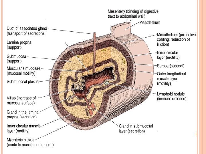
General Mucous plan: - membrane – lining epithelium lamina propria muscularis mucosa Sub mucosa Muscularis externa, Serosa or Adventitia.
Lining Epithelium: Pharynx Oesophagus Lower part of anal canal Stratified squamous non keratinized
Lamina propria : Loose connective tissue, lymphocytes & Blood vessels Supports epithelium and connects it with muscularis mucosa. § Muscularis mucosa: Double layered smooth muscle – Circular and longitudinal. Helps in localized movement of mucous membrane §
Sub mucosa: Loose connective tissue with elastic fibres, Blood vessels, lymphatics and nerves § Muscularis externa: Smooth muscle fibres – Inner circular and outer longitudinal. §
Serosa or adventitia: Serosa – Mesothelium Adventitia – connective tissue as in Oesophagus. § Nerve Plexus: Myenteric plexus. Sub mucosal Plexus. Mucosal plexus §
OESOPHAGUS Adventitia: Irregular dense connective tissue with many elastin fibres. § Muscularis externa: § Inner circular outer longitudenal Longitudinal coat is thicker than circular. upper 1/3 rd – Striated Middle 1/3 rd – striated and smooth lower 1/3 rd – smooth §
Sub mucosa: Loose connective tissue, mucous glands, blood vessels and nerves. Mucous glands open on to the epithelial surface and keep it moist & lubricated. Mucosa: Epithelium – non keratinized stratified squamous. Lamina propria – finger like papillae. Muscularis mucosa - Chiefly longitudinal muscle fibres.
Oesophagus