SPINAL CORD Histology of Spinal Cord The spinal


- Slides: 19
SPINAL CORD
Histology of Spinal Cord
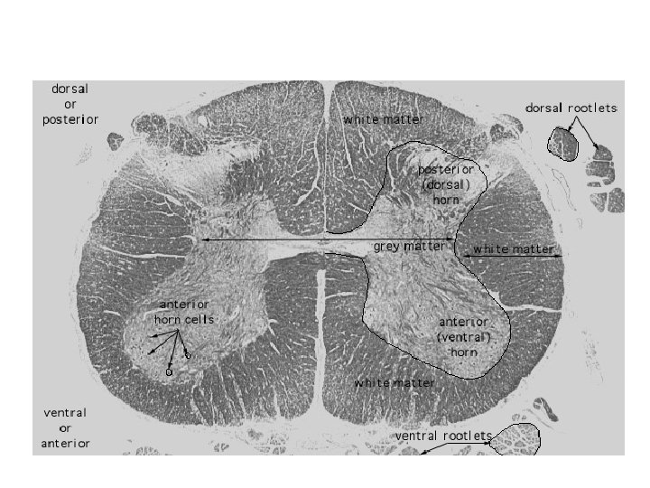
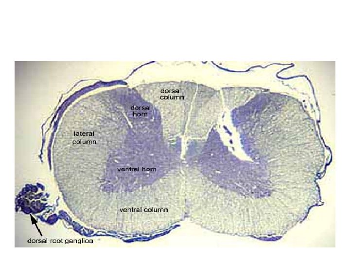
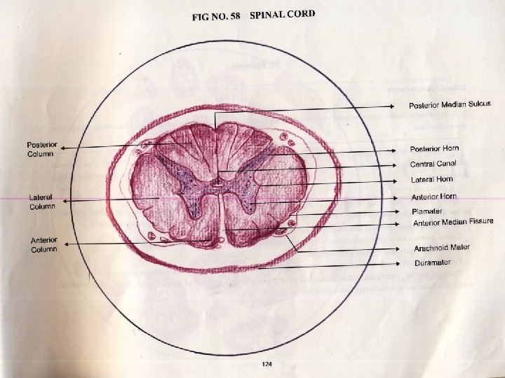
• The spinal cord is organized into two discrete parts. • The outer part contains ascending and descending nerve fibres. • These constitute white matter of the cord. • The inner part contains cell bodies of neurons and nerve fibres. • This is the gray matter of the spinal cord. • Neuroglial cells are in both white matter and gray matter.
• In the spinal cord , the gray matter roughly resembles H shape when in cross section. • Hence the gray matter is said to have two posterior horns and two anterior horns. • In the thoracic part, the cord shows lateral horns on each side as well.
• The pale staining fibrous material that surrounds the spinal cord is the piamater. • It follows the surface of the spinal cord intimately and dips into its fissure and sulci. • Blood vessels are present in the piamater.
• The white matter surrounds the H shaped region of gray matter. • White matter does not contain any cell bodies of neurons. • The fibers it contains originate from cell bodies lying either in gray matter of the brain or spinal cord or in the spinal ganglion. • The fibers are organized into tracts there are separate motor and sensory tracts.
O&A are neuroglial cells
Hand drawn diagram of spinal cord
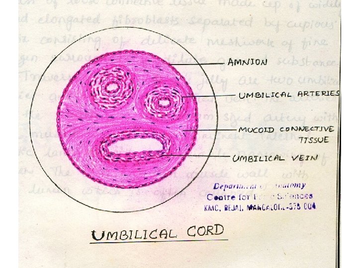
Umbilical cord • Outer covering of amniotic epithelial cells. • Inner mesenchyme or Wharton’s jelly. • Wharton’s jelly has • Glycosaminoglycans + Hyaluronic acid forming a liquid ground substance. • Collagen fibers are dispersed in this. • Stellate fibroblasts present.
• Two umbilical arteries and one umbilical vein present. • Arteries are medium sized. • Artery: • Prominent inner elastic lamina and thick tunica media with smooth muscles. • Vein: • Left umbilical vein is left. • Collapsed lumen is present. • Thin tunica media in vein.
Hand drawn diagram of umbilical cord
Slides
Umbilical cord
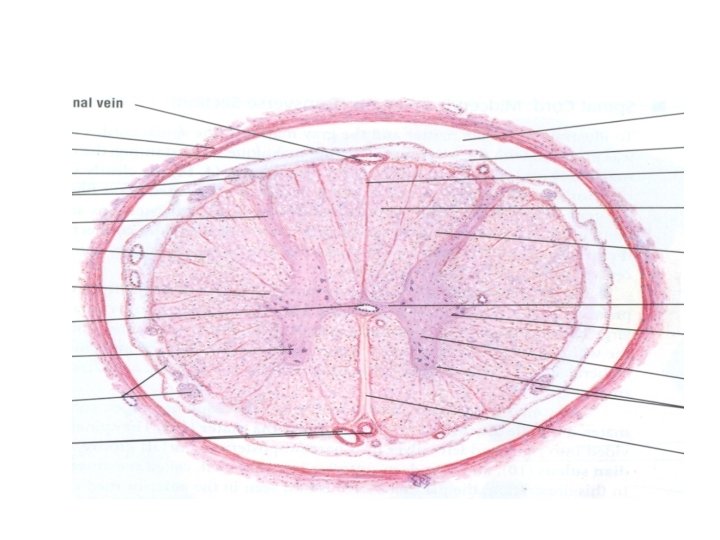
Spinal cord