Dr Andrea D Szkely Differentiation of the Neural

")
 - neuroectoderm NEURULATION formation of")


 N-Cadherin default")
")
 cavitation D 27 Below S 2 eminentia caudalis")


 (axon rich; white matter)")


")
")
")


 Oligodendrocyte nucleus (CNS) Basement membrane Nerve fiber")


 dorsolateral pathway (1) neural")


- Slides: 55
Dr. Andrea D. Székely Differentiation of the Neural Tube Development of the Spinal Cord Neural Crest Derivatives Acknowledgements to Dr Márk Kozsurek Dr Nándor Nagy
Levels of Neural Differentiation • ORGAN macroscopical changes in the neural tube (hemispheres, ventricles) • TISSUE functional regions form in the wall of the neural tube (motor, sensory, autonomous) • CELLULAR Different neuronal and glial types differentiate (histogenesis)
Milestones in Neural Tissue Differentiation INDUCTION (chorda dorsalis ) - neuroectoderm NEURULATION formation of the neural tube PROGENITOR CELLS form in the neural tube PROLIFERATION and DIFFERENTIATION of the progenitors • Neurone • Glia MIGRATION, AXONAL GROWTH Formation of SYNAPSES (communication between distant cells)
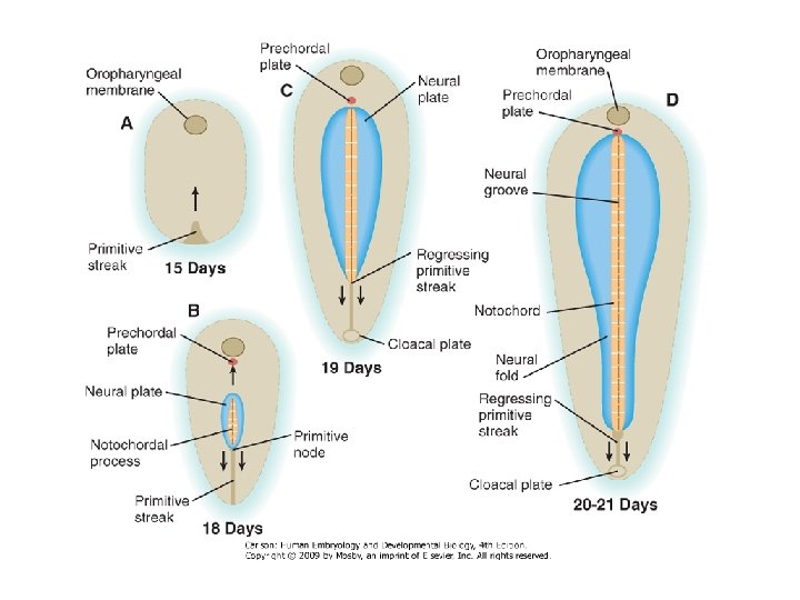
Primary Neurulation Neural induction and neurulation Specification of neural fate and formation of the neural tube. NEURAL INDUCTION : the delineation of ectodermal cells to the neural fate NEURULATION: the process in which the ectoderm of the future brain and spinal cord - the neural plate – develops, folds and forms the neural tube
Primary Neurulation
Induction of the Neural Tube epiblast BMP-2 BMP-4 follistatin chordin noggin (BMPinhibition) N-Cadherin default pathway inhibition of activin default mesoderm inhibition of default epidermis E-cadherin neural tube Numerous signal proteins inhibit the activity of BMP/Activin, thereby maintaining the default mechanism ( neural tube )
Closure of the neural tube in a „zip-lock” like manner © 2005 Elsevier )
Secondary Neurulation (described in avian embryos) cavitation D 27 Below S 2 eminentia caudalis neural tube fusion
Development of the CNS Craniocaudal patterning
Developmental Overview Neural Tube Primary Vesicles Secondary Vesicles Adult Structures Telencephalon Rhinencephalon, Amygdala, Hippocampus, Cerebrum (Cortex), Basal Ganglia, lateral ventricles Diencephalon Epithalamus, Thalamus , Hypothalamus, Subthalamus, Pituitary, Pineal, third ventricle Mesencephalon Tectum, Cerebral peduncle, Pretectum, cerebral aqueduct Metencephalon Pons, Cerebellum Myelencephalon Medulla Oblongata Prosencephalon Brain Mesencephalon Rhombencephalon Spinal Cord
Development of the Brain, Neuromeres Differentiation of the rostral end isthmus Rhombomeres midbrain i M r 1 r 3 P 6 Key element: r 2 r 4 r 5 P 5 Hox Genes P 4 r 7 -8 P 3 P 2 T 21 r 6 P 1 Prosomeres
Spinal Cord Development Composition of Neural Tube Internal limiting membrane Neuroepithelial cells Sclerotomal somitic mesoderm External limiting membrane
Neuroepithelial Cells EXTERNAL LAYER Pia mater Neural tube INTERNAL LAYER Ependyma Simple cuboidal epithelium ALSO covering the choroid plexus Neuroblast (Apolar) Glioblast Astroblast Oligodendroblast Neuroblast (Bipolar) Neuroblast (Unipolar) Astrocyte (Protoplasmic) Astrocyte (Fibrous) Oligodendrocyte Neuron
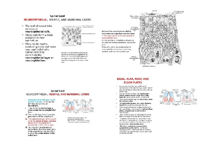
Differentiation in the Wall of the Neural Tube The wall of a recently closed neural tube consists of neuroepithelial cells (pseudostratified epithelium) Once the neural tube closes, neuroepithelial cells begin to give rise to primitive nerve cells, or neuroblasts forming the mantle layer. The outermost layer of the spinal cord, the marginal layer, contains nerve fibers emerging from neuroblasts in the mantle layer
Differentiation in the Wall of the Neural Tube Formation of neurones and glia G 1 Synth G 2 Mitosis Happens in the vicinity of the lumen Nuclear migration Germinal neuroepithelium (pseudostratified simple epithelium expressing multiple divisions) layers (cortex), neuronal clusters (nuclei) connections LUMEN G 3 Vertical divisioning
Development of the Spinal Cord (cell body; grey matter) (axon rich; white matter)
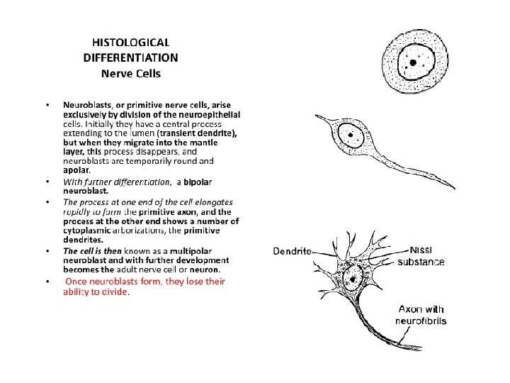
Development of Spinal Cord Neurogenesis Neuroblasts have a central process extending to the lumen (transient dendrite), but when they migrate into the mantle layer, this process disappears, and neuroblasts are temporarily round apolar. With further differentiation, two new cytoplasmic processes appear on opposite sides of the cell body, forming a bipolar neuroblast (primitive axon, primitive dendrites) The cell is then a multipolar neuroblast and with further development becomes a neuron Neuroepithelial cells Bipolar neuroblast Multipolar neuroblast
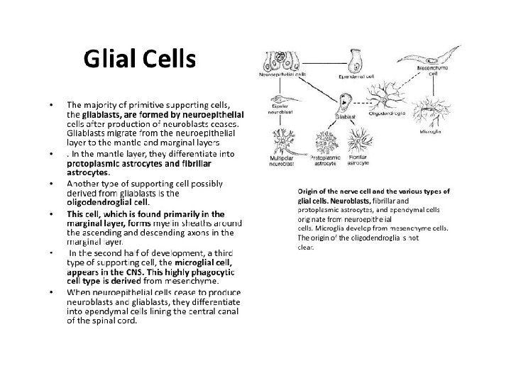
Development of Spinal Cord Gliogenesis Glioblasts migrate from the neuroepithelial layer to the mantle and marginal layers In the mantle layer they differentiate into protoplasmic and fibrillar astrocytes In the marginal layer oligodendroglial cells differentiate. This cell forms myelin sheaths around the ascending and descending axons Later a third type of supporting cell, the microglial cell, appears in the CNS. This highly phagocytic cell type is derived from vascular mesenchyme when blood vessels grow into the nervous system
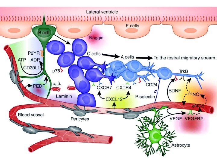
Migration along the Radial Glia in the CNS 40 μm/hour speed Glia vs neuron identification through astrotactin
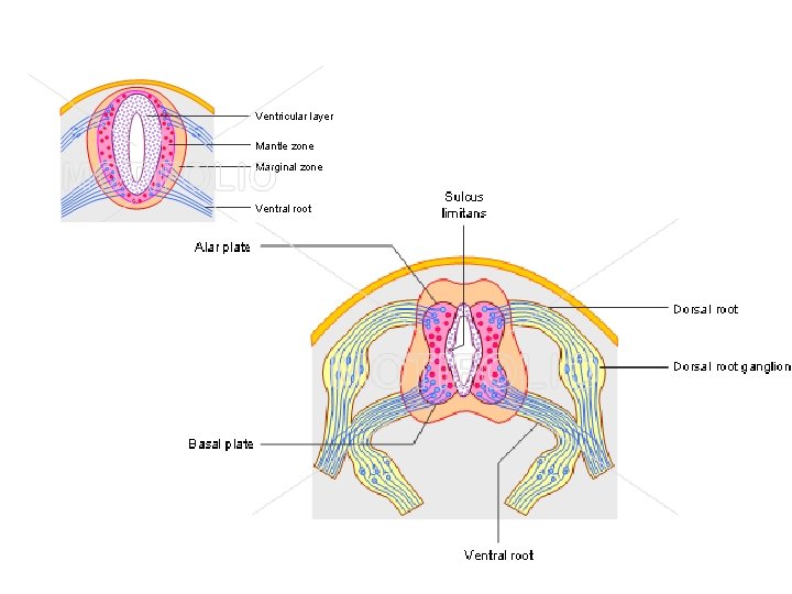
Spinal Cord Development A. Marginal Zone 1. Derivative of neural tube 2. White matter portion of central nervous system B. Alar plate 1. Derivative of neural tube 2. Portion of central nervous system 3. Associated with afferent (sensory) function C. Basal plate 1. Derivative of neural tube 2. Portion of central nervous system 3. Associated with efferent (motor) function D. Sulcus limitans (between B and C) 1. Derivative of neural tube 2. Portion of central nervous system 3. Separates alar plate from basal plate Intermediate zone
Development of Spinal Cord Compartmentalisation and Organization roof alar plate dorsal horn (sensory interneurons) Central canal sulcus limitans lateral horn (autonomic nuclei) intermediate zone (grey substance) ventricular zone (ependyma) ventral horn (motor neurons) marginal zone (white substance) basal plate Pax-6 floor plate commissure
Above the Sulcus Limitans A. Dorsal Columns 1. Derivative of alar plate (neural tube) 2. Portion of central nervous system 3. Neurons in the dorsal columns constitute afferent nuclei B. Dorsal rootlet 1. Derivative of neural crest 2. Portion of peripheral nervous system 3. Composed of Schwann cells and cell processes for sensory neuroblasts in spinal (dorsal root) ganglion C. Afferent sensory neuroblasts in spinal (dorsal root) ganglion 1. Derivative of neural crest 2. Portion of peripheral nervous system 3. Associated with afferent (sensory) function
Below the Sulcus Limitans A. Ventral Columns 1. Derivative of basal plate (neural tube) 2. Portion of central nervous system 3. Neurons in the dorsal columns constitute efferent nuclei B. Lateral Columns 1. Derivative of basal plate (neural tube) 2. Portion of central nervous system 3. Neurons in the lateral columns constitute efferent nuclei C. Ventral roots of spinal nerve* 1. Derivative of neural tube 2. Portion of central nervous system 3. Cell processes for motor neuroblasts in ventral horn
Spinal Cord Development Afferent sensory Sulcus neuroblasts limitans Roof Plate Marginal Zone Afferent sensory Dorsal column Lateral column neuroblasts in spinal ganglion Dorsal root of spinal nerve Efferent motor neuroblasts Floor Plate Ventral column Ventral root of spinal nerve
Compartmentalization of the Spinal Cord
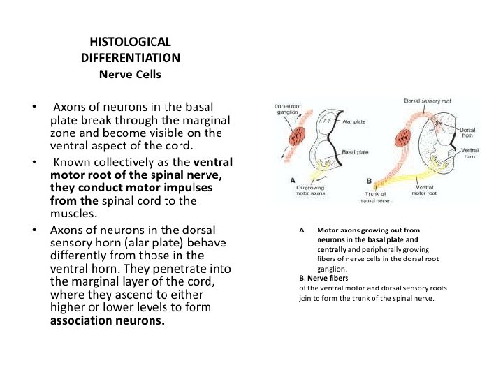
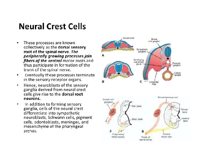
Development of Spinal Nerves Dorsal root ganglion Axons of the dorsal root multipolar Axons of the ventral root Motor nerve fibers begin to appear in the 4 th week, arising from cells in the basal plates. These fibers collect into bundles - ventral nerve roots Dorsal nerve roots form cells in dorsal root ganglia (spinal ganglia). Central processes form bundles that grow into the spinal cord opposite the dorsal horns. Distal processes join the ventral nerve roots to form a spinal nerve Almost immediately, spinal nerves divide into dorsal and ventral primary rami. Dorsal primary rami innervate dorsal axial musculature, vertebral joints, and the skin of the back. Ventral primary rami innervate the limbs and ventral body wall and form the major nerve plexuses (brachial and lumbosacral).
Development of Spinal Nerves Schwann cells myelinate the peripheral nerves with each cell myelinating only a single axon. These cells originate from neural crest, migrate peripherally, and wrap themselves around axons, forming the neurilemma sheath Beginning at the 4 th month of fetal life, many nerve fibers take on a whitish appearance as a result of deposition of myelin, which is formed by repeated coiling of the Schwann cell membrane around the axon. The myelin sheath surrounding nerve fibers in the spinal cord has a completely different origin, the oligodendroglial cells. Unlike Schwann cells, a single oligodendrocyte can myelinate up to 50 axons.
Nerve Fiber Coverings Schwann cell nucleus (PNS) Oligodendrocyte nucleus (CNS) Basement membrane Nerve fiber Unmyelinated axons Each Schwann cell is able to house multiple axons occupying invaginations of its cytoplasm. Myelinated axon Multiple wrapping of Schwann cell plasma membrane (“jelly - roll”) around single axon.
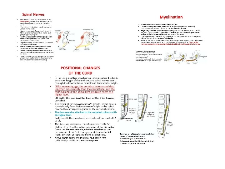
Myelination of Nerve Fibers • Myelin sheaths surrounding nerve processes in the CNS – – Begin to form during the late fetal period Continue during the first postnatal year. Formed by oligodendrocytes Derivative of neuroepithelial cells • Myelin sheaths surrounding nerve processes in the PNS – – – Begin about 20 weeks gestation Continue till puberty Formed by Schwann cells Derivative of neural crest Motor roots are myelinated before sensory roots
Positional Changes in the Spinal Cord 8 w 24 w newborn adult
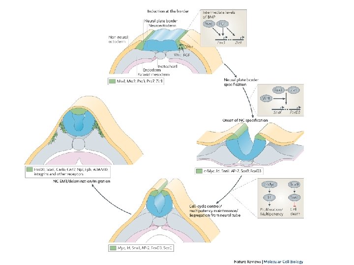
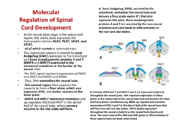
Molecular Events Dorsal and ventral regions of the developing spinal cord are dependent upon concentration gradients between members of the transforming growth factor beta (TGF-b) family of growth factors secreted in the dorsal neural tube and sonic hedgehog (SHH) secreted by the notochord and floor plate. Initially BMP 4 and 7 are secreted by ectoderm overlying the neural tube, then, BMP 4 induces a cascade of TGF-b proteins, including BMP 5, BMP 7, activin, and dorsalin in the roof plate and surrounding area. As a result, cells near the roof plate are exposed to the highest concentrations with more ventrally positioned cells seeing less and less of these factors. Similar events occur in the ventral region of the neural tube, only the signaling molecule is SHH. This factor is first expressed in the notochord followed by the establishment of a second signaling center in the floor plate. As a result, there is a diminishing concentration of SHH from the ventral to the dorsal region of the neural tube. These gradients then activate transcription factors that regulate differentiation of sensory and motor neurons.
Derivatives of the Neural Crest Migration into the mesenchymal layer Craniocaudal streams EMT - Epithelial – to - mesenchymal transformation Influenced by: ECM molecules, growth factors
Regions of the Neural Crest • Cranial (caudal to the forebrain until the 6 th rhombomere) • Vagal (between somites 1 -7) • Truncal (somites 8 -28) • Lumbosacral (caudal to somite 29)
Neural and Non-neural Derivatives of the Cranial Neural Crest
Neural Derivatives of the Truncal Neural Crest pigment cells (melanocytes) dorsolateral pathway (1) neural crest dorsal root, cranial nerve sensory ganglia ventrolateral pathway (2) parasympathic and sympathic ganglia mesoderm developing adrenal gland
Neural Derivatives of the Truncal Neural Crest ventrolateral pathway neural crest Schwann’s cells satellite cells unipolar sensory neurons multipolar ganglion cells in sympathetic ganglions FGF, NGF chromaffine cells in adrenal glands glomus caroticum, parafollicular (C) cells prevertebral plexus
Sympathetic prevertebral ganglia T 1 -T 4 Superior & middle cervical & stellate ganglion Cardiac and lung plexus T 5 -L 2 Celiac ganglion Sup. mesenteric ggl. Aorticorenal ggl. Inf. mesenteric ggl. Preganglionic fibers Postganglionic fibers
Mesenchymal Derivatives of Cephalic and Cardiac Crest
Neural Derivatives of Truncal and Vagal Crest cells
DERIVATIVES OF THE NEURAL CREST 1 a bipolar neuroblast 1 b differentiating neuroblast 1 c pseudounipolar sensory neuron 2 a unipolar neuroblast 2 b vegetative ganglion cells (multipolar) 2 c adrenal medulla - chromaffin cells 3 a glioblast 3 b Schwann cells 3 c satellite cells 4 a mesenchymal cells 4 b leptomeningeal cells 4 c ectomesenchymal cells 5 pigment cell 5 b melanocyte
Thank you for your attention!
EMBRYOLOGY SUMMARY The spinal cord develops from the NEURAL PLATE from above the notocord. With the folding of the NEURAL TUBE the central canal is formed (lining: ependyme). Neuroepithelial cells will give rise to neurons and glia. (Head mesenchyme) the DRG and the sympathetic neurones derive from the neural crest. The proneurones form a mantle zone (grey matter) with the axons growing towards the surface and so is the marginal zone formed (turns later into white matter). The notochord secrets a factor known as Sonic hedgehog or SHH. As a result, the floor plate then also begins to secrete SHH, and this will induce the basal plate to develop motoneurons. Meanwhile, the overlying ectoderm secretes bone morphogenetic protein (BMP). This induces the roof plate to begin to secrete BMP, which will induce the alar plate to develop sensory neurons. The alar plate and the basal plate are separated by the sulcus limitans. Additionally, the floor plate also secretes netrins. The netrins act as chemoattractants to decussation of pain and temperature sensory neurons in the alar plate across the anterior white commissure, where they then ascend towards the thalamus. The marginal zone develops into a massive passage of axans divided into 3 funiculi by the grey matter, posterior, lateral and anterior.
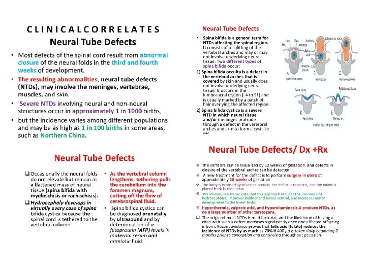
DEVELOPMENTAL MALFORMATIONS