Spinal cord and Peripheral nervous system Spinal cord


 ® Reflex center – ® Sensory receptor ® Sensory neuron")


 ® Innervate all")


 Farsightedness (hyperopia)")


- Slides: 27
Spinal cord and Peripheral nervous system
Spinal cord - Functions ® Sensory and motor pathway
Reflex arc (spinal cord) ® Reflex center – ® Sensory receptor ® Sensory neuron ® Interneuron (association neuron) ® Motor neuron (effector) ® An effector organ
Spinal Cord Anatomy ® Association neuron ® Motor http: //www. bayareapainmedical. com/wspin ecrd. html ® Gray Matter – “butterfly” interneurons ® White Matter – myelinated
Spinal cord Anatomy
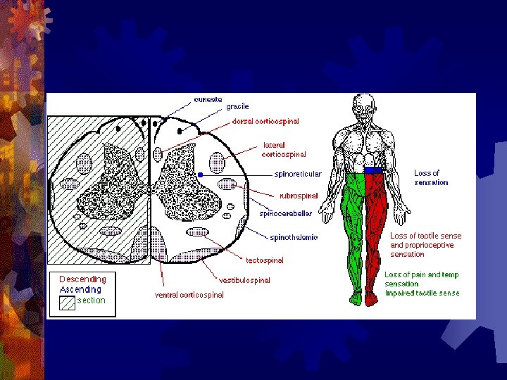
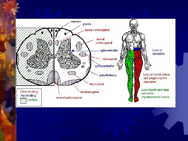
Spinal Cord tracts ® Sensory ® 1. Dorsal column 2. Spinothalamic ® Ascending tracts ® temperature, pressure, pain, light, touch
Spinal cord tracts continued Motor tracts 1. Corticospinal ® Decending ® Skeletal tone, voluntary muscle movement
Nerves attached to Sp. Cord ® Dorsal Root Ganglia – bundle of sensory nerves ® Ventral Root Ganglia – bundle of motor fibers
Peripheral Nervous system
Somatic Nervous System ® Includes all nerves in the musculoskeletal system, sense organs ® Receptor (receives impulse) to Effector (muscle fiber)
Autonomic Nervous System ® Motor neurons that control internal organs (involuntary) ® Innervate all organs ® Two divisions of
Autonomic Nervous System ® Sympathetic “Fight or flight response” ® Inhibits digestion ® Pupils dilate ® Accelerates heart rate ® Increase breathing rate. ® ® Parasympathetic Normal state ® Promotes digestion ® Pupils constrict ® Normal heartbeat ® “feed and breed” ®
The Eye: Photoreceptor Lens – refraction and focusing ® Iris – controls entrance of light into eye ® Pupil – window into the eye ® Choroid – blood vessels, absorbs stray light ®
Eye anatomy continued ® Sclera – white fiborous layer, protection ® Humors – Aqueous humor – between the cornea an lens ® Viterous humor – fills large cavity, gelatinous material ®
Eye Anatomy continued ® Ciliary body – holds lens in place ® Retina – contains receptors Cones – color vision ® Rods – black and white vision ® Optic Nerve ®
Rods and Cones Illustration
Eye Anatomy Continued Optic Nerve – picks up impulse ® Ciliary muscles – controls the shape of the lens ® Accommodation – ® ® ® Additional focusing power Near object – ciliary muscle contracts, lens becomes round
Physiology of sight ® Focusing – light rays bent by cornea, focus on the retina, refraction and inverted
Fields of Vision Illustration Refer to Lab on eye dissection
Cross section of head
Normal Vision 20/20 ® at a distance of 20 feet, you can read a certain line (labeled 20) on the chart and that your vision is normal. ® 20/40 -
Nearsightedness (myopic) Farsightedness (hyperopia)
Disorders of the Eye: Glaucoma – built up pressure in the eye due to lack of aqueous humor drainage
Vision of a person with Glaucoma
Cataracts- clouding of the lens