Spinal Cord Notes ANATOMY OF SPINAL CORD n


 n")

 gray horns n Lateral gray horns – cell bodies")
 is divided into columns n Anterior (ventral), posterior")


- Slides: 18
Spinal Cord Notes
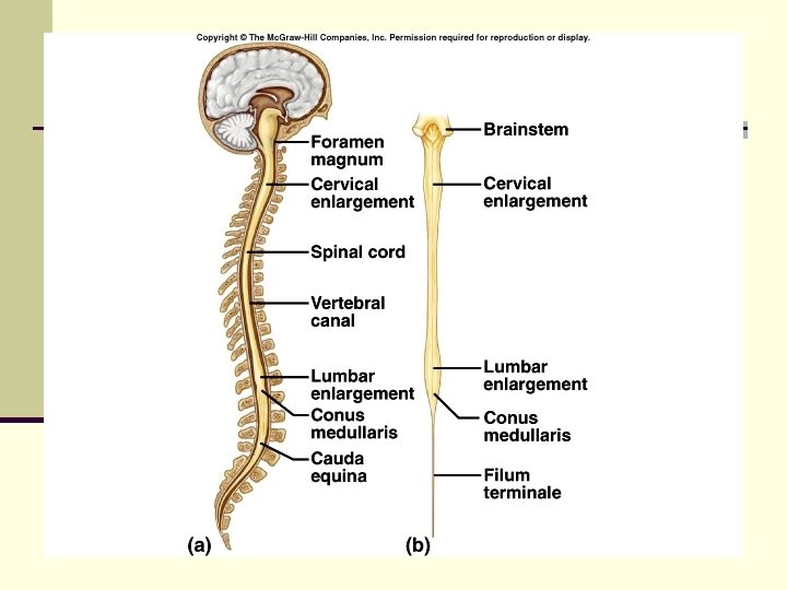
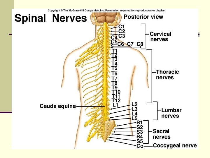
ANATOMY OF SPINAL CORD n Extends from medulla oblongata n Ends at second lumbar (L 2) vertebra in adults n 31 pairs of spinal nerves n Conus medularis – end of cord
n Two enlargements: n n Cervical enlargement - C 4 – T 1 – nerves to & from upper limbs Lumbar enlargement – T 9 -T 12 – nerves to & from lower limbs
n Cauda equina = “horse’s tail” n After conus medullaris, spinal cord divides into nerves that leave at lower levels
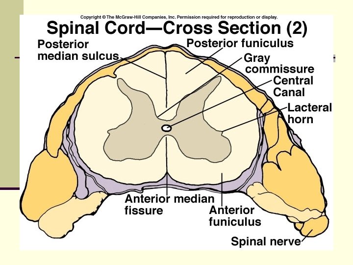
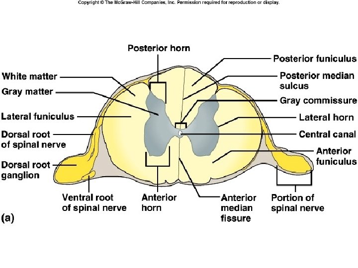
CROSS SECTION ANATOMY n Two grooves divide spinal cord: Anterior median fissure (deeper) n Posterior median sulcus n Gray matter (unmyelinated) forms an “H” in center n Gray commissure where fibers cross from side to side n
n Central canal – center of gray commisure; continuous with fourth ventricle n Gray matter is divided into horns: n Anterior (ventral) gray horns cell bodies of motor neurons to skeletal muscle
n Posterior ( dorsal ) gray horns n Lateral gray horns – cell bodies of motor neurons to cardiac, and smooth muscle & glands n Part of autonomic nervous system
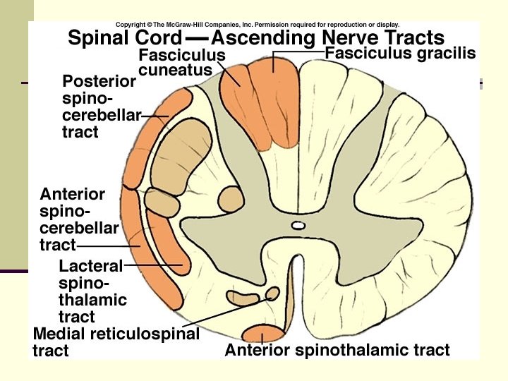
n White matter (myelinated nerve fibers) is divided into columns n Anterior (ventral), posterior (dorsal), lateral white columns
n Each column is divided into nerve tracts n Each tract carries one type of information (sensory or motor) n Ascending (sensory) tracts impulses toward brain n Descending (motor) tracts – impulses from brain
n Spinal cord has two functions: n n White matter tracts serve as information highways to and from brain Gray matter receives and integrates information, especially for spinal reflexes
n Name of tract indicate position in cord, where it begins and ends, and direction of impulses
DEVELOPMENT OF NS n Begins in third week of development n Ectoderm forms a neural tube n Neural tube defect due to low levels of a folic acid n Spina bifida – failure of laminae of spine to unite, caused by low levels of folic acid
n Portions of neural tube form fluid- filled vesicles n Hypothalamus is one of last areas of brain to develop n No new neurons form after birth, only grow and maturation n Brain reaches maximum weight as a young adult