Tissues of the Human Body Tissues groups of

Tissues of the Human Body Tissues: groups of cells closely associated that have a similar structure and perform a related function

Histology �The study of tissues is known as Histology. �People who study histology spend a lot of time looking in microscopes at the various body tissues.

4 Types of Tissues � 1. Epithelial � 2. Connective � 3. Muscular � 4. Nervous �Most organs contain all 4 types

1. Epithelial Tissue Description: coverings (0 ne side of epithelial tissue is always exposed to the outside **which could still be inside the body**) Location: lining and covering organs and body cavities, the secretory parts of organs and glands, the transport membranes of capillaries and alveolar sacs, and membranes which lubricate organs Names: according to structure, number of layers, arrangement, and shape

1. Epithelial Tissue �Functions �protection – epidermis �absorption – lining of intestines �secretion – ducts of glands �excretion – epidermis and lining of kidney capillaries �filtration – lining of kidney capillaries

Epithelial Structures Epithelium: layers of cells that line the cavity and cover flat surfaces Basement membrane: Basal Lamina (protein scaffolding) secreted by epithelial cells Reticular Lamina (crossed collagen fiber network) that support and anchor the epithelium Connective tissue: supports, connects, or separates different types of tissues *No blood supply. Nutrients and gases through diffusion. *Easily regenerates. Sheets of cells quickly regrow.

Microscopy Epithelial tissue Basement membrane Here is an example of an epithelial tissue. Note that one side of the tissue is exposed to the outside and the tissue is connected by a basement membrane.

Classes of Epithelia �Simple: just one layer or cell shape �Stratified: multiple layers and cell shapes
")
Shapes of Epithelia TYPE CELL SHAPE PICTURE EXAMPLE Squamous Squashed Endothelium (lines blood vessels) Mesothelium (serous lining of coelem) Cuboidal Cubed Walls of glands Columnar Columns *Cilia* *Goblet Cells* Lining of the gut tube; sometimes has cilia Pseudo-stratified Flat cells give rise to columns With cilia in respiratory tubes to move mucous/particles out of lungs. Transitional Stretches from 6 to 3 cells thick Lines urinary structures such as the bladder

Make a chart: Epithelium Type Locations in Body Function in Body


Simple Squamous Epithelium �Squamous cells are flat. From the side they look something like a fried egg.

Simple Cuboidal Epithelium �Secretory and absorptive tissue in glands as well as the liver and kidneys

Simple Columnar Epithelium � The secretory and absorptive lining of the Gastro-Intestinal tract

Stratified Squamous Epithelium �The epidermis of the body’s skin

Stratified Cuboidal Epithelium �Lines ducts �Found in the ducts of mammary glands, sweat glands, salivary glands, and the pancreas

Stratified Columnar Epithelium �Found in the vas deferens and pharynx. �Provides a thicker lining for some tubular structures in the body

Pseudostratified Columnar Epithelium �Located at trachea, bronchi, vas deferens �Help secrete mucus and used for absorption �Looks like it’s stratified, but it’s not. �The nuclei appear to be at different levels, however, there is really one layer of cells.

Transitional �Looks like stratified squamous but the cells are rounded at both the base of the tissue and the section exposed to the outside. �Found in the urinary bladder to allow it to expand contract.
 or into ducts (exocrine")
Glandular Epithelium �Can secrete substances into the bloodstream (endocrine glands) or into ducts (exocrine glands).

Endocrine Glands �Secrete products into the bloodstream �Products are stored in secretory cells or in follicle surrounding secretory cells �Hormones travel to target organs to increase response �No ducts

Exocrine Glands �Secrete their products into a systems of ducts. �Classified by how substances are excreted. � Merocrine: release fluid substances via exocytosis (salivary and sweat glands) � Apocrine: lose small portion of the cell body (mammary and ceruminous glands) � Holocrine: releases the entire cell (sebaceous glands)


Other Structures: Goblet Cells �Associated with columnar epithelium. �Secretes mucous.

Other Structures: Cilia �Associated with columnar cells. �Lines respiratory tract. �Works with goblet cells to move substances along the cells. Cilia


Quiz!! E Can You Identify the Classes of Epithelium? D A B C


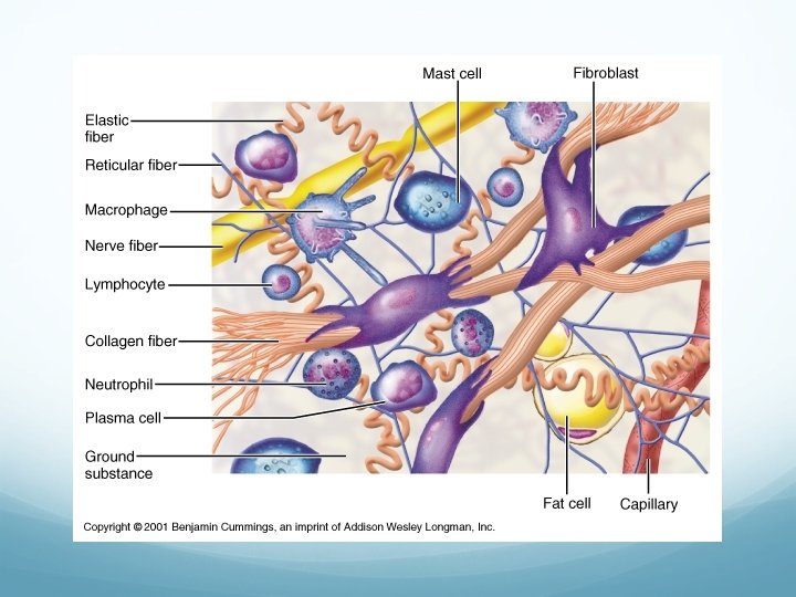
2. Connective Tissue � Description: support using fibers � Location: universal and most abundant tissue type in body � Functions: • • Provide support and protection Serve as frameworks Fill spaces Store fat Underlies epithelium Produce blood cells Protect against infections Help repair tissue damage � Always originates from mesenchyme (loosely organized undifferentiated mesodermal cells) � Have varying degrees of vascularity from cartilage (avascular) to bone (which has a rich blood supply) � Has non-living extracellular material/matrix (ground substance plus fibers) between its cells

Extracellular Matrix � Functions: � medium to dissolve solutes � transport � site of chemical reaction � Fibers � Collagen � Reticular fibers � Elastic �Ground substance � Jelly-like material made of sugar-protein molecules (proteoglycans) � Does not include the fibers
")
Cells of Connective Tissues �Fibroblasts �Star-shaped cells �Secretes protein into matrix producing fibers (phagocytosis) �Mast Cells �Immune cells that release heparin (an anticoagulant), histamine (promotes inflammatory reactions) �Osteocyte �a bone cell; a mature osteoblast that �Plasma Cells has become embedded in the bone �white blood cells that secrete large matrix volumes of antibodies �have an average half life of 25 years, �originate in the bone marrow they do not divide �Neutrophils �Chondrocyte �type of phagocyte and are normally �only cells found in healthy cartilage found in the bloodstream �produce and maintain the cartilaginous matrix, which consists �Lymphocytes mainly of collagen �any of 3 types of white blood cell (T Cells, B cells, Natural killer cells) �Macrophages
 • Composed of")
Fibers of Connective Tissues • Collagenous fibers • Thick (stain pink) • Composed of collagen • Great tensile strength • Most abundant in dense CT • Hold structures together • Tendons, ligaments • Elastic fibers • Bundles of protein elastin • All for response to stretch and distention • Elastic/rubbery (stain purple) • Vocal cords, air passages • Reticular fibers • Very thin collagenous fibers • Highly branched • Form supportive networks (stain purple) • Found in basement membranes and lymphatic tissues
: • Loose/Areolar connective")
Connective Tissue Categories • Fibrous/Proper Connective Tissue (with semi-fluid ground substance): • Loose/Areolar connective tissue • Adipose tissue • Reticular connective tissue • Dense (regular and irregular) connective tissue • Elastic connective tissue • Supporting/Specialized Connective Tissue: • Cartilage • Bone • Blood 34

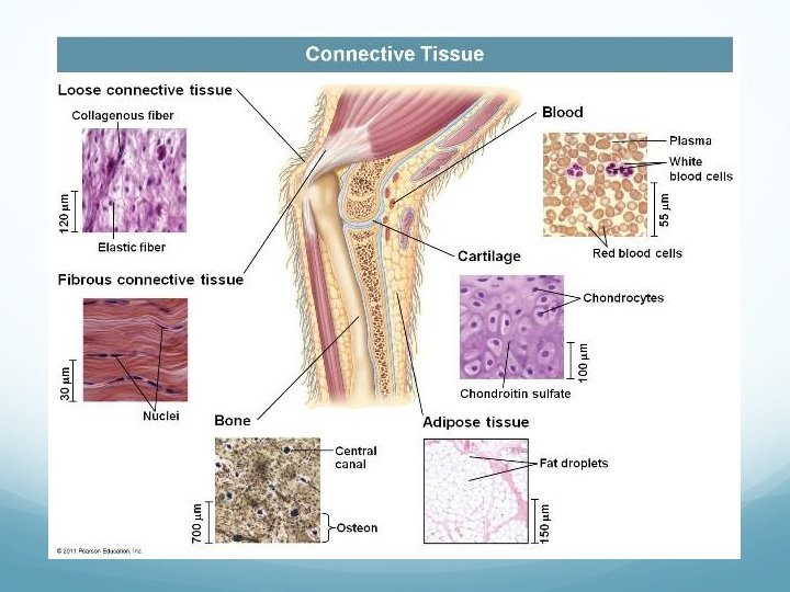

Connective Tissue Types • Loose Connective Tissue • Composition: fluid to gel-like matrix, fibroblasts, macrophages, collagen and elastic fibers • Location: beneath most epithelia, between muscles • Function: diffusion of nutrients; wrap and cushion organs 37
 and reticular fibers")
Connective Tissue Types • Adipose Tissue • Composition: Adipocytes (fat cells) and reticular fibers • Location: beneath skin (subcutaneous layer), behind eyeballs, around kidneys and heart • Function: Cushions, protects, insulates, energy storage

Connective Tissue Types • Reticular Connective Tissue • Composition: collagen fibers, fibroblasts, lymphocytes • Function: supports internal lymphatic organ walls • Location: walls of liver, spleen, lymphatic organs 39

Connective Tissue Types • Dense Regular Connective Tissue • Composition: packed collagen fibers, few fibroblasts • Location: tendons, ligaments • Function: attachment, tensile strength **Poor blood supply (slow healing)

Connective Tissue Types • Dense Irregular Connective Tissue • Composition: primarily collagen fibers randomly arranged • Location: dermis of skin, heart valves & capsules of organs • Function: tensile strength 41

Connective Tissue Types • Elastic Connective Tissue • Composition: elastic fibers, some collagenous fibers, fibroblasts • Location: walls of large arteries, airways, heart • Function: attachments between bones
 • Composition: Solid matrix, osteocytes in")
Specialized Connective Tissue: Bone • Bone (Osseous Tissue) • Composition: Solid matrix, osteocytes in lacunae • Location: skeleton • Function: supports and protects, forms blood cells, attachment for muscles **Highly vascular= fast healing 43

Specialized Connective Tissue: Cartilage • Cartilage • Composition: Rigid matrix; chondrocytes in lacunae; poor blood supply • Three (3) types: • Hyaline Cartilage • Location: ends of bones, nose, respiratory passages • Function: flexible support • Elastic Cartilage • Location: external ear, larynx • Function: provide flexibility • Fibrocartilage • Location: intervertebral discs, pads of knee and pelvic girdle • Function: shock absorber 44
 types of cartilage: Hyaline Cartilage Elastic Cartilage Fibrocartilage")
Specialized Connective Tissue: Cartilage Three (3) types of cartilage: Hyaline Cartilage Elastic Cartilage Fibrocartilage 45

Specialized Connective Tissue: Blood • Blood • Composition: fluid matrix called plasma; red blood cells; white blood cells; platelets • Location: throughout body in blood vessels; heart • Function: transports, defends (immune system), involved in clotting ( 46

Epithelial Tissues Quiz Review http: //www. biologycorner. com/anatomy/histology/


3. Muscular Tissue �Function: movement �Types: Skeletal, Cardiac, Smooth �Location: throughout body
 �Have a single, centrally located")
Cardiac Muscle �Striated (bands perpendicular to length of cell) �Have a single, centrally located nucleus and the muscle fibers branch often �Where two cardiac muscle cells meet, they form intercalated discs containing gap junctions and desmosomes, which bridge the two cells �Cardiac cells are the only cells that pulsate in rhythm…slow contractions and does not tire easily �Can only function under aerobic respiration

Cardiac Muscle

Cardiac Muscle

Smooth Muscle � Consists of cells with a single, centrally located nucleic � Cells are elongated/cylindrical with tapered ends and do not appear striated � Smooth muscle lines the walls of the blood vessels and certain organs such as the digestive and urogenital tracts, where it serves to advance the movement of substances � Called involuntary muscle because it is not under direct conscious control…slow and sustained contractions � Possess gap junctions � Mainly functions under aerobic respiration

Smooth Muscle

Smooth Muscle

Skeletal Muscle � Consist of long, cylindrical cells that, under a microscope, appear striated with bands perpendicular to the length of the cell � The many nuclei in each cell (multinucleated cells) are located near the outside along the plasma membrane, which is called the sarcolemma (muscle cell membrane) � Skeletal muscle is attached to bones and causes movements of the body � Because it is under conscious control, it is also called voluntary muscle…rapid contractions with great force and tire easily � No gap junctions present � Can function under aerobic and anaerobic respiration

Skeletal Muscle

Skeletal Muscle

REVIEW • Skeletal muscle • General characteristics: • Muscle cells are elongated cells = muscle fibers • Contractile • Three (3) types: • Skeletal muscle • Smooth muscle • Cardiac muscle • Attached to bones • Striated, multiple nuclei per cell • Voluntary movement of body • Smooth muscle • Walls of hollow organs, skin, walls of blood vessels • Non-striated, cells tapered at end, one nucleus per cell • Involuntary movement • Cardiac muscle • Heart wall • Striated, one nucleus per cell, branched ends with intercalated discs 59 • Involuntary movement


Skeletal Muscle Coverings � Endomysium: thin connective tissue covering muscle fiber � Perimysium: coarser fibrous membrane covering bundles of muscle fibers creating a fascicle (bundle of muscle fibers bound together by connective tissue) � Epimysium: tough fibrous connective tissue surrounding many fascicles; outer covering of entire skeletal muscle; blend into tendons or aponeurosis � Tendon: cord of dense fibrous tissue attaching a muscle to a bone � Aponeurosis: fibrous/membranous sheet connecting muscle and the part it moves � Fascia: layers of fibrous tissue covering and separating muscles
 Myofibril bundles Muscle fiber (cell)")
Skeletal Muscle Anatomy � Myofilament bundles (actin and myosin) Myofibril bundles Muscle fiber (cell) � Banding pattern: light and dark bands created by the arrangement of myofilaments (thick-myosin, thin-actin) in sarcomeres � Light (I) Bands: contain only actin filaments, parts of two adjacent sarcomeres � Has a darker area in the middle, Z disc (midline interruption between the connections of actin filaments � Dark (A) Bands: consists of actin and myosin; myosin filaments extend the entire length of A band; � has a lighter central area, H zone (H zone has a central line called M line (protein rods connecting myosin filaments))

Skeletal Muscle Anatomy


4. Nervous Tissue �Function: control and communication �Location: brain, spinal cord, nerves �Cells are called neurons �Support cells are called glia

Anatomy of a Generalized Neuron � Cell body: metabolic center (contains typical cell organelles except centrioles— amiotic) � Axon: one per cell; process of neuron, conduct impulses away from the cell body � Dendrites: many per cell; extension of neuron; conduct impulses toward the cell body � Axon hillock: axon arises from this cone-like region of cell body � Axon terminals: 100 s to 1000 s of brances at terminal end of axon; contains vesicles of neurotransmitters � Collateral branch: branch off of an axon � Synaptic cleft (synapse): separation between axon terminal and next neuron � Myelin: covering of most long neurons; whitish, fatty substance; protects, insulates, and speeds up neural transmission

Anatomy of a Generalized Neuron

Nervous Tissue

Nervous Tissue

Central vs. Peripheral Nervous System

Central Nervous System � Consists of: brain and spinal cord � Functions: integrating center (interpret incoming sensory information) and command center (issue instructions based on past experience and current conditions) � 4 types of cells: � Astrocytes: brace and anchor neurons to capillaries; control chemical environment in brain by picking up excess ions and recapturing released neurotransmitters � Microglia: phagocytes that dispose of dead brain cells and bacteria � Ependymal: help circulate cerebrospinal fluid that fills cavities and forms protective cushion around CNS � Oligodendrocytes: forms myelin sheath; protects and cushions nerves to speed up nerve transmission speed; produces white matter of brain
 and ganglia (group of")
Peripheral Nervous System � Consists of: nerves (cranial and spinal) and ganglia (group of nerve cell bodies) � Function: communication lines; linking all parts of the body � Functional Classifications: � Sensory/Afferent Division: nerve fibers that carry impulses to the CNS from receptors located throughout the body � Motor/Efferent Division: nerve fibers that carry impulses from the CNS to effector organs bringing about a motor response � 2 types of cells � Schwann cells: myelinate axons � Satellite cells: protects and cushions cells of PNS neurons


Practice Tissue Identification http: //www. wisconline. com/objects/index. asp? obj. ID=AP 1402 http: //www. gwc. maricopa. edu/class/bio 201/histop rc/prac 1 q. htm http: //teachers. olatheschools. com/~sschultzos/AP Histology/Epithelial. Slides. Practice. Quiz. A. pdf http: //spot. pcc. edu/anatomy/PDF/Q 3_Tissues. pdf http: //bio. rutgers. edu/~gb 102/lab_6/601 amepithelial. html
- Slides: 74