Tissues Body Tissues Groups of Cells are specialized

Tissues

Body Tissues · Groups of Cells are specialized for particular functions · Four primary types · Epithelium · Connective tissue · Nervous tissue · Muscle

Nervous tissue: Internal communication • Brain, spinal cord, and nerves Muscle tissue: Contracts to cause movement • Muscles attached to bones (skeletal) • Muscles of heart (cardiac) • Muscles of walls of hollow organs (smooth) Epithelial tissue: Forms boundaries between different environments, protects, secretes, absorbs, filters • Skin surface (epidermis) • Lining of GI tract organs and other hollow organs Connective tissue: Supports, protects, binds other tissues together • Bones • Tendons • Fat and other soft padding tissue Figure 4. 1

Epithelial Tissues · Body coverings · Body linings · Glandular tissue · Functions · Protection · Absorption · Filtration · Secretion Copyright © 2003 Pearson Education, Inc. publishing as Benjamin Cummings Slide 3. 42
 ·")
Epithelium Characteristics · Cells fit closely together · has one free surface (apical) · The lower surface is bound by a basement membrane (glycoprotein-Collagen) · Avascular (have no blood supply) · Regenerate easily if well nourished

Classification of Epithelium · Number of cell layers

Classification of Epithelium · Shape of cells

Simple Epithelium · Simple squamous · Usually forms membranes · Lines body cavities. Lines lungs and capillaries

Simple Epithelium · Simple cuboidal · Common in glands and their ducts · Forms walls of kidney tubules · Covers the ovaries
 Simple columnar epithelium Description: Single layer of tall cells with round to oval")
(c) Simple columnar epithelium Description: Single layer of tall cells with round to oval nuclei; some cells bear cilia; layer may contain mucussecreting unicellular glands (goblet cells). Simple columnar epithelial cell Function: Absorption; secretion of mucus, enzymes, and other substances; ciliated type propels mucus (or reproductive cells) by ciliary action. Location: Nonciliated type lines most of the digestive tract (stomach to anal canal), gallbladder, and excretory ducts of some glands; ciliated variety lines small bronchi, uterine tubes, and some regions of the uterus. Basement membrane Photomicrograph: Simple columnar epithelium of the stomach mucosa (860 X). Figure 4. 3 c
 Pseudostratified columnar epithelium Description: Single layer of cells of differing heights, some not")
(d) Pseudostratified columnar epithelium Description: Single layer of cells of differing heights, some not reaching the free surface; nuclei seen at different levels; may contain mucussecreting cells and bear cilia. Cilia Mucus of mucous cell Pseudostratified epithelial layer Function: Secretion, particularly of mucus; propulsion of mucus by ciliary action. Location: Nonciliated type in male’s sperm-carrying ducts and ducts of large glands; ciliated variety lines the trachea, most of the upper respiratory tract. Trachea Photomicrograph: Pseudostratified ciliated columnar epithelium lining the human trachea (570 x). Basement membrane Figure 4. 3 d

Stratified Epithelium · Stratified squamous · Found as a protective covering where friction is common · Locations: Skin, Mouth, Esophagus oral cavity, throat, vagina, and anal canal · can accumulate keratin

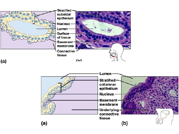
Stratified Epithelium · Stratified cuboidal- ducts of mammary glands, sweat glands, salivary glands, and the pancreas · Stratified columnar · line vas deferens, male urethra, and part of pharynx · Rare in our body comparatively

Stratified Epithelium · Transitional epithelium · Both cube and elongated cells · Lines organs of the urinary system

Glandular Epithelium · Gland – one or more cells that secretes a particular product · Two major gland types · Endocrine gland · Ductless · Secretions are hormones · Exocrine gland · Empty through ducts to the epithelial surface · Include sweat and oil glands

Types of Glands
 Microvilli Secretory vesicles containing mucin Rough ER Golgi apparatus (a)")
Unicellular Gland (Goblet Cell) Microvilli Secretory vesicles containing mucin Rough ER Golgi apparatus (a) Nucleus (b) Figure 4. 4

Connective Tissue · Found everywhere in the body · Functions · Binds body tissues together · Supports the body · Provides protection · store fat · produce blood cells · protect against infections · help repair tissue damage

Table 4. 1

Connective Tissue Characteristics · Variations in blood supply · Extracellular matrix · Non-living material that surrounds living cells (protein sugars water etc)

Extracellular Matrix · Two main elements · Ground substance – mostly water along with adhesion proteins and polysaccharide molecules · Proteoglycans Protein core + large polysaccharides (chrondroitin sulfate and hyaluronic acid) · Fibers · Produced by the cells · Three types · Collagen fibers · Elastic fibers · Reticular fibers

Connective Tissue Proper • Types: – Loose connective tissue • Areolar • Adipose • Reticular – Dense connective tissue • Dense regular • Dense irregular • Elastic
 Connective tissue proper: loose connective tissue, areolar Description: Gel-like matrix with all three")
(a) Connective tissue proper: loose connective tissue, areolar Description: Gel-like matrix with all three fiber types; cells: fibroblasts, macrophages, mast cells, and some white blood cells. Elastic fibers Function: Wraps and cushions organs; its macrophages phagocytize bacteria; plays important role in inflammation; holds and conveys tissue fluid. Collagen fibers Location: Widely distributed under epithelia of body, e. g. , forms lamina propria of mucous membranes; packages organs; surrounds capillaries. Fibroblast nuclei Epithelium Lamina propria Photomicrograph: Areolar connective tissue, a soft packaging tissue of the body (300 x). Figure 4. 8 a
 Connective tissue proper: loose connective tissue, adipose Description: Matrix as in areolar, but")
(b) Connective tissue proper: loose connective tissue, adipose Description: Matrix as in areolar, but very sparse; closely packed adipocytes, or fat cells, have nucleus pushed to the side by large fat droplet. Function: Provides reserve food fuel; insulates against heat loss; supports and protects organs. Nucleus of fat cell Location: Under skin in the hypodermis; around kidneys and eyeballs; within abdomen; in breasts. Vacuole containing fat droplet Adipose tissue Mammary glands Photomicrograph: Adipose tissue from the subcutaneous layer under the skin (350 x). Figure 4. 8 b
 Connective tissue proper: loose connective tissue, reticular Description: Network of reticular fibers in")
(c) Connective tissue proper: loose connective tissue, reticular Description: Network of reticular fibers in a typical loose ground substance; reticular cells lie on the network. Function: Fibers form a soft internal skeleton (stroma) that supports other cell types including white blood cells, mast cells, and macrophages. Location: Lymphoid organs (lymph nodes, bone marrow, and spleen). White blood cell (lymphocyte) Reticular fibers Spleen Photomicrograph: Dark-staining network of reticular connective tissue fibers forming the internal skeleton of the spleen (350 x). Figure 4. 8 c
 Connective tissue proper: dense connective tissue, dense regular Description: Primarily parallel collagen fibers;")
(d) Connective tissue proper: dense connective tissue, dense regular Description: Primarily parallel collagen fibers; a few elastic fibers; major cell type is the fibroblast. Collagen fibers Function: Attaches muscles to bones or to muscles; attaches bones to bones; withstands great tensile stress when pulling force is applied in one direction. Location: Tendons, most ligaments, aponeuroses. Nuclei of fibroblasts Shoulder joint Ligament Photomicrograph: Dense regular connective tissue from a tendon (500 x). Tendon Figure 4. 8 d
 Connective tissue proper: dense connective tissue, dense irregular Description: Primarily irregularly arranged collagen")
(e) Connective tissue proper: dense connective tissue, dense irregular Description: Primarily irregularly arranged collagen fibers; some elastic fibers; major cell type is the fibroblast. Nuclei of fibroblasts Function: Able to withstand tension exerted in many directions; provides structural strength. Location: Fibrous capsules of organs and of joints; dermis of the skin; submucosa of digestive tract. Fibrous joint capsule Collagen fibers Photomicrograph: Dense irregular connective tissue from the dermis of the skin (400 x). Figure 4. 8 e
 Connective tissue proper: dense connective tissue, elastic Description: Dense regular connective tissue containing")
(f) Connective tissue proper: dense connective tissue, elastic Description: Dense regular connective tissue containing a high proportion of elastic fibers. Function: Allows recoil of tissue following stretching; maintains pulsatile flow of blood through arteries; aids passive recoil of lungs following inspiration. Elastic fibers Location: Walls of large arteries; within certain ligaments associated with the vertebral column; within the walls of the bronchial tubes. Aorta Heart Photomicrograph: Elastic connective tissue in the wall of the aorta (250 x). Figure 4. 8 f
- Slides: 29