Cirrosi Epatica Definizione Meccanismi della fibrogenesi epatica Fisiopatologia

Cirrosi Epatica • Definizione • Meccanismi della fibrogenesi epatica • Fisiopatologia dell’ipertensione portale • Complicanze maggiori della cirrosi • Cenni di terapia delle complicanze • Management della cirrosi compensata/scompensata

CIRROSI Alterazione dell’architettura del fegato caratterizzata da noduli e formazione di tessuto collagene CAUSE: Tutte le cause di danno epatico cronico

Progressione del danno epatico cronico Causa iniziale Stadio finale Complicanze ● anni Cirrosi -Fibrosi -Distorsione dell’architettura epatica

Portal Vein Hepatic Artery Sinusoids Bile Duct Central Veins

Normal Cirrhosis Changes of hepatic microcirculation in cirrhosis

Activation of HSC

LIVER CIRRHOSIS : from activation of HSC to cirrhotic nodules

Fibrosi virale Spazio portale 1. – Necrosi a ponte porto-centrale 2. – Epatite da interfaccia e sviluppo di setti che circondano il parechima 3. – Perdita di connessioni vascolari con il sistema portale

fibrosi biliare Modello BDL CBP CB secondaria CSP Formazione di setti porto-portali La vena centrolobulare è conservata Spazio portale

Centrolobular fibrosis portal tract 1. – Secondary to venous outflow problems (e. g. chronic heart failure) 2. 2. - Chronic ETOH consumption 3. – Development of central to central septa and “reversed lobulation”

CONSEQUENCES OF DEVELOPING PROGRESSIVE HEPATIC FIBROSIS structural & functional intrahepatic resistance splanchnic blood flow (cirrhosis) HCC systemic hyperdynamic circulation portal hypertension variceal bleeding portosystemic collaterals hepatic encephalopathy hepatopulmonary syndrome decrease central volume ascites cardiac output cardiomyopathy HRS spontaneous bacterial peritonitis

Ipertensione portale • Condizione fisiopatologica causata dall’aumento della pressione venosa nel distretto portale • Principale conseguenza della cirrosi epatica e principale meccanismo delle sue principali complicanze cliniche • Riconosce anche cause extraepatiche, in cui le manifestazioni cliniche dominanti dipendono dalla sede di origine della ipertensione portale

Anatomia e Fisiologia della circolazione portale Sistema venoso ad alta portata e bassa resistenza che drena il sangue dagli organi addominali per convogliarlo al fegato; da questo, attraverso i sinusoidi e le vene sovrepatiche, raggiunge la vena cava inferiore e la circolazione sistemica • Flusso epatico: 1. 5 l/min (80% venoso, portale) • Pressione portale: 7 mm. Hg (diretta o wedge) • Pressione sovra-epatiche/atrio dx: 3 -5 mm. Hg • Gradiente porto-epatico (HVPG): ≤ 5

Classificazione e cause • Cause Pre-epatiche • Trombosi portale • Trombosi splenica • Cause Intraepatiche • Presinusoidali • Sinusoidali • Postsinusoidali • Cause post-epatiche • • Membrana cavale Pericardite costrittiva Insufficienza tricuspidale Grave scompenso cardiaco destro

Cause di ipertensione portale intraepatica • Pre-sinusoidali • Schistosomiasi, sarcoidosi, tossici, fibrosi epatica congenita, malattie mielo proliferative • Sinusoidali o miste • Cirrosi, epatite alcolica, iperplasia nodulare rigenerativa • Post-sinusoidali • Malattia veno-occlusiva • Sindrome di Budd-Chiari
 HCC")
PORTAL HYPERTENSION: pathophysiological sequelae structural & functional splanchnic blood flow intrahepatic resistance (cirrhosis) HCC systemic hyperdynamic circulation portal hypertension variceal bleeding portosystemic collaterals hepatic encephalopathy hepatopulmonary syndrome decrease central volume ascites cardiac output cardiomyopathy HRS spontaneous bacterial peritonitis

Conseguenze dell’ ipertensione portale • Splenomegalia ed ipersplenismo • Circoli collaterali ed emorragie digestive • Shunt porto-sistemici ed encefalopatia porto -sistemica • Vasodilatazione splancnica ed alterazioni della emodinamica sistemica

Circoli collaterali Ligamento falciforme Gastro-esofagei Parete addome e retro peritoneo Gastro-esofagei Vene emorroidarie

Conseguenze degli shunts portosistemici • Riducono il flusso portale • Aumentano insufficienza epatica • NON riducono significativamente la pressione portale • Favoriscono la circolazione iperdinamica • Favoriscono iperammoniemia • Endotossinemia • Encefalopatia porto sistemica

Complicanze maggiori della cirrosi


Complicanze maggiori della cirrosi

Natural History of Chronic Liver Disease Development of cirrhosis Chronic liver disease Compensated cirrhosis Decompensated cirrhosis Death Development of complications: · Variceal hemorrhage · Ascites · Encephalopathy · HRS Orthotopic liver transplant (OLT)

Mortalità per complicanze di cirrosi epatica - 384 Pts follow-up: 5 anni Fattovich G et al, Gastroenterology 1997; 112: 463
 Score 1 Encephalopathy None Points 2 Grade 1 -2 Grade 3 -4")
Child-Turcotte-Pugh (CTP) Score 1 Encephalopathy None Points 2 Grade 1 -2 Grade 3 -4 (precipitant) Ascites 3 (chronic) None Mild Moderate <2 2 -3 >3 >3. 5 2. 8 -3. 5 <2. 8 PT (seconds prolonged) <4 or INR <1. 7 4 -6 1. 7 -2. 3 >6 >2. 3 Bilirubin (mg/dl) Albumin (g/dl) Child A: 5 -6 pts Child B: 7 -9 pts Child C: 10 -15 pts MINIMAL LISTING CRITERIA: CTP SCORE 7 POINTS
 · Predicts 3 -month mortality among patients with")
MELD (Model for End-Stage Liver Disease) · Predicts 3 -month mortality among patients with chronic liver disease on the liver waiting list · MELD = (0. 957 x LN (creatinine) + 0. 378 x LN (bilirubin) + 1. 12 x LN (INR) + 0. 643) x 10 · Minimum score = 6 (risk of death on WL 20%) · Maximum score = 40 (risk of death on WL 100%)

Diagnostica della cirrosi con ipertensione portale • • Clinico-semeiologica Misurazione invasiva emodinamica portale Eco-doppler Endoscopia digestiva

Diagnosi clinico-semeiologica • • Splenomegalia Circoli collaterali superficiali Gavoccioli emorroidari Spider Naevi Edema perimalleolare Ascite Asterixis

Semeiotica dell’ipertensione portale
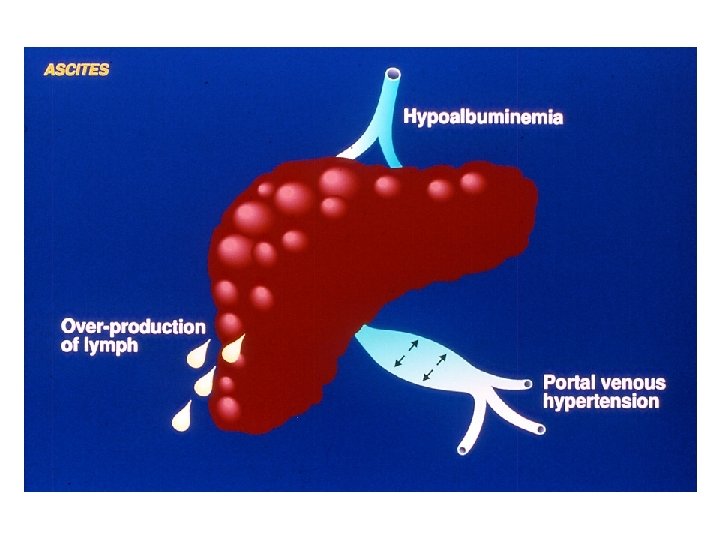
 detectable only by US Ø")
Definition of ascites • Uncomplicated Ø Grade 1 (mild) detectable only by US Ø Grade 2 (moderate) moderate symmetrical abdominal distension Ø Grade 3 (large) marked abdominal distension • Refractory Ascites that cannot be mobilized or early recurrence of ascites not prevented by medical therapy Ø Diuretic-resistant Ø Diuretic-intractable

Treatment of Ascites • Bed rest unproven benefit • Dietary sodium restriction to 5. 2 g/d (90 mmol) • Diuretics Ø Anti-Mineralocorticoids Spironolactone, Canrenoate, Canrenone Ø Loop diuretics Furosemide, Torasemide, Ethacrynic Acid Ø Other potassium-sparing diuretics Amiloride, Triamterene

Diagnostic Criteria of Refractory Ascites • Treatment duration Intensive diuretic therapy (spironolactone 400 mg/d + furosemide 160 mg/d) for at least 1 wk Salt-restricted diet (< 90 mmol or 5. 2 g of salt/d) • Lack of response Weight loss < 0. 8 kg over 4 days Urinary sodium output < sodium intake • Early ascites recurrence Grade 2 or 3 ascites within 4 wks of initial mobilization • Diuretic-induced complications Hepatic Encephalopathy, Renal Impairment, Hyponatremia, Hypo/Hyperkalemia

PERITONITE BATTERICA SPONTANEA: definizione ed epidemiologia · Infezione del liquido ascitico · PMN > 250/mm 3 e colturale + su liquido ascitico · Microbiologia: G-: E. Coli, K. Pneumoniae (60%) G+: Strepto (25%) Anaerobi: rari · Non evidente sorgente di infezione addominale trattabile chirurgicamente · 10 -25% dei cirrotici ospedalizzati con ascite · Mortalità elevata (17 -50%) · Alto rischio di recurrence: 43 -70% (6 -12 mesi)
 3.")
PERITONITE BATTERICA SPONTANEA: forme cliniche 1. Classic 2. CNNA (culture negative neutrocytic ascites) 3. MNB (monomicrobial non-neutrocytic bacterascites) carcinosi peritoneale, pancreatite, peritonite TBC Classica CNNA MNB PMN > 250 < 250 Colturale + - +

PERITONITE BATTERICA SPONTANEA: terapia e profilassi · Cefotaxime: 2 g/12 hrs per 5 -10 gg · Ciprofloxacina: 400 mg/12 hrs per 5 gg, poi orale · Se non risposta: Stafilo? · + Albumina: 1. 5 g/kg alla diagnosi, 1 g/kg dopo 48 hrs (prevenire HRS!) · Chinolonico long-term (Norfloxacina 400 mg/die) soprattutto se proteine < 1 g/dl nel liquido ascitico
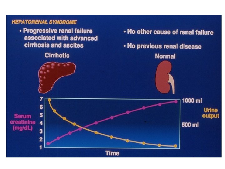
 • Type I HRS Rapid and progressive renal failure")
Definition of Hepatorenal Syndrome (HRS) • Type I HRS Rapid and progressive renal failure with a doubling of serum creatinine to a level greater than 2. 5 mg/dl or a halving of the creatinine clearance to less than 20 ml/min in less than 2 wks • Type II HRS More chronic form with a slowly progressive increase in serum creatinine level to greater than 1. 5 mg/dl or a creatinine clearance of less than 40 ml/min

DIFFERENCES BETWEEN TYPE-1 AND TYPE-2 HRS Type-2 Type-1 Spontaneous Precipitated Renal failure Moderate and steady Severe and progressive Consequence Refractory ascites Terminal hepatorenal failure Months Days Onset Survival

Definition of Hepatorenal Syndrome: Major Criteria • Chronic or acute liver disease with liver failure and portal hypertension • Low glomerular filtration rate (serum creatinine > 1. 5 mg/dl or creatinine clearance < 40 ml/min) • Absence of shock, excessive fluid loss, ongoing bacterial infection and recent treatment with nephrotoxic drugs • No sustained improvement in renal function following expansion with 1. 5 l of isotonic saline • Proteinuria < 0. 5 g/dl • No US evidence of renal tract disease

Definition of Hepatorenal Syndrome: Minor Criteria • • • Urine volume < 500 ml/d Urine sodium < 10 mmol/d Urine osmolality < plasma osmolality Urine red cell count < 50/high power field Serum sodium < 130 mmol/l

The Pathophysiology of HRS

Diagnosi e classificazione endoscopica Varici esofagee Varici fondo

Diagnosi e classificazione endoscopica Gastropatia ipertensiva GAVE

Dilation and Rupture of Varices Varix P Flow Risk increases in portal pressure and collateral blood flow caused by: • Meals • Alcohol • Exercise • Increased intra-abdominal pressure

Acute variceal bleeding Sclerotherapy + Drugs vs Sclerotherapy alone Mortality
 BB + nitrati fallimento successo continuare LEV(+ BB) Malattia epatica")
PREVENZIONE RISANGUINAMENTO (profilassi secondaria) BB + nitrati fallimento successo continuare LEV(+ BB) Malattia epatica avanzata TIPS OLT successo Malattia epatica stabile Continuare + follow up TIPS (DSSR) D´Amico, 2004

HEPATIC ENCEPHALOPATHY – NOMENCLATURE Hepatic Encephalopathy Nomenclature · Type A Associated with Acute liver failure · Type B Associated with porto-systemic Bypass without intrinsic hepatocellular disease · Type C Associated with Cirrhosis and portosystemic shunting Ferenci et al. , Hepatology 2002; 35: 716
 · Rapid deterioration in the level of")
Encephalopathy of acute liver failure (Type A) · Rapid deterioration in the level of consciousness · Increased intracranial pressure (ICP) · Reduced cerebral perfusion pressure · Neuropathologically, there is brain edema · Pathogenesis is multifactorial with ammonia playing a major role

TYPE C HEPATIC ENCEPHALOPATHY IS THE ENCEPHALOPATHY OF CIRRHOSIS Type C Hepatic Encephalopathy is the Encephalopathy of Cirrhosis · Neuropsychiatric complication of cirrhosis · Results from spontaneous or surgical / radiological portal-systemic shunt + chronic liver failure · Failure to metabolize neurotoxic substances · Alterations of astrocyte morphology and function (Alzheimer type II astrocytosis)

CHARACTERISTICS OF TYPE A VS. TYPE C ENCEPHALOPATHY Characteristics of Type A vs. Type C Hepatic Encephalopathy Type A Type C · Rapid onset · Gradual onset · Frequently fatal · Rarely fatal · Main cause: cerebral edema · Main cause: shunting / toxin · Precipitant · Treatment: rarely effective short of liver transplant · Treatment: usually effective

PATHOPHYSIOLOGY OF HEPATIC ENCEPHALOPATHY Pathophysiology of Hepatic Encephalopathy Ammonia Upregulation of astrocytic peripheral benzodiazepine receptors (PBR) Neurosteroid production Modulation of GABAA receptor Hepatic encephalopathy

PATHOPHYSIOLOGY OF HEPATIC ENCEPHALOPATHY Hepatic Encephalopathy Pathogenesis Toxins NH 3 Shunting Failure to metabolize NH 3 Bacterial action Protein load GABA-BD receptors

Pathogenesis of Hepatic Encephalopathy: GABA-BD receptors role of

HEPATIC ENCEPHALOPATHY IS A CLINICAL DIAGNOSIS Hepatic Encephalopathy Is A Clinical Diagnosis · Clinical findings and history important · Ammonia levels are unreliable · Ammonia has poor correlation with diagnosis · Measurement of ammonia not necessary · Number connection test · Slow dominant rhythm on EEG

STAGES OF HEPATIC ENCEPHALOPATHY Stages of Hepatic Encephalopathy Stage Mental state Neurologic signs 1 Mild confusion: limited attention span, irritability, inverted sleep pattern Incoordination, tremor, impaired handwriting 2 Drowsiness, personality changes, intermittent disorientation Asterixis, ataxia, dysarthria 3 Somnolent, gross disorientation, marked confusion, slurred speech Hyperreflexia, muscle rigidity, Babinski sign 4 Coma No response to pain, decerebrate posture

Poor Correlation of Ammonia Levels With Presence or Severity of Encephalopathy 400 350 300 250 Venous total 200 ammonia mmol/L 150 100 50 0 Grade 1 Grade 2 Grade 3 Grade 4 Severity of Hepatic Encephalopathy Ong et al. , Am J Med 2003; 114: 188

BLOOD AMMONIA LEVELS ONLY LEAD TO CONFUSION “Blood ammonia levels cause as much confusion in those requesting the measurement as in the patients in whom they are being measured” Adrian Reuben Hepatology 2002; 35: 983

MINIMAL HEPATIC ENCEPHALOPATHY Minimal Hepatic Encephalopathy · Occurs in 30 -70% of cirrhotic patients without overt hepatic encephalopathy · Detected by psychometric and neuropsychological testing · May improve with lactulose or synbiotics (probiotics and fermentable fiber) · Predicts overt encephalopathy

TREATMENT OF HEPATIC ENCEPHALOPATHY Treatment of Hepatic Encephalopathy · Identify and treat precipitating factor · Infection · GI hemorrhage · Prerenal azotemia · Sedatives · Constipation · Lactulose (adjust to 2 -3 bowel movements/day) · Protein restriction, short-term (if at all)

ACTIONS OF LACTULOSE Actions of Lactulose NH 3 Decreased p. H NH 4+ Lactic acid NH 3 Lactulose Ureaseproducing bacteria Increase cathartic effect

PROTEIN RESTRICTION IS NOT NECESSARY IN HEPATIC ENCEPHALOPATHY Protein Restriction Is Not Necessary in Hepatic Encephalopathy 4 Hypoproteic diet Normoproteic diet 3 2 Hepatic encephalopath y stage 1 0 0 1 2 3 4 5 6 7 Day Córdoba, J Hepatology 2004; 91: 38 8 9 10 11 12 13 14

Treatment Recommendations - Cirrhosis Decompensated Compensated Hepatoma Surveillance U/S, AFP q 6 months Variceal Bleed SBP Varices Surveillance Ascites Monitor Liver Function PT, Alb, Bili q 3 -6 months HRS Encephalopathy (Garcia-Tsao G, 2003)

Management of cirrhosis · Maintain nutrition/reduce salt intake · Prevent bone loss · Prevent bleeding · Prevent encephalopathy · High index suspicion for sepsis · Close control diabetes · Management of complications · Early diagnosis and therapy of HCC · (Selected antiviral therapy) · Appropriate referral to transplant centres

Natural History of Cirrhosis in 2008: Altered by What We Do · More aggressive screening · HCC identified earlier · Ablative therapies for HCC (downstaging) · Obliteration of varices/beta-blockade · TIPSS · Liver Transplantation


HEPATIC ENCEPHALOPATHY – TREATMENT SUMMARY Hepatic Encephalopathy Treatment: Summary Increase ammonia fixation in liver: · Ornithine aspartate · Benzoate Shunt occlusion or reduction Decrease ammonia production in gut: · Lactulose · Antibiotics · Adjustment in dietary protein

Liver Transplant Indications Contraindications · Decompensated · Extrahepatic cirrhosis - all causes malignancy (hepatitis C most common indication) · Active infection · Intrahepatic malignancy · Active substance abuse · Acute liver failure · Advanced cardiopulmon-ary disease · Metabolic disease

Child score and survival in 424 patients 1. 0 CPT - A. 8 . 6 CPT - B Cum Survival . 4 CPT - C . 2 0. 0 0 200 400 OLT 1 -yr survival Heumann et al 600 800 1000 1200 OLT 2 -yr survival 1400 1600 Days

MELD and Survival on Transplant Waiting List 100 92. 3% 90. 7% 80 Probabilit y of survival (%) <15 15 - 20 66. 0% 60 20 - 29 40 33. 8% 30+ 20 0 0 2 4 6 8 Months from listing 10 12

Organ Allocation for Liver Transplant · Fulminant hepatic failure has highest priority · MELD score determines priority in cirrhosis · Amongst patients with same blood type, highest MELD score determines priority · Waiting time used only to break ties with identical MELD scores · MELD scores are updated at regular intervals
 Regions 1 7 6 9 2 10 8")
United Network for Organ Sharing (UNOS) Regions 1 7 6 9 2 10 8 11 5 3 4

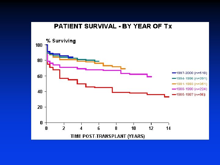
Liver Transplantation in the U. S. · Current 1 - and 3 -year survival rates are 90% and 80%, respectively · During 2005, ~6, 000 liver transplantations were performed · During 2005, ~17, 000 patients were on the waiting list and ~2, 000 died on it or were removed from it because they became too sick for transplant · Main problem is the shortage of donors · Expansion of donor pool: marginal livers, split-livers, live donors

NATURAL HISTORY OF CHRONIC LIVER DISEASE – SUMMARY Natural History of Chronic Liver Disease Development of cirrhosis Chronic liver disease Compensated cirrhosis Decompensated cirrhosis Median survival ~ 9 years Median survival ~ 1. 6 years Development of complications · Variceal : hemorrhage · Ascites · Encephalopathy · Jaundice Death Orthotopic liver transplant (OLT)

MANAGEMENT OF COMPENSATED CIRRHOSIS – SUMMARY Management of Compensated Cirrhosis Chronic liver disease Compensated cirrhosis Decompensated cirrhosis Death Diagnosis: · Liver biopsy · Clinical/LSS Screen for varices (EGD): · Large varices beta-blockers · Small varices EGD in 1 -2 yrs · No varices EGD in 2 -3 yrs Screen for HCC: · Ultrasound and AFP q 6 mos · Measures to stop alcohol use · Hep A and B vaccination Orthotopic liver transplant (OLT)

MANAGEMENT OF ASCITES – SUMMARY Management of Ascites Chronic liver disease Compensated cirrhosis Decompensated cirrhosis Death Ascites Diagnosis: · Clinical · US or CAT scan Treatment: · Spironolactone-based · No NSAIDs Early diagnosis of SBP: · Paracentesis q admission or with symptoms · No aminoglycosides in SBP Orthotopic liver transplant (OLT)

MANAGEMENT OF VARICEAL BLEEDING – SUMMARY Management of Variceal Bleeding Chronic liver disease Compensated cirrhosis Decompensated cirrhosis Death Variceal Bleed Diagnosis / Treatment: · Endoscopy within 12 hrs Other Treatment: · Prophylactic antibiotics Prophylaxis of rebleed: · Beta-blockers prior to d/c or · Serial ligation post d/c Orthotopic liver transplant (OLT)

MANAGEMENT OF HEPATIC ENCEPHALOPATHY - SUMMARY Management of Encephalopathy Chronic liver disease Compensated cirrhosis Decompensated cirrhosis Death Encephalopathy Diagnosis · Clinical Treatment: · D/C diuretics · D/C sedatives · Dx paracentesis · No long-term protein restriction Orthotopic liver transplant (OLT)


Cenni di terapia dell’ipertensione portale • Farmaci antifibrotici: una promessa ancora non mantenuta • L’unica terapia in grado di modificare la progressione è, al momento, il trattamento del fattore etiologico di base (es no Et-OH) • Il target della terapia è quello di ridurre, farmacologicamente o “chirurgicamente” il gradiente venoso porto-epatico • L’end-point clinico è la prevenzione primaria o secondaria del sanguinamento da varici

Terapia dell’ipertensione portale • Vasocostrittori arteriolari splancnici • Beta bloccanti non selettivi • Vasopressina ed analoghi • Somatostatina ed analoghi • Terapia Endoscopica • Scleroterapia • Legatura • Terapia Chirurgica • H shunt (mesocavale) • Splenorenale distale • TIPS

Mediatori vasoattivi nella ipertensione portale VASODILATATORI • Glucagone • Prostaciclina • Adenosina • Fattore Natriuretico Atriale • VIP • Endotossine • TNF • NO VASOCOSTRITTORI • Norepinefrina • Serotonina • Endotelina • Angiotensina II • Vasopressina

Treatment of ascites: Grade 1 Ascites • No specific treatment, only reduction of dietary sodium intake

Treatment of ascites: Grade 2 Ascites • Bed rest unproven benefit • Dietary sodium restriction to 5. 2 g/d (90 mmol) • Diuretics Anti-Mineralocorticoids Spironolactone, Canrenoate, Canrenone Loop diuretics Furosemide, Torasemide, Ethacrynic Acid Other potassium-sparing diuretics Amiloride, Triamterene

Treatment of ascites: use of Diuretics in Grade 2 Ascites • Spironolactone: alone at first, 100 -200 mg/d once • Adequate response: weight loss = - 0. 5 kg/d (1 kg/d if peripheral edema) • If not adequate response: stepwise increase • If failure to spironolactone 200 mg/d after 2 -3 wks: + Furosemide (20 -40 mg/d) • If failure: maximal doses of spironolactone 400 mg/d + furosemide 160 mg/d • Canrenoate, Amiloride (5 -30 mg/d), or Torasemide may be used alternatively

Treatment of ascites: Complications of diuretic therapy • Electrolyte imbalance (hyponatremia, hypo/hyperkalemia, metabolic acidosis, metabolic hypochloremic alkalosis) if hyperkalemia > 6 mmol/l: stop spironolactone if hypokalemia < 3. 5 mmol/l: stop furosemide • • • Renal impairment Hepatic Encephalopathy Gynecomastia Testis hypotrophia Muscle cramps (also related to effective hypovolemia): may benefit from albumin, Zn sulfate
 •")
Treatment of ascites: Contraindications of diuretic therapy • Severe hyponatremia (< 120 mmol/l) • Renal impairment (serum creatinine > 150 umol/l) • Active bacterial infection

Treatment of ascites: Grade 3 Ascites • Paracentesis, followed by • Sodium restriction • Diuretic therapy to reduce the risk of recurrence within 4 wks (90% vs 20%)

Treatment of ascites: use of Paracentesis in Grade 3 Ascites • Effective and safe in single session • All ascitic fluid may be removed, even though in large amount • Plasma volume expansion once paracentesis has been completed Ø Plasma substitute if paracentesis < 5 l Ø Albumin if paracentesis > 5 l (6 -8 g/l of ascites removed)

Treatment of ascites: Contraindications and complications of paracentesis • Severe coagulopathy or marked thrombocytopenia (< 50. 000/ml) • Previous surgery or peritoneal adhesions (risk of bowel perforations) • Bleeding (may be fatal) • Leakage of ascitic fluid • Renal impairment

Treatment of ascites: Refractory Ascites • Repeated total paracentesis, followed by • Sodium restriction • Diuretic therapy (if tolerated; stop when urine sodium output < 30 mmol/d) • TIPS (if paracentesis are not tolerated any more)

Treatment of dilutional hyponatremia • No published controlled trials • Water restriction is ineffective • Plasma volume expansion with colloids may have some benefit • Vasopressin-2 receptor antagonists seem to be promising

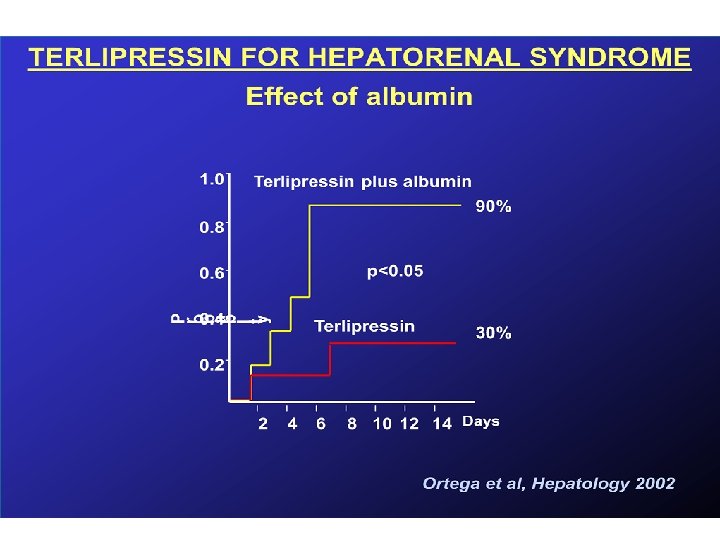
Therapy of Hepatorenal Syndrome: present and future • • Liver Transplantation Vasopressin Analogues α-Adrenergic Agonists Molecular Adsorbent Recirculating System
 • Rationale: increase of splanchnic and")
Therapy of Hepatorenal Syndrome: Vasopressin Analogues (Ornipressin, Terlipressin) • Rationale: increase of splanchnic and systemic vascular resistance, resulting in a redistribution of the circulating blood volume, via suppression of the RAA and sympathetic activities • Useful in combination with albumin and low-dose dopamin
 • Rationale: improvement of systemic vasoconstriction and")
Therapy of Hepatorenal Syndrome: α-Adrenergic Agonists (midodrine) • Rationale: improvement of systemic vasoconstriction and urinary sodium excretion • Effective in combination with plasma volume expansion and octreotide (an inhibitor of the release of endogenous vasodilators)
 INFEZIONI INTERCORRENTI")
HE: FATTORI PRECIPITANTI • • • ABUSO DI FARMACI (diuretici, sedativi, oppiati) INFEZIONI INTERCORRENTI ECCESSO DI ALCOL / PROTEINE EMORRAGIA DIGESTIVA INTERVENTI CHIRURGICI COPROSTASI IPERAZOTEMIA ALCALOSI IPOKALIEMICA HCC
")
HE: TERAPIA MEDICA • • IDENTIFICARE FATTORE PRECIPITANTE LIMITARE INTROITO PROTEICO LATTULOSIO (attenzione meteorismo!) ANTIBIOTICI TOPICI (Paromomicina, Rifaximina) • CLISTERINI MEDICATI sistematicamente!!!
- Slides: 102