Parts of the brain Sanjaya Adikari Department of

Parts of the brain Sanjaya Adikari Department of Anatomy
 Spinal cord Foramen magnum")
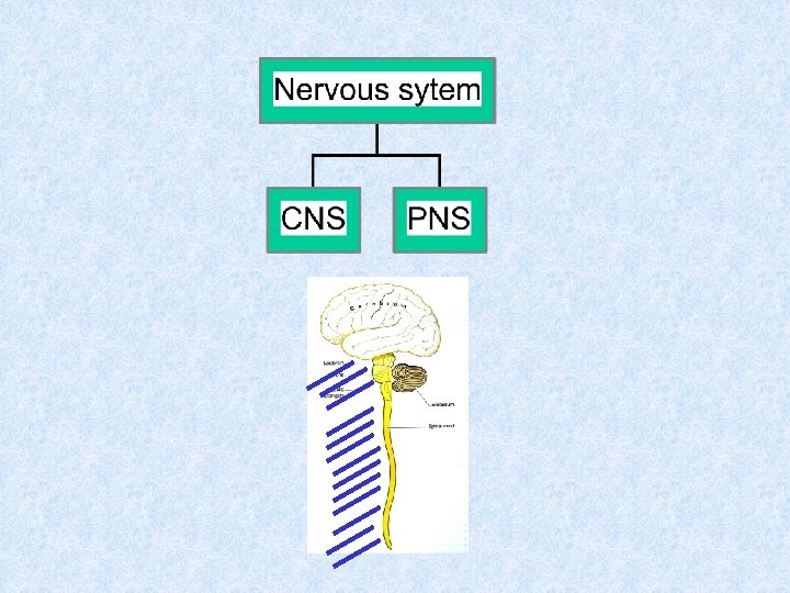
Central Nervous System (CNS) Spinal cord Foramen magnum

Skull Vertebral column

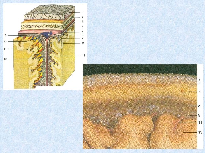
Meninges Dura mater Arachnoid mater Pia mater

Dura mater Arachnoid mater Ventricle Pia mater Ependymal cell layer

Main divisions of the brain 1 1 2 3 4 = forebrain + 2 6 5 = midbrain 3 4 6 5 = hindbrain

1 + 2 Cerebrum Diencephalon midbrain 3 pons 4 medulla = brainstem 5 6 Cerebellum

Cerebrum is the largest part of the brain. It is situated in the anterior and middle cranial fossae and the whole concavity of the vault of the skull Has two parts; • Cerebral hemispheres – Left & right cerebral hemispheres • Diencephalon – Consists of thalamus, hypothalamus etc.

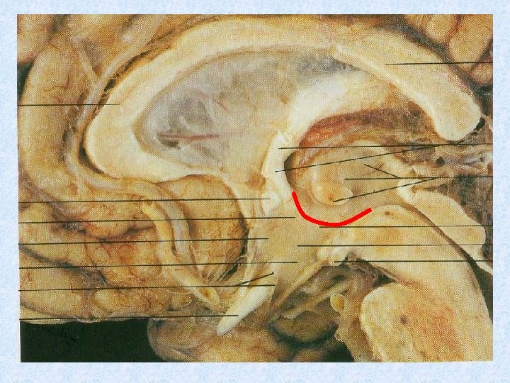
Mid-sagittal section of brain Diencephalon thalamus hypothalamus Spinal cord

Diencephalon thalamus hypothalamus Spinal cord

Cerebral hemispheres • Largest part of the brain • Separated by a deep mid-sagittal fisure called longitudinal cerebral fissure • The fissure contains the falx cerebri and anterior cerebral arteries • Tentorium cerebelli separates cerebral hemispheres from the cerebellum

Falx cerebri Anterior cerebral arteries Dura mater Tentorium cerebelli Corpus callosum

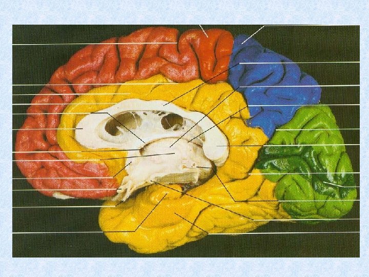
Sulci and Gyri • To increase the surface area of the brain the cerebral hemispheres are thrown into folds - gyri • The gyri are separated from each other by fissures - sulci • Hemispheres are divided into lobes (named according to the cranial bones under which they lie) by main sulci – Central – Parieto-occipital – Lateral
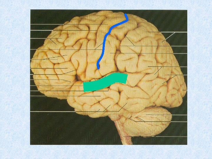

Central sulcus Frontal Lobe Parieto. Lateral sulcus Temporal Lobe occipital sulcus Occipital Lobe

Cranial Fossae

Main gyri • Precentral gyrus • Postcentral gyrus • Superior/middle/inferior frontal gyri • Superior/middle/inferior temporal gyri • Cingulate gyrus • Parahippocampal gyrus

Precentral gyrus Postcentral gyrus Superior, middle, inferior frontal gyri Superior, middle, inferior temporal gyri

Cingulate gyrus Parahippocampal gyrus

Functional areas of the brain

Homunculus

Homework • Draw a labelled line diagram to illustrate a midsagittal section of the brain including the brain stem. • Draw a labelled line diagram to illustrate a horizontal section of the cerebrum through the head of the caudate nucleus. Study the above diagrams before you come for the next lecture on parts of the brain

Lecture II

Gray matter and white matter Gray matter White matter • Gray matter consists of nerve cells • White matter consists of nerve fibres

Gray matter of the cerebral cortex • Five types of cells are organized into six cortical layers

Molecular layer External granular layer External pyramidal layer Internal granular layer Ganglionic layer (Internal pyramidal layer) Multiform layer

White matter • Composed of myelinated nerve fibres • Supported by neuroglia • Classified into three groups according to their connections – Commissural fibers – Association fibers – Projection fibers

Commissural fibers • Connects corresponding regions of the two hemispheres • Corpus callosum, fornix, anterior and posterior commissures • Corpus callosum, the largest commissure of the brain, is divided into rostrum, genu, body and the splenium

Corpus callosum Body Genu Splenium Rostrum

Association fibers • Connects various cortical regions within the same hemispheres • Divided into short and long groups • Short association fibers lie immediately beneath the cortex and connect adjacent gyri • Long association fibers are arranged into named bundles - fasciculi

Projection fibers • Afferent and efferent nerve fibers passing to and from the brain stem to the cerebral cortex • Internal capsule, corona radiata, optic radiation

Internal capsule and corona radiata Corona radiata Internal capsule Optic radiation Optic tract Cerebral peduncle Pyramidal tract

Internal capsule Putamen Cerebral peduncle Amygdala

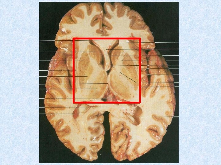
Basal ganglia • Basal ganglia are collection of masses of gray matter within the white matter of cerebral hemispheres Gray matter of cerebral cortex Basal ganglia White matter of cerebrum

Basal ganglia…. . cont. • Corpus striatum – Divided into two by internal capsule of white matter • Caudate nucleus • Lentiform nucleus (putamen & globus pallidus) • Amygdaloid • Claustrum

Basal ganglia…. . cont.
 Internal")
Thalamus Anterior horn of lateral ventricle Head of caudate nucleus Lentiform nucleus (putamen) Internal capsule Claustrum Lentiform nucleus (globus pallidus) Tail of caudate nucleus External capsule

Fiber tracts in the internal capsule Frontopontine Corticobulbar Corticospinal Thalamocortical Parieto/temporo/occipito pontine Visual & auditory

Basal ganglia…. . cont. • Some definitions include the following also under basal ganglia – Subthalamic nucleus – Substantia nigra Midbrain

Diencephalon • Consists of the following – Thalamus – Subthalamus – Hypothalamus – Epithalamus • Habenular nucleus • Pineal gland

Corpus callosum Fornix Thalamus Mamillary body

Thalamus Epithalamus Subthalamus Hypothalamus

Fornix Roof of 3 rd ventricle Interthalamic connection Lentiform nucleus Thalamus Internal capsule Hypothalamus Optic chiasma Pituitary Mammillary body

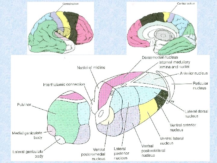
Thalamus • Large ovoid mass of gray matter • Forms large part of diencephalon • Very important cell station • Receives main sensory tracts (except olfactory pathway) • Integrates information it receives and relays to the cerebral cortex and subcortical regions • Integrates visceral and somatic functions

Thalamic nuclei

Hypothalamus • Part of the diencephalon that extends from the optic chiasma to the posterior border of the mammillary bodies • Almost all physiological activities of the body are influenced by hypothalamus – Integration of autonomic functions – Regulation of endocrine functions – Maintaining body homiostasis – Regulation of body temperature and body fluids – Sexual behaviour, emosions, drive to eat and drink

Hypothalamus…. • Contains some important cell groups – Supraoptic nucleus – Paraventricular nucleus • These have axons running down into the posterior lobe of the pituitary gland • Other cell groups deliver their neurosecretions into the hypothalamo-hypophyseal poryal system leading to the anterior lobe of the pituitary gland
 • Projects backwards from the diencephalon to lie posterior to")
Pineal gland (Pineal body) • Projects backwards from the diencephalon to lie posterior to the midbrain • Progressive calcification with age becoming visible in x-ray • Produces metatonin hormone in a circadian rhythm influenced by light • Indirectly controls the function of other endocrine organs, including the pituitary

Lecture III

Development of the central nervous system Sanjaya Adikari Department of Anatomy

Neural tube & cavity
 2 Mesencephalon (midbrain) 3 Rhombencephalon (hindbrain) 1 a")
Development of brain 1 Prosencephalon (forebrain) 2 Mesencephalon (midbrain) 3 Rhombencephalon (hindbrain) 1 a 1 b 2 3 a 3 b

1 a 1 b Telencephalon Cerebral hemispheres Diencephalon Thalamus, hypothalamus subthalamus, epithalamus 2 3 a 3 b Midbrain Metencephalon Myelencephalon tectum, tegmentum and cerebral pedunculi pons, upper medulla and cerebellum lower medulla

Lateral ventricle Foramen of Monro 3 rd ventricle Aqueduct 4 th ventricle Central canal of SC

Development of spinal cord and brain stem Mantle layer Marginal layer Neuroepithelial layer

1 Neuroblast cells Neurons Neuroepithelial cells 2 Gliablast cells Glial cells Ependymal cells Oligodendroglia Astroglia Microglia
 Marginal layer Sulcus limitans Neuroepithelial layer Basal plate (motor)")
Mantle layer Alar plate (sensory) Marginal layer Sulcus limitans Neuroepithelial layer Basal plate (motor)

Sensory Motor Posterior horn Lateral horn Sensory Motor Anterior horn

SE BE VE VA BA GSA

Brainstem • Consists of midbrain, pons and medulla oblongata • Broad functions – Conduit for ascending and descending tracts – Contains reflex control centers of respiratory and cardiovascular systems – Associated with control of consciousness – Contains nuclei of cranial nerves III to XII

Midbrain • About 2 cm in length • Connects the pons and cerebellum to forebrain by passing through the opening of tentorium cerebelli • On the posterior surface are superior and inferior colliculi – SC: centers for visual reflexes. Connected to lateral geniculate body – IC: lower auditory centers. Connected to medial geniculate body • On the anterior surface are crus cerebri. Contains cerebral aqueduct • Contains cranial nerve nuclei III and IV

Transverse section of midbrain Tectum Tegmentum Crus cerebri or cerebral peduncles

Transverse sections of midbrain Superior colliculus Cerebral aqueduct Red nuclues Substantia nigra Cerebral peduncle

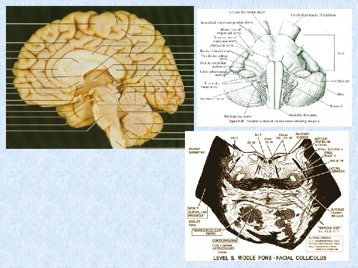
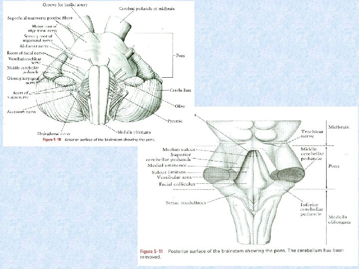
Pons • Anterior to the cerebellum • Bridges the two hemispheres of the cerebellum • On the anterior surface many transverse fibers that go into the middle cerebellar peduncle • Basilar groove in the midline anteriorly • Motor and sensory roots of trigeminal nerve emerge from anterior surface • Posterior surface contains the upper part of the floor of the 4 th ventricle above

Medulla oblongata • Connects to the spinal cord at the level of the foramen magnum, where the first cervical spinal nerve roots start • Anterior surface contains the pyramids and the olives • Posterior surface contains cuneate and gracile tubercles below and the lower part of the floor of the 4 th ventricle above • Laterally are the inferior cerebellar peduncles

Motor and sensory decussations of medulla sensory Posterior surface motor Anterior surface

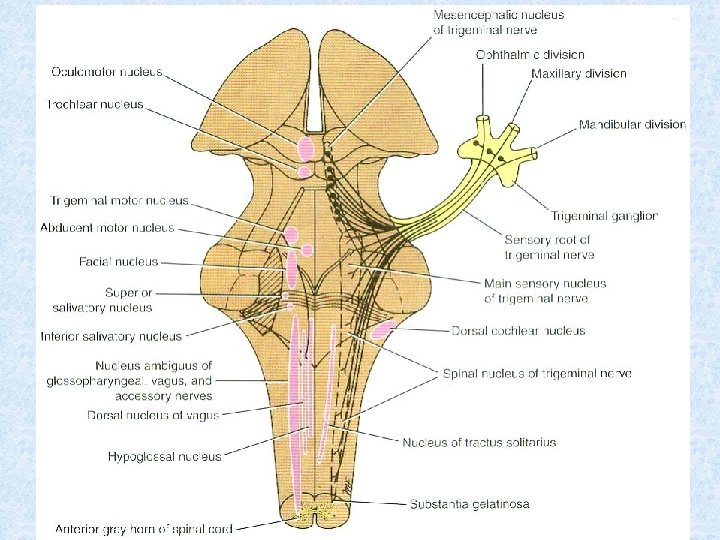
Arrangement of cranial nerve nuclei in the flow of the 4 th ventricle • Extensive lateral spread of 4 th ventricle during embryonic development has caused the alar plates to lie lateral to the basal plates • Somatic motor (efferent) nuclei lie closer to the midline and somatic sensory (afferent) nuclei lie away from the midline • Visceral nuclei lie in between, with visceral motor more closer and visceral sensory more distant from the midline

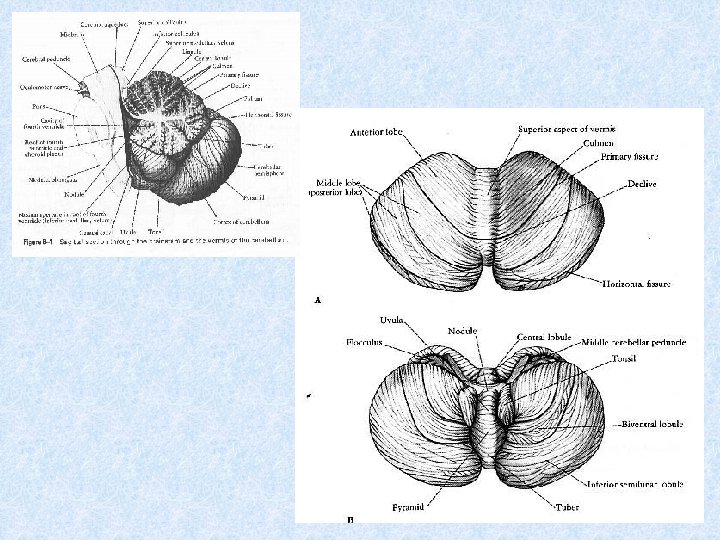
Cerebellum • Lies posterior to the 4 th ventricle, pons and medulla • Two cerebellar hemispheres joined by a narrow median vermis • Connected to brain stem by superior, middle and inferior cerebellar peduncles

Cerebellum…. . • Has 3 lobes – anterior, middle and flocculonodular • Primary fissure lies between anterior and middle lobes • Uvulonodular fissure lies between middle and flocculonodular lobes • Horizontal fissure lies within the middle lobe separating superior and inferior surfaces

Cerebellum…cont. • Has an outer cortex of cells. It has 3 layers – granular, purkinje and molecular layers • Four intracerebellar nuclei – dentate, emboliform, globose and fastigial • Afferent fibers enter through middle and inferior cerebellar peduncles • Efferent fibers (axons of purkinje cells) synapse with cerebellar nuclei. Efferents from the nuclei leave through the superior cerebellar peduncle

Granular cell layer Purkinje cell layer Molecular cell layer

Emboliform nucleus Dentate nucleus Middle cerebellar peduncle

Function of the cerebellum

Cerebellar ataxia • Diseases of the lateral cerebellar lobes – Limb ataxia – Gait broad-based and veered towards the side of the lesion • Diseases of the cerebellar vermis – Truncal ataxia – Tendency to fall backwards/sideways – No limb ataxia

Limbic system • Functionally important but not well defined anatomically • Surrounds the corpus callosum and the diencephalon • Deals with behaviour, imotion and memory

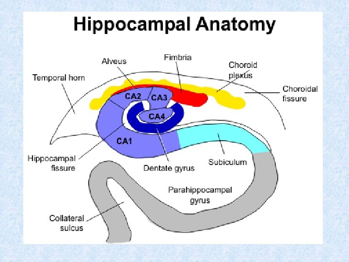
Components of the limbic system • Cingulate gyrus • Subcallosal gyrus • Septal area and olfactory bulb • Hippocampal formation – Hippocampal gyrus – Dentate gyrus – Parahippocampal gyrus • Amygdaloid body and mammillary body • Anterior nucleus of thalamus • Hypothalamus

Parts and connecting pathways

Ventricular system of the brain

Formation of CSF 1. Formed mainly in choroid plexus of the ventricles 2. Some is formed from the ependymal cells lining the ventricles and at perivascular spaces Blood-CSF barrier

Choroid plexus of lateral ventricle

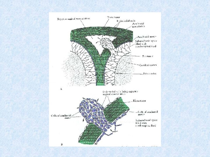
Circulation of CSF Arachnoid granulations Foramina of Luschka & Magendie

Lecture IV

How to draw different sections of the brain stem

Posterior surface Anterior surface

Main nerve tracts in the spinal cord B A C G F E D

A. Posterior white columns B. Lateral corticospinal tract C. Anterior & posterior spinocerebellar tracts D. Anterior & posterior spinothalamic tracts E. Olivospinal, vestibulospinal, tectospinal tracts F. Anterior corticospinal tract G. Rubrospinal tract

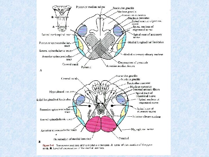
Closed medulla at the level of decussation of pyramids Spinal tract of trigeminal nerve Fasciculus gracilis Nucleus gracilis Fasciculus cuneatus Nucleus cuneatus Spinal nucleus of trigeminal nerve Spinal root of the accessory nerve Posterior spinocerebellar tract Central canal Spinothalamic tract Medial longitudinal fasciculus Anterior spinocerebellar tract Decussation of pyramids Pyramid

Closed medulla at the level of decussation of medial lemnisci Fasciculus gracilis Spinal tract of trigeminal nerve Nucleus gracilis Fasciculus cuneatus Nucleus cuneatus Spinal nucleus of trigeminal nerve Central canal Medial longitudinal fasciculus Posterior spinocerebellar tract Spinal root of the accessory nerve Spinothalamic tract Anterior spinocerebellar tract Decussation of medial lemnisci Hypoglossal nerve Pyramid

Medulla Oblongata at the level of middle of the olivary nuclei Inferior medullary velum Cavity of 4 th ventricle Medial longitudinal fasciculus Spinal tract & nucleus of trigeminal nerve Vestibular & cochlear nuclei Inferior cerebellar peduncle Vagus nerve Anterior spinocerebellar tract Reticular formation Spinothalamic tract Olivary nucleus Tectospinal tract Olive Medial lemniscus Pyramid Hypoglossal nerve

Pons at the level of facial colliculus Cavity of 4 th ventricle Superior medullary velum Medial longitudinal fasciculus Superior cerebellar peduncle Facial colliculus Reticular formation Vestibular nuclei Inferior cerebellar peduncle Spinal lemniscus Spinal tract & nucleus of trigeminal nerve Medial lemniscus Transverse pontine fibres Facial nerve Abducent nerve Corticospinal & corticonuclear fibres Pontine nuclei

Midbrain at the level of inferior colliculus Periaqueductal Gray Matter Cerebral aqueduct Trigeminal lemniscus Spinal lemniscus Trochlear nerve Inferior colliculus Mesencephalic nucleus of trigeminal nerve Medial longitudinal fasciculus Medial lemniscus Substantia nigra Reticular formation Cerebral peduncle Interpeduncular fossa Decussation of superior cerebellar peduncles

Midbrain at the level of superior colliculus Periaqueductal Gray Matter Cerebral aqueduct Superior colliculus Mesencephalic nucleus of trigeminal nerve Trigeminal lemniscus Spinal lemniscus Medial longitudinal fasciculus Medial lemniscus Substantia nigra Reticular formation Cerebral peduncle Red nucleus Oculomotor nerve

Medial longitudinal fasciculus PPRF = Paramedian pontine reticular formation
- Slides: 106